. 2008 Dec;82(23):11568-76.
doi: 10.1128/JVI.01318-08. Epub 2008 Sep 25.

Cell-restricted immortalization by human papillomavirus correlates with telomerase activation and engagement of the hTERT promoter by Myc

Affiliations

Cell-restricted immortalization by human papillomavirus correlates with telomerase activation and engagement of the hTERT promoter by Myc

Xuefeng Liu et al. J Virol. 2008 Dec.

Abstract

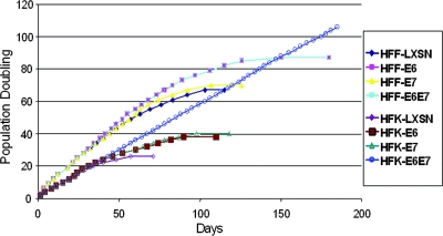
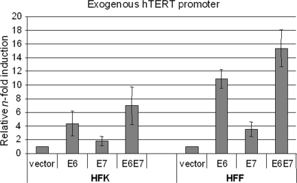
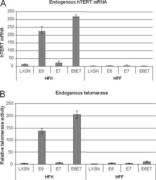
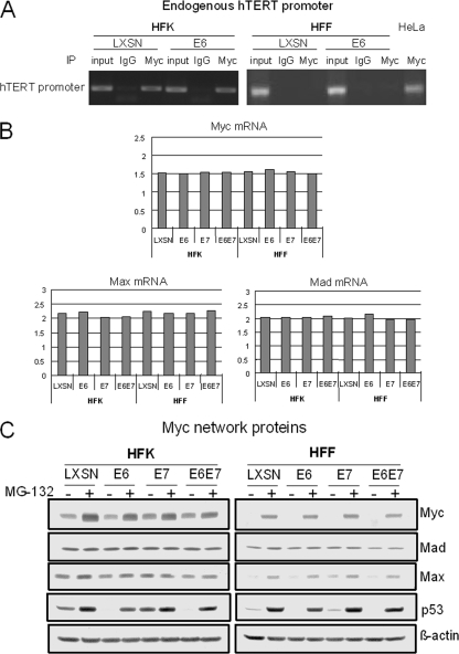
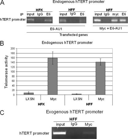
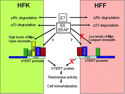
The high-risk human papillomaviruses (HPVs) are the causative agents of nearly all cervical cancers and are etiologically linked to additional human cancers, including those of anal, oral, and laryngeal origin. The main transforming genes of the high-risk HPVs are E6 and E7. E6, in addition to its role in p53 degradation, induces hTERT mRNA transcription in genital keratinocytes via interactions with Myc protein, thereby increasing cellular telomerase activity. While the HPV type 16 E6 and E7 genes efficiently immortalize human keratinocytes, they appear to only prolong the life span of human fibroblasts. To examine the molecular basis for this cell-type dependency, we examined the correlation between the ability of E6 to transactivate endogenous and exogenous hTERT promoters and to immortalize genital keratinocytes and fibroblasts. Confirming earlier studies, the E6 and E7 genes were incapable of immortalizing human fibroblasts but did delay senescence. Despite the lack of immortalization, E6 was functional in the fibroblasts, mediating p53 degradation and strongly transactivating an exogenous hTERT promoter. However, E6 failed to transactivate the endogenous hTERT promoter. Coordinately with this failure, we observed that Myc protein was not associated with the endogenous hTERT promoter, most likely due to the extremely low level of Myc expression in these cells and/or to differences in chromatin structure, in contrast with hTERT promoters that we found to be activated by E6 (i.e., the endogenous hTERT promoter in primary keratinoctyes and the exogenous hTERT core promoter in fibroblasts), where Myc is associated with the promoter in either a quiescent or an E6-induced state. These findings are consistent with those of our previous studies on mutagenesis and the knockdown of small interfering RNA, which demonstrated a requirement for Myc in the induction of the hTERT promoter by E6 and suggested that occupancy of the promoter by Myc determines the responsiveness of E6 and the downstream induction of telomerase and cell immortalization.


Figures

FIG. 1.
FIG. 1.
HPV E6 and E7 oncoproteins are sufficient to immortalize primary HFKs but not primary HFFs. Primary HFKs and HFFs were transduced with the indicated pLXSN-based retroviruses with E6, E7, E6E7 or an empty vector and selected as previously described. Cultures were passed continuously in vitro as described in the text, and the number of cell doublings was calculated and plotted versus the time in culture. Cultures that did not proliferate and expand in 20 days for HFKs and 30 days for HFFs were considered senescent and were terminated at the indicated times. This experiment was repeated a second time with similar results.
FIG. 2.
FIG. 2.
Characterization of HFK and HFF strains expressing HPV E6 and E7 proteins. (A) Diagram of HPV-16 E6 and E7 mRNA expression and locations of primers used in this study. The E6 and E7 open reading frames are shown as open boxes. The dotted lines flanking the open boxes represent vector sequences. The alternative splicing sites in E6 are depicted as dotted lines. The numbers above the E6E7 transcripts are the nucleotide positions of the first nucleotide of the start codon and the last nucleotide of the stop codon of both E6 and E7 or the first nucleotide of the 5′ splicing site (5′ ss) or the last nucleotide of the 3′ splicing sites (3′ ss) in the HPV-16 genome. The primers used in this study are depicted below the transcript lines as arrows and numbers showing the locations (as nucleotide [nt] positions) of primers in the genome. (B) Confirmation of E6 and E7 mRNA expression. Primary HFKs and HFFs were transduced with pLXSN expressing 16E6, E7, E6E7, or an empty vector as previously described. Following antibiotic selection, the cell strains were analyzed for E6 and E7 mRNAs. Total cellular RNA was isolated from the transduced cell strains and treated with a DNA-free kit (Ambion), and RT-PCR was performed with the sets of HPV-16 unspliced E6- and E7-specific primers described in Materials and Methods. GAPDH mRNA was detected as an internal control. PCR products were analyzed on 2% agarose gels. (C) Expression of p53 and pRb proteins. The stable cell lines described above were lysed in electrophoresis sample buffer. The proteins were separated on 4- to 20%-gradient gels, transferred to a PVDF membrane, and reacted with mouse anti-p53 monoclonal antibody or rabbit anti-pRb. Anti-β-actin antibody was used to verify equal loading of samples. Low amounts of p53 protein were observed in E6- and E6E7-expressing cells, and a decreased level of pRb protein was noted in E7-expressing cells.
FIG. 3.
FIG. 3.
E6 and E7 induce an exogenous core hTERT promoter in both HFKs and HFFs. Primary HFKs and HFFs were cotransfected with wild-type hTERT core promoter (pGL3B-hTERT) and either E6, E7, or E6E7. The pRL-CMV R. reniformis reporter plasmid was also transfected into the cells to standardize for transfection efficiency. Luciferase activity was measured 24 h after transfection, using the Dual-Luciferase Reporter assay system (Promega). Relative n-fold activation reflects normalized luciferase activity induced by E6 and E7 compared to the normalized activity of the control vector. The value of pGL3B-hTERT activity with the empty vector was set to 1. Error bars show standard deviations for at least three independent experiments. E6 is sufficient to induce the hTERT promoter in both HFK and HFF cells.
FIG. 4.
FIG. 4.
E6 is sufficient to induce endogenous hTERT transcription and telomerase activity in primary HFKs but not in HFFs. (A) hTERT mRNA expression in stable keratinocyte and fibroblast cell lines. RNAs were used for detection of hTERT mRNA by qRT-PCR as described in Materials and Methods. A considerable amount of hTERT mRNA was detected in HFKs expressing E6 or both E6 and E7, but there was no detectable mRNA in HFFs expressing either the control vector (LXSN), E6, E7, or both E6 and E7. (B) Telomerase activity. A q-TRAP assay was done as described in Materials and Methods. Telomerase activity was observed in HFKs expressing E6 or both E6 and E7. There was no detectable telomerase activity in HFFs expressing either the control vector, E6, E7, or both E6 and E7.
FIG. 5.
FIG. 5.
Myc expression and occupancy on the hTERT promoter differ in HFKs and HFFs. (A) Myc binds to endogenous hTERT promoter in primary HFKs but not in primary HFFs. Myc binding to the endogenous hTERT promoter was assayed by ChIP. Myc binding to the hTERT promoter was evaluated in the absence and presence of E6. HeLa cells were used as positive controls for Myc binding to the endogenous hTERT promoter. Myc binds to the endogenous hTERT promoter in HFKs with or without E6, but it does not bind to the promoter in primary HFFs. (B) Quantitation of Myc, Mad, and Max mRNAs. RNAs were subjected to Sybr green-based real-time RT-PCR on a Bio-Rad iQ5 system according to the manufacturer's instructions. GAPDH was used as an internal control, and data were analyzed using the normalized expression (ΔΔCT) method according to the manufacturer's (Bio-Rad's) guidelines. (C) Expression of Myc, Max, Mad, and p53 proteins. HFKs and HFFs expressing either the control vector, E6, E7, or both E6 and E7 were plated into 100-mm dishes in duplicate. After cells reached 80 to 90% confluence, a set of cells was treated with MG-132 for 4 h. All cell extracts were harvested with 2× SDS sample buffer. The same amounts of cell extracts were loaded onto SDS-4 to 20% polyacrylamide gels for electrophoresis, and protein was transferred to a PVDF membrane and blotted with anti-Myc, anti-Max, anti-Mad, and β-actin. Expression of both Myc and Max was lower in HFFs than in HFKs. There was no significant difference in the levels of Mad in HFKs and HFFs.
FIG. 6.
FIG. 6.
Myc and E6 associate with the hTERT promoter in HFFs. (A) Myc-mediated E6 binding to hTERT promoter. HFKs or HFFs transfected with E6-AU1 or both Myc and E6-AU1 were subjected to ChIP assays as described above, using rabbit anti-Myc antibody (N262; Santa Cruz Biotechnology) and monoclonal anti-AU1 antibody (Covance). (B) Myc induces telomerase activity in both HFKs and HFFs. HFKs and HFFs were transduced with pLXSN-Myc retrovirus, and cell lysates were analyzed with q-TRAP assays. Myc alone induces a similar level of telomerase activity in both HFKs and HFFs. (C) Endogenous Myc binds to exogenous hTERT promoter in HFFs. The plasmid pGL3B-hTERT was transfected to HFFs, and a Myc antibody or rabbit IgG was used for IP.
FIG. 7.
FIG. 7.
A proposed model for E6 regulation of the hTERT promoter and cell immortalization. Using information from this and previous studies (12, 14, 30, 57, 58, 62, 64), we propose a model to explain how HPV E6 might regulate the hTERT promoter in a cell-type-specific manner. The ability of E6 and E7 to target p53 and Rb in HFF and HFK cells does not differ. However, the low levels of Myc protein in fibroblasts (HFF) relative to keratinoctyes (HFK) is probably a major determinant for the lack of Myc on the endogenous HFF hTERT promoter. Contributions of chromatin structure, however, might also contribute to the limited access of Myc and other regulatory proteins to the HFF hTERT promoter. Since E6 can bind to the HFF hTERT promoter in the absence of detectable Myc, it appears that other proteins resident on this site might mediate its binding. In separate studies, for example, it has been documented that E6 interacts with Myc, BRCA1, and NFX-1.

References

    1. Androphy, E. J., N. L. Hubbert, J. T. Schiller, and D. R. Lowy. 1987. Identification of the HPV-16 E6 protein from transformed mouse cells and human cervical carcinoma cell lines. EMBO J. 6989-992. - PMC - PubMed
    1. Banks, L., P. Spence, E. Androphy, N. Hubbert, G. Matlashewski, A. Murray, and L. Crawford. 1987. Identification of human papillomavirus type 18 E6 polypeptide in cells derived from human cervical carcinomas. J. Gen. Virol. 681351-1359. - PubMed
    1. Bedard, K. M., M. P. Underbrink, H. L. Howie, and D. A. Galloway. 2008. The E6 oncoproteins from human betapapillomaviruses differentially activate telomerase through an E6AP-dependent mechanism and prolong the lifespan of primary keratinocytes. J. Virol. 823894-3902. - PMC - PubMed
    1. Benanti, J. A., M. L. Wang, H. E. Myers, K. L. Robinson, C. Grandori, and D. A. Galloway. 2007. Epigenetic down-regulation of ARF expression is a selection step in immortalization of human fibroblasts by c-Myc. Mol. Cancer Res. 51181-1189. - PubMed
    1. Casillas, M. A., S. L. Brotherton, L. G. Andrews, J. M. Ruppert, and T. O. Tollefsbol. 2003. Induction of endogenous telomerase (hTERT) by c-Myc in WI-38 fibroblasts transformed with specific genetic elements. Gene 31657-65. - PubMed

Publication types

MeSH terms