. 2008 Sep 26;321(5897):1837-41.
doi: 10.1126/science.1163600.

Disruption of the CFTR gene produces a model of cystic fibrosis in newborn pigs

Affiliations

Disruption of the CFTR gene produces a model of cystic fibrosis in newborn pigs

Christopher S Rogers et al. Science. .

Abstract

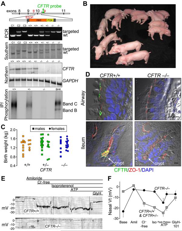
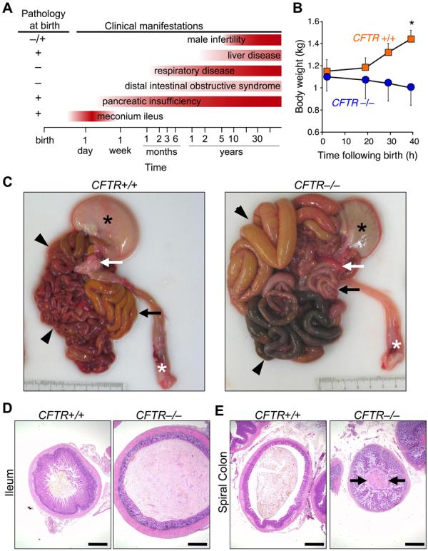
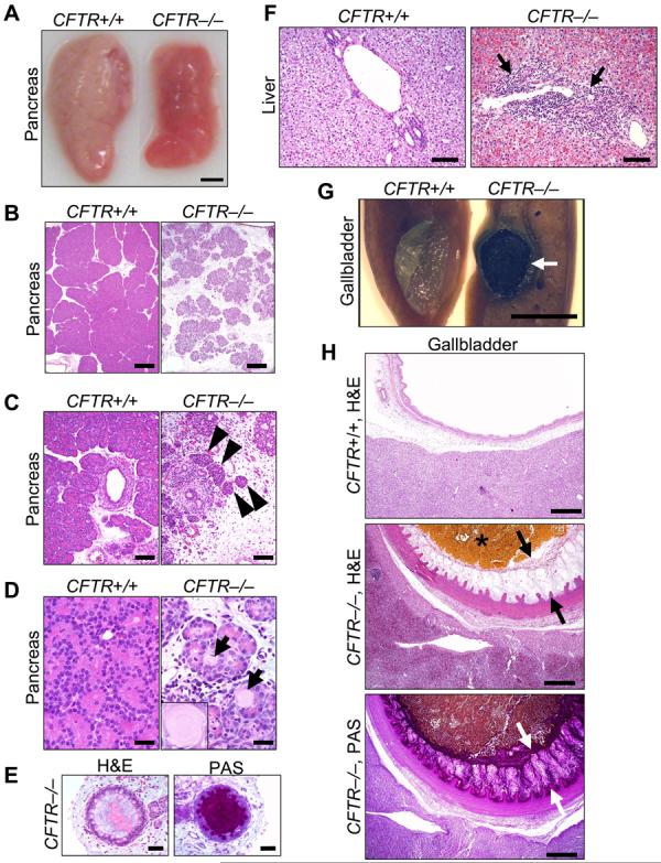
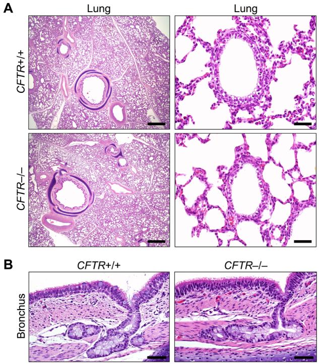
Almost two decades after CFTR was identified as the gene responsible for cystic fibrosis (CF), we still lack answers to many questions about the pathogenesis of the disease, and it remains incurable. Mice with a disrupted CFTR gene have greatly facilitated CF studies, but the mutant mice do not develop the characteristic manifestations of human CF, including abnormalities of the pancreas, lung, intestine, liver, and other organs. Because pigs share many anatomical and physiological features with humans, we generated pigs with a targeted disruption of both CFTR alleles. Newborn pigs lacking CFTR exhibited defective chloride transport and developed meconium ileus, exocrine pancreatic destruction, and focal biliary cirrhosis, replicating abnormalities seen in newborn humans with CF. The pig model may provide opportunities to address persistent questions about CF pathogenesis and accelerate discovery of strategies for prevention and treatment.


Figures

Figure 1
Figure 1. CFTR−/− piglets appear normal at birth
A) Upper panel depicts insertion into porcine CFTR exon 10 of a PGK promoter (yellow) driving a neomycin resistance cDNA (orange), and an engineered stop codon. Position of probe (green), PCR primers (arrowheads) and BglII sites (B) are indicated. Second and third panels show genotyping by PCR and Southern blot of genomic DNA. Lanes C1, C2, and C3 contain controls of CFTR+/+, +/− and −/− DNA. Fourth panel shows northern blot of ileal CFTR and GAPDH mRNA. Consistent with the northern blot, quantitative RT-PCR of exon 10, the targeted site, detected <0.1% of CFTR transcripts in CFTR −/− ileum relative to CFTR+/+ (n=6 and 4). Fifth panel shows immunoprecipitation and phosphorylation of CFTR plus recombinant CFTR in BHK cells. B) First litter containing piglets of all three genotypes. C) Birth weights. Mean±SD of weights: 1.31±0.24 kg for CFTR+/+, 1.35±0.28 kg CFTR+/−,and 1.31±0.23 kg CFTR−/−. D) Immunocytochemistry of CFTR in airway epithelia (top) and ileum (bottom). Figures are differential interference contrast with staining for ZO-1 (a component of tight junctions, red), CFTR (green), and nuclei (DAPI, blue). See also Fig. S1. Bars, 10 μm. E) Tracings of in vivo nasal voltage (Vt) measured in newborn piglets. After baseline measurements, the following agents/solutions were sequentially added to the epithelial perfusate: amiloride (100 μM), Cl-free solution, isoproterenol (10 μM), ATP (100 μM), and GlyH-101 (100 μM). F) Average nasal Vt measurements as indicated in panel E. Data from 4 CFTR+/+ and 4 CFTR+/− piglets were not statistically different and were combined and compared to data from 5 CFTR−/− piglets. Values of baseline nasal Vt for CFTR−/− piglets differed from the controls, as did the changes in Vt induced by adding amiloride, a Cl-free solution, and GlyH-101 (all P<0.05). Data are mean±SEM.
Figure 2
Figure 2. CFTR−/− piglets develop meconium ileus
A) Schematic shows some clinical and histopathological CF manifestations. Note that pathological abnormalities are present before clinical disease becomes apparent. B) Weight following birth. Animals were fed colostrum and milk-replacer. n = 7 CFTR+/+ and 4 CFTR−/−. Data are mean±SEM. *P<0.05. C) Gross appearance of gastrointestinal tract. Piglets were fed colostrum and milk-replacer for 30-40 h and then euthanized. Stomach (black *), small intestine (arrowheads), pancreas (white arrow), rectum (white *), and spiral colon (black arrow). Of 16 CFTR−/− piglets, the obstruction occurred in small intestine in 7 and spiral colon in 9. D, E) Microscopic appearance of the ileum (D) and colon (E). H&E stain. Bars, 1 mm. Images are representative of severe meconium ileus occurring in 16 of 16 CFTR−/− piglets.
Figure 3
Figure 3. CFTR−/− piglets have exocrine pancreatic destruction and liver and gallbladder abnormalities
A) Gross appearance of pancreas. Bar, 0.5 cm. B) Loss of parenchyma in the CFTR−/− pancreas. H&E stain. Bars, 500 µm. C) Pancreatic ducts and islets of Langerhans (arrowheads). Bars, 100 µm. D) CFTR−/− ductules and acini dilated by eosinophilic inspissated material that formed concentrically lamellar concretions (arrows and insert). H&E stain. Bars, 33 µm. E) Ducts within the CFTR−/− pancreas. H&E stain, left; PAS stain, right. Bars, 50 µm. F) Microscopic appearance of liver. H&E stain. Arrows indicate focal expansion of portal areas by chronic cellular inflammation. Bars, 100 µm. G) Gross appearance of gallbladder. When the CFTR+/+ gallbladder was sectioned, bile drained away rapidly with collapse of the mucosal wall. CFTR−/− bile was congealed (arrow) and retained in the lumen of a smaller gallbladder. Bar, 0.5 cm. H) Microscopic appearance of gallbladder. CFTR−/− gallbladders had congealed, inspissated bile with variable mucus production (arrows, H&E stain) highlighted as a magenta color in periodic acid-Schiff (PAS) stained tissue. Bars, 500 µm. Images are representative of severe pancreatic lesions (15/15 CFTR−/− piglets), mild to moderate liver lesions (3/15), and mild to severe gall bladder/duct lesions (15/15).
Figure 4
Figure 4. The lungs of newborn CFTR−/− and CFTR+/+ piglets appear normal
A) Microscopic appearance of lung from piglets <12 hr old. H&E staining. Bars, 1 mm (left) and 50 µm (right). B) Bronchial epithelia and submucosal glands. H&E staining. Bars, 50 µm. Images are representative of lack of lesions in 15 of 15 CFTR−/−.

Comment in

  • Are pigs more human than mice?
    Elferink RO, Beuers U. Elferink RO, et al. J Hepatol. 2009 Apr;50(4):838-41. doi: 10.1016/j.jhep.2008.12.014. Epub 2009 Jan 9. J Hepatol. 2009. PMID: 19231004 No abstract available.

References

    1. Andersen DH. Am. J. Dis. Child. 1938;56:344.
    1. Quinton P. Physiol. Rev. 1999;79:S3. - PubMed
    1. Welsh MJ, Ramsey BW, Accurso F, Cutting GR. In: The Metabolic and Molecular Basis of Inherited Disease. Scriver CR, et al., editors. McGraw-Hill; New York: 2001. pp. 5121–5189.
    1. Rowe SM, Miller S, Sorscher EJ. N. Engl. J. Med. 2005 May 12;352:1992. - PubMed
    1. Riordan JR, et al. Science. 1989;245:1066. - PubMed

Publication types

MeSH terms

Substances