. 2008 Nov;173(5):1361-8.
doi: 10.2353/ajpath.2008.080444. Epub 2008 Sep 25.

Hyaluronan-mediated leukocyte adhesion and dextran sulfate sodium-induced colitis are attenuated in the absence of signal transducer and activator of transcription 1

Affiliations

Hyaluronan-mediated leukocyte adhesion and dextran sulfate sodium-induced colitis are attenuated in the absence of signal transducer and activator of transcription 1

Sudip K Bandyopadhyay et al. Am J Pathol. 2008 Nov.

Abstract

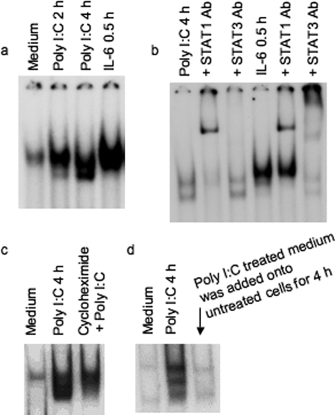
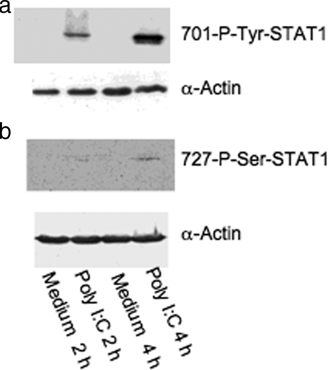
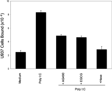
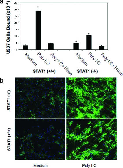
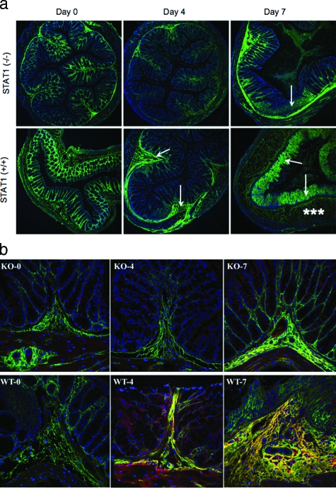
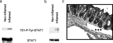
Inflammatory bowel disease is a chronic inflammatory condition of the intestinal mucosa whose etiology is unclear but is likely to be multifactorial. We have shown previously that an increased amount of hyaluronan (HA) is present both in the inflamed mucosa of inflammatory bowel disease patients and in isolated human cells after polyI:C treatment. The signal transducer and activator of transcription (STAT)1 protein plays an important role in many signaling pathways that are associated with inflammation. We therefore investigated the role of STAT1 in adhesive interactions that occur between leukocytes and polyI:C-induced mucosal smooth muscle cells (M-SMCs). Activation of STAT1 was observed after the polyI:C treatment of M-SMCs. Specific phosphorylation of tyrosine and serine residues of STAT1 was observed in polyI:C-treated, but not untreated, M-SMC cultures. To evaluate further the role of STAT1, a corresponding STAT-1-null mouse was used. PolyI:C-induced, HA-mediated leukocyte adhesion to colon SMCs from STAT1-null mice was significantly decreased compared with that from wild-type control mice. In vivo, using the dextran sulfate sodium-induced model of colon inflammation, both tissue damage and HA deposition were attenuated in STAT1-null mice compared with that in wild-type control mice. Additionally, the inter-alpha-trypsin inhibitor (IalphaI), a proteoglycan essential for facilitating leukocyte binding to the HA matrix, was reduced in STAT1-null mice. Together, these results demonstrate that STAT1 plays an important role in HA-mediated inflammatory processes.


Figures

Figure 1
Figure 1
STAT1 activation by poly I:C treatment in M-SMCs. Electrophoretic mobility shift assays using labeled hSIE as a probe with equal amounts of whole cell extracts (15 μg) prepared from cells in the presence of 2% fetal bovine serum as indicated in figures. a: Activation of STAT protein in 2- or 4-hour poly I:C-treated and 30-minute IL-6-treated extracts. b: Supershift analyses with 4-hour poly I:C-treated extract or with 30-minute IL-6-treated extract were performed after pre-incubation with STAT1- or STAT3-specific antibody. c: Cells treated with and without poly I:C and cycloheximide 30 minutes before the poly I:C treatment. d: Cultured M-SMCs were treated for 4 hours with medium obtained from 4-hour poly I:C-treated M-SMCs. Several exposures were taken from the autoradiogram and one of the exposures was used for this figure.
Figure 2
Figure 2
Phosphorylation of STAT1 by Western blots. M-SMCs were serum-starved for 18 hours and then treated for 2 or 4 hours with poly I:C in 2% serum-containing medium as indicated and equal protein (30 μg) amount of cellular extracts were used for Western blot analyses using phosphor-specific antibodies 701-tyrosine (a) and 727-serine (b) residues of STAT1 protein. The same blot was then reused with α-actin antibody to compare equal loading of each sample.
Figure 3
Figure 3
Reduced U937 cell attachment to poly I:C-stimulated M-SMCs with Jak or STAT inhibitors. M-SMCs were untreated or treated with poly I:C for 18 hours in the presence or absence of 100 μmol/L of AG490 or EGCG as indicated. Labeled U937 cells were used for the binding assay as described in Materials and Methods section.
Figure 4
Figure 4
Poly I:C-stimulated HA-mediated cell attachment is less in STAT1-null SMCs compared to wild-type cells. a: Isolated SMCs from STAT1 wild-type (+/+) and null (−/−) mice were treated with or without poly I:C for 18 hours and a quantitative assay was performed using labeled U937 cells for attachment as described in the Materials and Methods. b: Parallel cultures on coverslips were treated similarly, methanol-fixed, and stained for HA (green) and nuclei (blue, 4,6-diamidino-2-phenylindole) as described in the Materials and Methods section.
Figure 5
Figure 5
Reduced HA deposition and less inflammatory changes in STAT1−/− mice after DSS treatment. a: Confocal image demonstrates HA polymers (green) in mouse colon sections after DSS treatment in STAT1−/−-null (top) and STAT1+/+ wild-type animals (bottom). The null mice section demonstrates minimal staining on day 4 and moderate staining on day 7 that is localized to the submucosa (arrow), whereas the wild-type mice have moderate staining of the submucosa (arrow) on day 4 and heavy staining of the lamina propria (asterisks) on day 7; cells are identified by their nuclei (blue). b: HA (green) and IαI (red) staining in colon designated as wild-type STAT1 (WT) and STAT1-null mice (KO) sections after a 4- or 7-day treatment with DSS.
Figure 6
Figure 6
Tyrosine phosphorylation of STAT1 in inflamed whole tissue extracts and specifically in muscularis mucosa. Western blot analyses using equal amounts of protein from whole cell extracts (50 μg) (a) and equal volume of laser-dissected muscularis mucosa extracts (b) from both noninflamed and inflamed area from same patient sample are shown with 701-phospho-specific tyrosine antibody. c: Microscopic picture demonstrated by asterisks that the microdissected cells derived from the muscularis mucosa.

References

    1. Beagley KW, Elson CO. Cells and cytokines in mucosal immunity and inflammation. Gastroenterol Clin North Am. 1992;21:347–366. - PubMed
    1. Becker C, Dornhoff H, Neufert C, Fantini MC, Wirtz S, Huebner S, Nikolaev A, Lehr HA, Murphy AJ, Valenzuela DM, Yancopoulos GD, Galle PR, Karow M, Neurath MF. Cutting edge: IL-23 cross-regulates IL-12 production in T cell-dependent experimental colitis. J Immunol. 2006;177:2760–2764. - PubMed
    1. Podolsky DK. Inflammatory bowel disease. N Engl J Med. 2002;347:417–429. - PubMed
    1. Fuss IJ, Neurath M, Boirivant M, Klein JS, de la Motte C, Strong SA, Fiocchi C, Strober W. Disparate CD4+ lamina propria (LP) lymphokine secretion profiles in inflammatory bowel disease. Crohn’s disease LP cells manifest increased secretion of IFN-gamma, whereas ulcerative colitis LP cells manifest increased secretion of IL-5. J Immunol. 1996;157:1261–1270. - PubMed
    1. Fiocchi C, Fukushima K, Strong SA, Ina K. Pitfalls in cytokine analysis in inflammatory bowel disease. Aliment Pharmacol Ther. 1996;10(Suppl 2):61–70. - PubMed

Publication types

MeSH terms