. 2008 Dec;26(12):3139-49.
doi: 10.1634/stemcells.2008-0519. Epub 2008 Sep 25.

Endogenous matrix metalloproteinase (MMP)-3 and MMP-9 promote the differentiation and migration of adult neural progenitor cells in response to chemokines

Affiliations

Endogenous matrix metalloproteinase (MMP)-3 and MMP-9 promote the differentiation and migration of adult neural progenitor cells in response to chemokines

Basam Z Barkho et al. Stem Cells. 2008 Dec.

Abstract

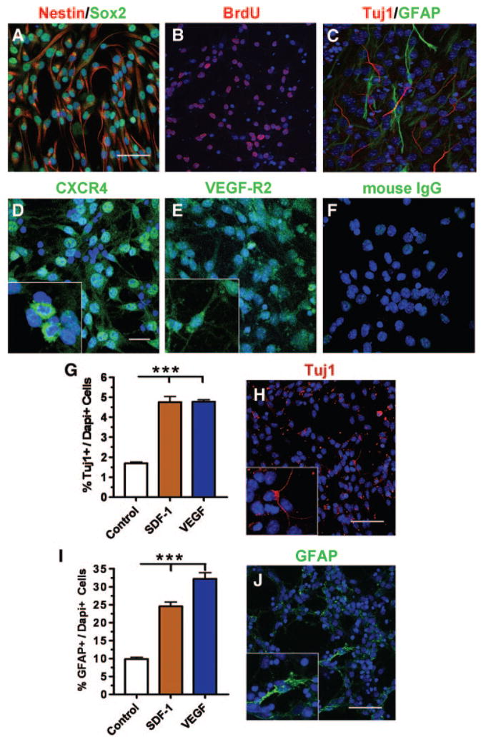
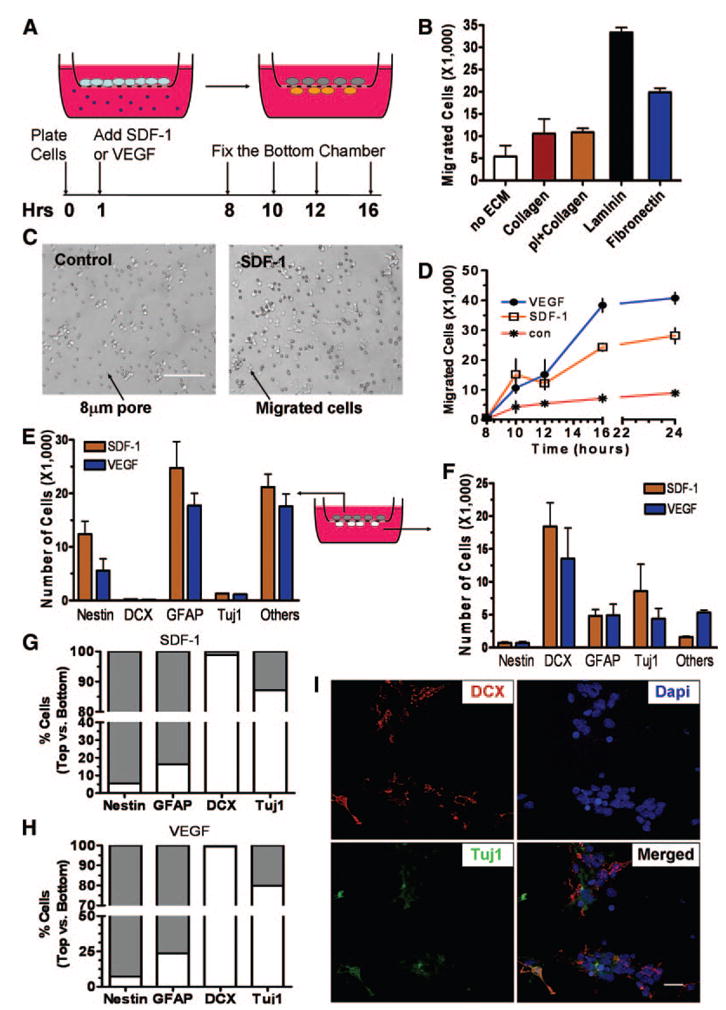
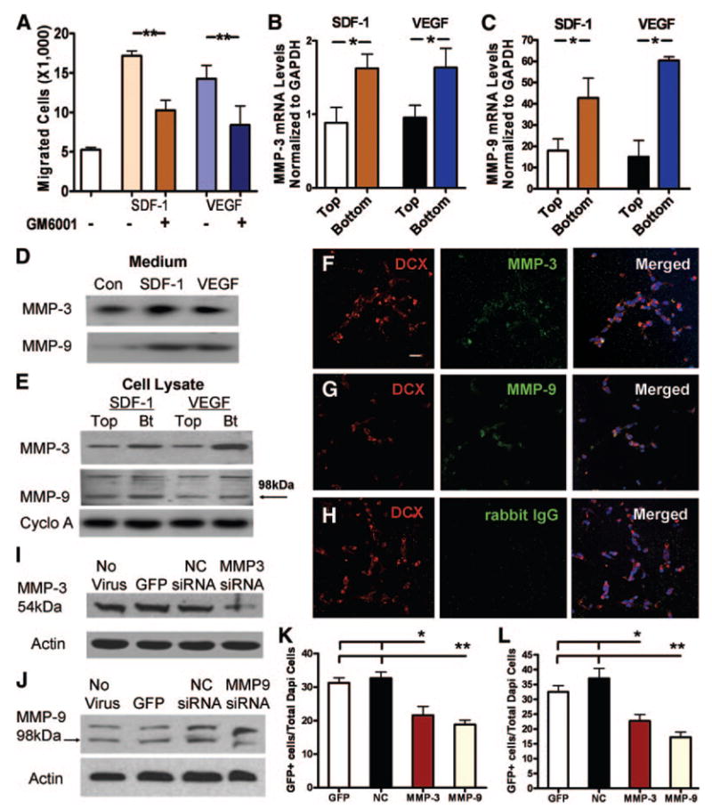
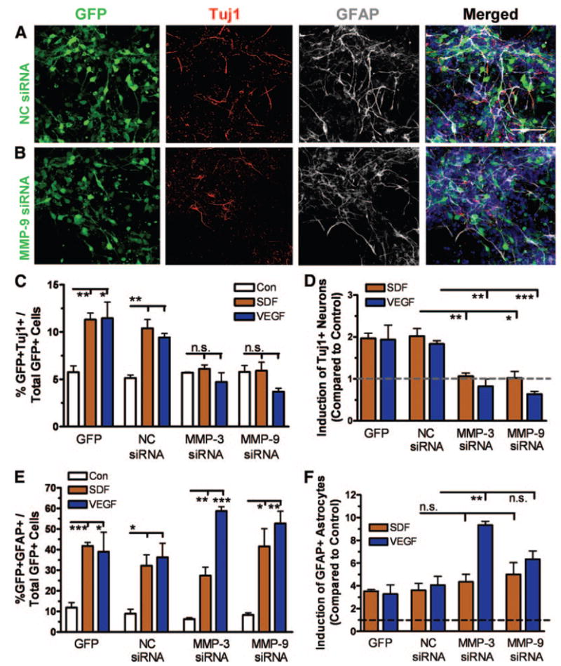
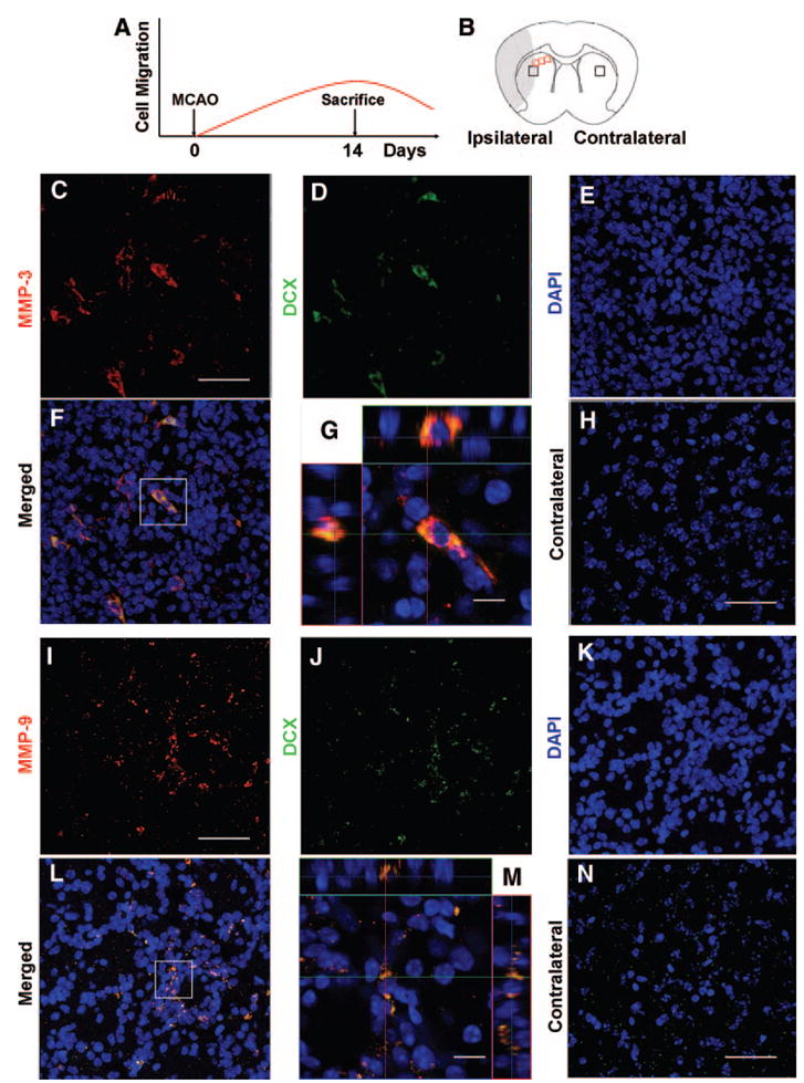
Adult neurogenesis is regulated by both intrinsic programs and extrinsic stimuli. The enhanced proliferation of adult neural stem/progenitor cells (aNPCs) in the subventricular zone and the migration of neuroblasts toward the ischemic region in adult brains present a unique challenge as well as an opportunity to understand the molecular mechanisms underlying the extrinsic cue-induced neurogenic responses. Matrix metalloproteinases (MMPs) are a family of proteinases known to play a role in extracellular matrix remodeling and cell migration. However, their presence in aNPCs and their potential function in injury-induced aNPC migration remain largely unexplored. Here we demonstrate that in response to two injury-induced chemokines, stromal cell-derived factor 1 (SDF-1) and vascular endothelial growth factor, aNPCs differentiated into migratory cells that expressed increased levels of MMP-3 and MMP-9. Whereas differentiated neuroblasts and a subpopulation of astrocytes migrated toward the chemokines, undifferentiated progenitors did not migrate. Blocking the expression of MMP-3 or MMP-9 in aNPCs interfered with both the differentiation of aNPCs and chemokine-induced cell migration. Thus, endogenous MMPs expressed by aNPCs are important for mediating their neurogenic response to extrinsic signals.


Conflict of interest statement

Disclosure of Potential Conflicts of Interest

The authors indicate no potential conflicts of interest.

Figures

Figure 1
Figure 1
Multipotent adult neural stem/progenitor cells (aNPCs) differentiate in response to SDF-1 and VEGF. (A): Cultured aNPCs expressed immature stem cell markers Sox2 (green) and Nestin (red). (B): Under proliferating conditions, cultured aNPCs incorporated BrdU (red). Blue, Dapi. (C): In the absence of growth factors, aNPCs differentiated into neurons (Tuj1, red) and astrocytes (GFAP, green). (D–F): Proliferating aNPCs expressed the receptors for SDF-1 and VEGF. (D): CXCR4 (green). (E): VEGF-R2 (green). (F): Control immunostaining using mouse IgG. Scale bar = 50 μm. Blue, Dapi. Insets in (D, E) show higher-magnification images. (G): Both SDF-1 and VEGF promoted neuronal differentiation of aNPCs after 16 hours of treatment (***, p < .001; n = 3). (H): Example of Tuj1+ neurons quantified in (G). Scale bar = 50 μm. (I): Both SDF-1 and VEGF led to increased astrocyte differentiation of aNPCs after 16 hours of treatment (***, p < .001; n = 3). (J): Example of GFAP+ astrocytes quantified in (I). Scale bar = 50 μm. Abbreviations: BrdU, bromodeoxyuridine; CXCR4, chemokine (C-X-C motif) receptor 4; Dapi, 4,6-diamidino-2-phenylindole; GFAP, glial fibrillary acidic protein; SDF-1, stromal cell-derived factor 1; Tuj1, β-tubulin; VEGF, vascular endothelial growth factor; VEGF-R2, vascular endothelial growth factor receptor 2.
Figure 2
Figure 2
Adult neural stem/progenitor cells (aNPCs) differentiate and migrate in response to SDF-1 and VEGF. (A): Experimental schematics showing in vitro migration assay. Tissue culture inserts with an 8.0-μm pore were used to separate the top and bottom chambers. aNPCs were plated onto the membrane of the top chamber, and either SDF-1 or VEGF was administered to the bottom chamber. At specific time points, the migratory and stationary cells were analyzed. (B): Quantification of cell migration on different ECM substrates indicated that laminin was the most effective ECM at promoting cell migration (n = 4). (C): Bright-field image of cells migrated to the bottom of chambers on laminin-coated inserts. con: no SDF-1 was added. Scale bar = 100 μm. (D): Time course of adult aNPC migration toward SDF-1 and VEGF. The cell migration was initially observed at 10 Hrs and plateaus at 16 Hrs (n = 3). (E): The majority of stationary cells on the top chamber were Nestin+ immature cells and GFAP+ astrocytes (n = 3). (F): The majority of migratory cells at the bottom were DCX+ and Tuj1+ neuroblasts and some GFAP+ astrocytes (n = 3). (G): The percentage distribution of different lineages of cells between the top chamber and the bottom chamber after SDF-1 treatment. (H): The percentage distribution of different lineages of cells between the top chamber and the bottom chamber after VEGF treatment. (I): Example of migratory DCX+ (red) and Tuj1+ (green) cells in the bottom chamber. Merged image shows the colocalization of DCX- and Tuj1-expressing cells. Blue, Dapi. Scale = 20 μm. Abbreviations: con, control; Dapi, 4,6-diamidino-2-phenylindole; ECM, extracellular matrix; GFAP, glial fibrillary acidic protein; Hrs, hours; pI, polylysine; SDF-1, stromal cell-derived factor 1; Tuj1, β-tubulin; VEGF, vascular endothelial growth factor.
Figure 3
Figure 3
MMP-3 and MMP-9 expressed by adult neural stem/progenitor cells (aNPCs) are important for their migration response to SDF-1 and VEGF. (A): A broad-spectrum MMP inhibitor, GM6001, inhibited SDF-1- or VEGF-induced cell migration (**, p < .01; n = 4). (B, C): Quantitative polymerase chain reaction analysis of migratory and stationary cells demonstrated that mRNA levels of MMP-3 (B) and MMP-9 (C) were significantly higher in migratory cells (Bt) compared with stationary cells (top chamber) (n = 4). (D, E): The protein levels of MMP-3 and MMP-9 in the culture medium (D) and cell lysate (E) of the Bt were higher than those in the top chamber (Cyclophillin A antibody used as a loading Con). (F–H): Migrated DCX+ neuroblasts (red) expressed MMP-3 ([F], green) and MMP-9 ([G], green). Mouse IgG, instead of MMP antibodies, was used as negative Con ([H], green). Scale bar = 20 μm. (I): Western blot analysis showing that lentivirus-MMP-3-siRNA could efficiently knockdown endogenous MMP-3 (54 kDa) in aNPCs compared with the Con lentivirus (lentivirus-NC-siRNA and lentivirus-GFP)-infected aNPCs and uninfected aNPCs (β-actin antibody used as a loading Con). (J): Western blot analysis showing that lentivirus-MMP-9-siRNA could efficiently knock down endogenous MMP-9 (98 kDa) in aNPCs compared with Con lentivirus-infected aNPCs and uninfected aNPCs. (K, L): The knockdown of MMP-3 and MMP-9 led to reduced cell migration in response to either SDF-1 (K) or VEGF (L) (**, p < .01; n = 3). Abbreviations: Bt, bottom chamber; Con, control; GAPDH, glyceraldehyde-3-phosphate dehydrogenase; GFP, green fluorescent protein; MMP, matrix metalloproteinase; NC, control nonsilencing; SDF-1, stromal cell-derived factor 1; siRNA, small interfering RNA; VEGF, vascular endothelial growth factor.
Figure 4
Figure 4
Knockdown of MMP-3 and MMP-9 interferes with SDF-1- and VEGF-induced adult neural stem/progenitor cell (aNPC) differentiation. (A, B): Sample images showing that lentivirus-NC-siRNA-infected (A) and lentivirus-MMP-9-siRNA-infected (B) cells differentiated into Tuj1+ neurons (red) or GFAP+ astrocytes (white) in the presence of SDF-1. Blue, 4,6-diamidino-2-phenylindole. Green, GFP. Scale bar = 50 μm. (C): Acute knockdown of MMP-3 or MMP-9 using lentivirus-siRNA abolished SDF-1- and VEGF-induced neuronal differentiation compared with lentivirus-NC-siRNA- and lentivirus-GFP-infected aNPCs. (D): The neuronal induction by SDF-1 or VEGF was abolished by acute knockdown of MMP-3 or MMP-9. The data shown in (E) was normalized to a no-chemokine control condition (shown in dotted line). (E): Acute knockdown of MMP-3 (**, p < .01) using lentivirus-siRNA potentiated VEGF- but not SDF-1-induced astrocyte differentiation compared with controls. MMP-9-siRNA did not have a similar effect (p = .1) (F): The data shown in (F) was normalized to a no-chemokine condition (shown in dotted line). *, p < .05; p < .01; ***, p < .001. Abbreviations: GFAP, glial fibrillary acidic protein; GFP, green fluorescent protein; MMP, matrix metalloproteinase; NC, control nonsilencing; n.s., nonsignificant; SDF, stromal cell-derived factor; siRNA, small interfering RNA; Tuj1, β-tubulin; VEGF, vascular endothelial growth factor.
Figure 5
Figure 5
Migrating DCX+ cells in an MCAO mouse model express MMP-3 and MMP-9 mRNA. (A): Mice were analyzed at 14 days post-MCAO, corresponding to the peak of neuroblast migration (based on literature [1]). (B): Schematic diagram showing mouse brains subjected to unilateral MCAO. Both ipsilateral and contralateral sides were analyzed. The gray boxes indicate the brain regions where images were taken (shown in [C–M]). The red boxes indicate the regions subjected to quantification of DCX+ and MMP+ cells. (C–G): Riboprobes detected increased MMP-3 mRNA ([C], red) in DCX mRNA expressing neuroblast (D) in the ipsilateral side compared with the contralateral side (H). Scale bar = 50 μm. (G): Image of a single cell from (F) (white box) showing that MMP-3 and DCX mRNA are expressed in the same cells. Scale bar = 20 μm. (I–M): Riboprobes detected increased MMP-9 mRNA ([I], red) in DCX mRNA expressing neuroblast (J) in the ipsilateral side but not the contralateral side (N). Scale bar = 50 μm. (N): Image of a single cell from (L) (white box) showing that MMP-9 and DCX mRNA are expressed in the same cells. Scale bar = 20 μm. Abbreviations: DAPI, 4,6-diamidino-2-phenylindole; MCAO, middle cerebral artery occlusion; MMP, matrix metalloproteinase.

References

    1. Kokaia Z, Lindvall O. Neurogenesis after ischaemic brain insults. Curr Opin Neurobiol. 2003;13:127–132. - PubMed
    1. Thored P, Aridsson A, Cacci E, et al. Persistent production of neurons from adult brain stem cells during recovery after stroke. Stem Cells. 2006;24:739–747. - PubMed
    1. Zhang ZG, Zhang L, Tsang W, et al. Correlation of VEGF and angio-poietin expression with disruption of blood-brain barrier and angiogenesis after focal cerebral ischemia. J Cereb Blood Flow Metab. 2002;22:379–392. - PubMed
    1. Robin AM, Zhang ZG, Wang L, et al. Stromal cell-derived factor 1alpha mediates neural progenitor cell motility after focal cerebral ischemia. J Cereb Blood Flow Metab. 2006;26:125–134. - PubMed
    1. Zhang H, Vutskits L, Pepper MS, et al. VEGF is a chemoattractant for FGF-2-stimulated neural progenitors. J Cell Biol. 2003;163:1375–1384. - PMC - PubMed

Publication types