. 2008 Sep 26;4(9):e1000201.
doi: 10.1371/journal.pgen.1000201.

FtsK-dependent dimer resolution on multiple chromosomes in the pathogen Vibrio cholerae

Affiliations

FtsK-dependent dimer resolution on multiple chromosomes in the pathogen Vibrio cholerae

Marie-Eve Val et al. PLoS Genet. .

Abstract

Unlike most bacteria, Vibrio cholerae harbors two distinct, nonhomologous circular chromosomes (chromosome I and II). Many features of chromosome II are plasmid-like, which raised questions concerning its chromosomal nature. Plasmid replication and segregation are generally not coordinated with the bacterial cell cycle, further calling into question the mechanisms ensuring the synchronous management of chromosome I and II. Maintenance of circular replicons requires the resolution of dimers created by homologous recombination events. In Escherichia coli, chromosome dimers are resolved by the addition of a crossover at a specific site, dif, by two tyrosine recombinases, XerC and XerD. The process is coordinated with cell division through the activity of a DNA translocase, FtsK. Many E. coli plasmids also use XerCD for dimer resolution. However, the process is FtsK-independent. The two chromosomes of the V. cholerae N16961 strain carry divergent dimer resolution sites, dif1 and dif2. Here, we show that V. cholerae FtsK controls the addition of a crossover at dif1 and dif2 by a common pair of Xer recombinases. In addition, we show that specific DNA motifs dictate its orientation of translocation, the distribution of these motifs on chromosome I and chromosome II supporting the idea that FtsK translocation serves to bring together the resolution sites carried by a dimer at the time of cell division. Taken together, these results suggest that the same FtsK-dependent mechanism coordinates dimer resolution with cell division for each of the two V. cholerae chromosomes. Chromosome II dimer resolution thus stands as a bona fide chromosomal process.


Conflict of interest statement

The authors have declared that no competing interests exist.

Figures

Figure 1
Figure 1. FtsK-dependent and FtsK-independent Xer recombination.
A. Chromosome dimer formation and resolution in E. coli. The two homologous chromosomes are depicted by thick and thin lines, to allow for the visualization of crossovers. B. Link between the central region of XerCD-target sites (right) and the recombination pathway adopted at these sites (left). The XerCD-dif recombination complex is viewed from the C-terminal side of the recombinases, to show the C-terminal interactions of XerC and XerD. Strands cleaved by XerC and XerD in E. coli are shown with thick and thin lines, respectively. Positions of strand cleavages by E. coli XerC and XerD are indicated by white and black triangles, respectively. The WebLogo was generated using the alignment of putative dif sites from the larger chromosome of 27 γ-Proteobacteria (Text S1). The XerC-binding site, XerD-binding site and central region of dif Ec are indicated below the alignment.
Figure 2
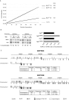
Figure 2. Growth competition of V. cholerae deficient in CDR strains against their parent.
f: frequency of cells that the mutant strains fail to produce at each generation compared to their parent.
Figure 3
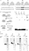
Figure 3. In vitro cleavage of dif1 and dif2 by the V. cholerae recombinases.
A. Putative XerCVc and XerDVc cleavage sites on dif1 and dif2 and scheme of the suicide substrates used in this study. The top and bottom strands of dif1 and dif2 are depicted as black and grey strands. Their equivalents in dif Ec are cleaved by XerCEc and XerDEc, respectively. Grey triangles further indicate the positions equivalent to these where XerCEc and XerDEc cleave dif Ec. A white triangle indicates the XerDVc-cleavage position reported for dif1 . Top and bottom strand suicide substrates contain a nick opposite the position expected to be cleaved by XerCVc and XerDVc if the E. coli paradigm is followed, respectively. T1, B1, T2, B2: suicide substrates on dif1 and dif2, respectively. B. Scheme of a XerC-suicide cleavage reaction. C. Covalent complex formation by MBPXerCVc and XerDVc on suicide substrates. Schemes of substrates and products are shown on the top and on the right of the gel, respectively. Suicide substrates were labeled on the 5′ side of the continuous strand, as indicated (5′*). D. Cleavage sites of XerCVc and XerDVc on dif1 and dif2. Schemes of substrates are shown on the top of the gels. Suicide substrates were labeled on the 3′ side of the continuous strand, as indicated (3′*). PNK: phosphorylation with T4 polynucleotide kinase; G+A: chemical cleavage ladder. Sequences resulting from the chemical cleavage are indicated beside the gels. Bases of the central region and of the XerCD-binding sites are indicated in black and grey, respectively. The deduced cleavage points are indicated by black triangles.
Figure 4
Figure 4. FtsKVc-dependent recombination at dif1 and dif2.
A. Reconstitution of V. cholerae Xer recombination at plasmid-borne dif1 and dif2 sites in E. coli cells. Top panel: gel showing a typical result. A scheme of the substrate and product bands is shown beside the gel. dif sites are represented by triangles. Bottom panel: quantification plot displaying the mean and standard deviations of at least three independent experiments. B. Recombination by wild-type (+) and catalytically inactive (YF) recombinases. HJ: HJ intermediate.
Figure 5
Figure 5. Species specificity in Xer recombination.
A. Amino acid residue conservation in the γ-domain of FtsK and in the C-terminal tail of XerD. Numbers indicate the position of the first and of the last residues of the alignments in the amino acid sequence of the V. cholerae proteins. Positions of full conservation and of strong or weaker groups of conservation are indicated by stars, semi-colons or dots, respectively, following the Clustal 1.83 scheme. Black bars and vertical arrows indicate residues implicated in FtsK-XerD interaction and in KOPS recognition in E. coli, respectively. B. Species-specificity in Xer recombination on plasmid-borne dif1, dif2 and dif Ec sites. The mean and standard deviation of at least three independent experiments are plotted. ND: not determined. C. FtsK-independent recombination at dif Ec by wild-type (+) and catalytically inactive (YF) V. cholerae recombinases. D. FtsK-independent recombination by the V. cholerae recombinases on hybrid dif sites.
Figure 6
Figure 6. V. cholerae FtsK Orienting Polar Sequences.
A. Growth competition of E. coli cells encoding FtsK hybrids. N: cells carrying a complete deletion of the C-terminal domain and linker region of FtsKEc; NLCEc: cells carrying full length FtsKEc; NLCVc and NLCHi: cells in which the C-terminal domain FtsKEc was replaced by the one of FtsKVc and FtsKHi, respectively. f: frequency of cells that the parental N strain fails to produce at each generation compared to the FtsK hybrids. B. 5′-GGGCAGGG-3′ inhibits recombination activation by FtsKVc. Plasmid recombination at E. coli dif by XerCDEc was induced with 0.5% arabinose. Ec[NRE]: FtsK50C Ec[NRE]; Vc[NRE]: FtsKVc[NRE]. KOPS-0: substrate without GGGCAGGG sequences; KOPS-2: substrate with triple overlapping GGGCAGGG sequences in the non-permissive orientation on both sides of the two dif sites. C. Scheme of the two V. cholerae chromosomes showing the distributions of the GGGCAGGG and GGGNAGGG motifs. Upper bars: motifs found in the leading strand; Lower bars: motifs found in the lagging strand. Number, frequency, skew and skew significance (p-skew) are indicated for each motif. Recently acquired genomic regions are indicated (superintegron, CTX and TLC prophages and the Vibrio Pathogenicity Island VPI).
Figure 7
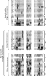
Figure 7. The non-homologous chromosomes of Proteobacteria with multipartite genomes carry divergent dif sites.
Alignment of the chromosome dimer resolution sites of a few Proteobacteria harboring a single or multiple chromosomes. Bases identical to the γ-Proteobacteria dif consensus are shaded in black. Species abbreviations follow the KEGG convention.

References

    1. Heidelberg JF, Eisen JA, Nelson WC, Clayton RA, Gwinn ML, et al. DNA sequence of both chromosomes of the cholera pathogen Vibrio cholerae. Nature. 2000;406:477–483. - PMC - PubMed
    1. Xu Q, Dziejman M, Mekalanos JJ. Determination of the transcriptome of Vibrio cholerae during intraintestinal growth and midexponential phase in vitro. Proc Natl Acad Sci U S A. 2003;100:1286–1291. - PMC - PubMed
    1. Casjens S. The diverse and dynamic structure of bacterial genomes. Annu Rev Genet. 1998;32:339–377. - PubMed
    1. Yamaichi Y, Fogel MA, Waldor MK. par genes and the pathology of chromosome loss in Vibrio cholerae. Proc Natl Acad Sci U S A. 2007;104:630–635. - PMC - PubMed
    1. Srivastava P, Chattoraj DK. Selective chromosome amplification in Vibrio cholerae. Mol Microbiol. 2007;66:1016–1028. - PubMed

Publication types