. 2008 Sep 26;4(9):e1000165.
doi: 10.1371/journal.ppat.1000165.

Substrate binding mode and its implication on drug design for botulinum neurotoxin A

Affiliations

Substrate binding mode and its implication on drug design for botulinum neurotoxin A

Desigan Kumaran et al. PLoS Pathog. .

Abstract

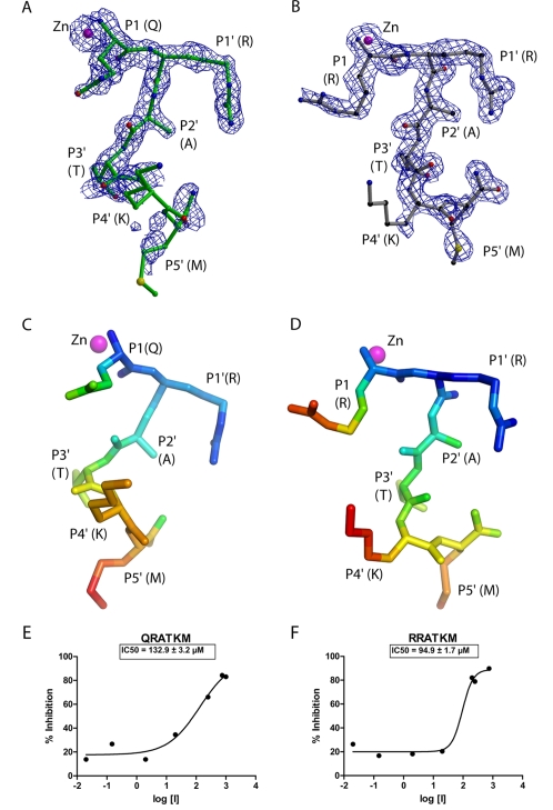
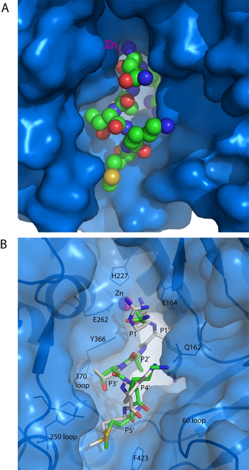
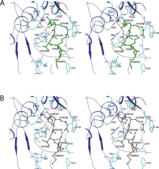
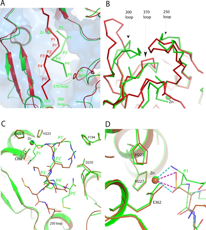
The seven antigenically distinct serotypes of Clostridium botulinum neurotoxins, the causative agents of botulism, block the neurotransmitter release by specifically cleaving one of the three SNARE proteins and induce flaccid paralysis. The Centers for Disease Control and Prevention (CDC) has declared them as Category A biowarfare agents. The most potent among them, botulinum neurotoxin type A (BoNT/A), cleaves its substrate synaptosome-associated protein of 25 kDa (SNAP-25). An efficient drug for botulism can be developed only with the knowledge of interactions between the substrate and enzyme at the active site. Here, we report the crystal structures of the catalytic domain of BoNT/A with its uncleavable SNAP-25 peptide (197)QRATKM(202) and its variant (197)RRATKM(202) to 1.5 A and 1.6 A, respectively. This is the first time the structure of an uncleavable substrate bound to an active botulinum neurotoxin is reported and it has helped in unequivocally defining S1 to S5' sites. These substrate peptides make interactions with the enzyme predominantly by the residues from 160, 200, 250 and 370 loops. Most notably, the amino nitrogen and carbonyl oxygen of P1 residue (Gln197) chelate the zinc ion and replace the nucleophilic water. The P1'-Arg198, occupies the S1' site formed by Arg363, Thr220, Asp370, Thr215, Ile161, Phe163 and Phe194. The S2' subsite is formed by Arg363, Asn368 and Asp370, while S3' subsite is formed by Tyr251, Leu256, Val258, Tyr366, Phe369 and Asn388. P4'-Lys201 makes hydrogen bond with Gln162. P5'-Met202 binds in the hydrophobic pocket formed by the residues from the 250 and 200 loop. Knowledge of interactions between the enzyme and substrate peptide from these complex structures should form the basis for design of potent inhibitors for this neurotoxin.


Conflict of interest statement

The authors have declared that no competing interests exist.

Figures

Figure 1
Figure 1. Hexapeptides and inhibition plots.
Electron density maps (blue mesh) for bound substrate (QRATKM) and its variant (RRATKM) are shown in A and B, respectively. The electron density is from composite omit maps (2|Fo|-|Fc|) and contoured at 1σ level. QRATKM (green) and RRATKM (gray) peptides are shown in ball and stick model. Zinc, oxygen and nitrogen atoms are shown as magenta, red and blue spheres, respectively. Carbon atoms are shown in green (QRATKM) and black (RRATKM). Figures were prepared using Molscript, Raster3D and Bobscript –. Distribution of B factors for the QRATKM and RRATKM are shown in C and D, respectively. The peptide atoms are colored according to B factor with RGB (Red-Green-Blue) color ramp with blue and red corresponding to the lowest (17 Å2) and highest (50 Å2). Pymol (DeLano, W.L. The PyMOL Molecular Graphics System (2002) on World Wide Web http://www.pymol.org) was used to prepare C and D figures. Inhibition of Balc424 catalytic activity at increasing concentrations (µM) of QRATKML (E) and of RRATKML (F).
Figure 2
Figure 2. Binding of the substrate peptide in the active site of Balc424.
(A) Figure shows the substrate peptide (QRATKM) binding in the active site of Balc424. Balc424 is shown in solid blue-colored surface. Substrate peptide is shown in sphere (CPK) model. Carbon, oxygen, nitrogen, sulfur and zinc atoms are shown in green, red, blue, yellow and magenta, respectively. (B). Superposed stick models of QRATKM and RRATKM are shown in green, and gray, respectively. Carbon atoms are shown in green (QRATKM) and gray (RRATKM). Balc424 is shown in semi-transparent surface (blue) representation with secondary elements at the active site pocket. Only a few substrate-binding residues are shown as markers. Pymol (DeLano, W.L. The PyMOL Molecular Graphics System (2002) on World Wide Web http://www.pymol.org) was used to prepare these figures.
Figure 3
Figure 3. Structure of Balc424 in complex with the QRATKM, a segment of substrate SNAP-25.
(A). Stereo view of the active site shows the molecular interactions between the substrate peptide (QRATKM) and protease (Balc424). Protease and substrate residues are shown in cyan and green, respectively. Blue color ribbon represents the protease secondary elements at the vicinity of the active site. (B). Stereo view of the active site center of Balc424 (cyan stick) with RRATKM (gray ball and stick). Oxygen, nitrogen, sulfur and zinc atoms are shown in red, blue, yellow and magenta, respectively. Hydrogen bonds are depicted as black dash lines while zinc co-ordination is shown in solid line.
Figure 4
Figure 4. Schematic diagram of the molecular interactions between the Balc424 and substrate peptides.
The interactions between Balc424 active site residues and the substrate peptides are shown. (A) QRATKM and (B) RRATKM. Black, red, blue, and yellow colored circles represent carbon, oxygen, nitrogen, and sulfur atoms, respectively. For clarity, zinc co-ordination and water molecules involved in the interactions at the active site are not shown. (C). A Schematic diagram representing S1 to S5′sites. Residues of the enzyme forming the subsites and substrate peptide are shown in red and blue, respectively. Proteolytic site is shown in cyan colored double-headed arrow. Figures A and B were prepared with Ligplot . ChemDraw ultra (CambridgeSoft, Inc) was used to prepare figure C.
Figure 5
Figure 5. Comparison of Balc424 complexed with segments of SNAP-25 (197–202) and (146–204) and an inhibitor (N-Ac-CRATKML).
(A) and (B) show the superposition of Balc424 with SNAP-25 (197–202) and (146–204). Green and red represent SNAP-25 (197–202) and SNAP-25 (146–204), respectively. Superposition of Balc424 with substrate peptide (SNAP-25 [197–202]) and N-Ac-CRATKML peptide at the vicinity of the active site are shown in C and D. N-Ac-CRATKML peptide complex is shown in brown color. Zinc binding and selective substrate binding residues are in stick model.

References

    1. Lacy DB, Tepp W, Cohen AC, DasGupta BR, Stevens RC. Crystal structure of botulinum neurotoxin type A and implications for toxicity. Nat Struct Biol. 1998;5(10):898–902. - PubMed
    1. Simpson LL. Molecular pharmacology of botulinum toxin and tetanus toxin. Annu Rev Pharmacol Toxicol. 1986;26:427–453. - PubMed
    1. Swaminathan S, Eswaramoorthy S. Structural analysis of the catalytic and binding sites of Clostridium botulinum neurotoxin B. Nat Struct Biol. 2000;7(8):693–699. - PubMed
    1. DasGupta BR, Rasmussen S. Amino acid composition of Clostridium botulinum type F neurotoxin. Toxicon. 1983;21(4):566–569. - PubMed
    1. DasGupta BR, Rasmussen S. Purification and amino acid composition of type E botulinum neurotoxin. Toxicon. 1983;21(4):535–545. - PubMed

Publication types

MeSH terms

Substances