. 2008 Sep 26;3(9):e3289.
doi: 10.1371/journal.pone.0003289.

Distinct effects of IL-18 on the engraftment and function of human effector CD8 T cells and regulatory T cells

Affiliations

Distinct effects of IL-18 on the engraftment and function of human effector CD8 T cells and regulatory T cells

Richard G Carroll et al. PLoS One. .

Abstract

IL-18 has pleotropic effects on the activation of T cells during antigen presentation. We investigated the effects of human IL-18 on the engraftment and function of human T cell subsets in xenograft mouse models. IL-18 enhanced the engraftment of human CD8(+) effector T cells and promoted the development of xenogeneic graft versus host disease (GVHD). In marked contrast, IL-18 had reciprocal effects on the engraftment of CD4(+)CD25(+)Foxp3(+) regulatory T cells (Tregs) in the xenografted mice. Adoptive transfer experiments indicated that IL-18 prevented the suppressive effects of Tregs on the development of xenogeneic GVHD. The IL-18 results were robust as they were observed in two different mouse strains. In addition, the effects of IL-18 were systemic as IL-18 promoted engraftment and persistence of human effector T cells and decreased Tregs in peripheral blood, peritoneal cavity, spleen and liver. In vitro experiments indicated that the expression of the IL-18Ralpha was induced on both CD4 and CD8 effector T cells and Tregs, and that the duration of expression was less sustained on Tregs. These preclinical data suggest that human IL-18 may have use as an adjuvant for immune reconstitution after cytotoxic therapies, and to augment adoptive immunotherapy, donor leukocyte infusions, and vaccine strategies.


Conflict of interest statement

Competing Interests: Z.L.J. is an employee of GlaxoSmithKline; the other authors have no competing financial interests to declare.

Figures

Figure 1
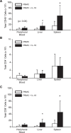
Figure 1. Human IL-18 Promotes Systemic Increases in Human CD8 T Cell Numbers in NOD/scid/β2m null mice injected with human PBMCs.
NOD/scid/β2mnull mice were injected intraperitoneally with 5×10⁷ human PBMCs and 24 hours later, a three week course of daily subcutaneous injections of 15 µg recombinant human IL-18 was initiated. At the end of the injection time course, the animals were sacrificed, peripheral blood was collected, and liver and spleen homogenates were prepared. Human cell populations were detected by flow cytometry. Absolute cell numbers in the peripheral blood were determined using Tru-Count tubes; absolute cell numbers from spleen and liver were determined using a Coulter Multisizer 3. The mean±s.d. is shown for (A) human CD45+ cells, (B) human CD4+ cells, and (C) human CD8+ cells. Open bars represent values from mock-treated animals; filled bars represent values from IL-18 treated animals. These data are a representative single experiment of seven different experiments. Asterisks indicate significant differences between mock- and IL-18-treated animals (n = 6 to 10 mice/group; p<0.05).
Figure 2
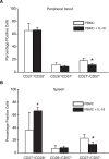
Figure 2. Human IL-18 Mediates a Decrease in Tregs in NOD/scid/β2mnull mice Injected with Human PBMCs.
5×10⁷ PBMCs were transferred into NOD/scid /β2Mnull mice. The day following cell injection, a three week course of subcutaneous huIL-18 injections was initiated. Animals in the experiment depicted in panel A received twice-weekly doses of 15 µg pegylated human IL-18, while the animals in the experiments depicted in panel B received daily doses of 15 µg recombinant human IL-18. (A) At the end of IL-18 treatment, cells isolated from the peripheral blood, peritoneal cavity, spleen and liver were stained with antibodies to human CD45, CD4, and CD25 and analyzed by flow cytometry. The mean percentage of CD+CD25+ cells from each tissue is depicted. Filled bars represent values obtained from IL-18-treated animals; open bars represent values obtained from mock-treated animals. (B) Paraffin sections from the spleens of mock- or IL-18-treated animals were reacted with antibodies specific for human CD25 or Foxp3 . Antibody-reactive cells per 400X field were enumerated blindly, and the mean±s.d. depicted. Solid bars depict sections from IL-18-treated animals, while open bars represent sections from mock-treated animals. The experiment in panel A was performed 2 times, and the experiment in panel B was performed 6 times. Asterisks indicate significant differences between mock- and IL-18-treated animals (n = 8/group; p<0.05).
Figure 3
Figure 3. The Human IL-18-Mediated Decrease in Tregs is Not Restricted to NOD/scid/β2mnull Mice.
NOD/scid/IL-2rγnull (NOG) mice were injected with 5×10⁶ human PBMCs, and 24 hours later, a three week course of daily subcutaneous injections of 15 µg recombinant human IL-18 was initiated. The absolute numbers of human CD8+ cells (A) and CD4+CD25+Foxp3+ cells (B) in the peripheral blood were determined using TruCount tubes. Values from individual mice are depicted. (C) The ratio of CD8 cell number to CD4+25+Foxp3+ cell number for each animal in panels A and B is plotted on a log scale. In panels A and B, asterisks (*) indicate significant differences between mock- and IL-18-treated animals (n = 6 to 8/group; p<0.05). In panel C, asterisks (**) indicate p<0.001.
Figure 4
Figure 4. Effects of Human IL-18 on CD8 T Cell Differentiation in NOG mice.
NOG mice were injected intraperitoneally with 5×10⁶ human PBMCs, and beginning 1 day later, were injected with 15 µg recombinant human IL-18 daily for three weeks. Spleen tissue and peripheral blood were collected and analyzed for the presence of different CD8+ T cell subsets by flow cytometry. Cell populations were initially gated on human CD45 and human CD8. Filled bars represent values mean±s.d. obtained from IL-18-treated animals; open bars represent values obtained from mock-treated animals. The data in this figure represent results from one of two independent experiments. Asterisks indicate significant differences between mock- and IL-18-treated animals (p<0.05).
Figure 5
Figure 5. Human IL-18 Accelerates Xenogeneic GVHD in NOG mice.
5×10⁶ PBMC were transferred into NOG mice on day 0, and IL-18 or PBS injected daily starting on day 1. On the x-axis are days after transfer of cells. On the y-axis is the proportion of recipients surviving (n = 8/group; P<.02).
Figure 6
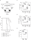
Figure 6. Effect of Human IL-18 on Treg-Induced Delay of Xenogeneic GVHD in NOG Mice.
NOG mice were injected with 2×10⁷ Treg-depleted human PBMCs supplemented with either 4 million autologous CD4+CD25 T cells (“CD4 T cells”) or 4 million autologous, ex vivo-expanded CD4+CD25+ cells (“Tregs”). One cohort of each group received daily injections of 15 µg recombinant human IL-18 for three weeks. Human cell engraftment in the peripheral blood was measured 11, 22, and 30 days post-injection. The animals were followed until the onset of xenogeneic GVHD. (A) Experimental overview. (B) Kaplan-Meier Survival Analysis (Log-Rank) of the indicated cohorts of mice. The Holm-Sidak method for multiple comparisons (significance level 0.05) was performed, and showed significant differences between all groups. (C–E) Peripheral blood levels of human CD8+ T cells were measured at Day 11 (C), Day 22 (D), and Day 30 (E) post-injection. Note that all IL-18-treated animals are not represented in panel E due to their death from acute GVHD.
Figure 7
Figure 7. Human IL-18 Does Not Prevent Treg Suppressive Function in Vitro.
Purified Tregs were expanded ex vivo for 18 days, and then tested in an in vitro suppression assay. Autologous PBMCs were labeled with CFSE and stimulated using anti-CD3 Ab coated beads and mixed with either no Tregs (top panel) or expanded Tregs at the indicated ratio (Treg:PBMC) in the presence or absence of recombinant human IL-18 (500 ng/ml). Histograms show the expansion of CD8+ cells on day 4 of culture.
Figure 8
Figure 8. Rapid Down Regulation of IL18-Rα on Tregs Following Cell Activation.
Purified populations of Tregs, as well as bulk CD4 and CD8 cells, were activated with either αCD3/αCD28 beads, PHA (5 µg/ml), or PHA+recombinant human IL-18 (500 ng/ml). IL-18Rα expression was measured by flow cytometry. IL-18Rα MFI on effector CD4 cells (A), Tregs (B) and effector CD8 cells (C) is plotted before stimulation and 1, 2, and 3 days following stimulation.

References

    1. Okamura H, Tsutsi H, Komatsu T, Yutsudo M, Hakura A, et al. Cloning of a new cytokine that induces IFN-gamma production by T cells. Nature. 1995;378:88–91. - PubMed
    1. Ushio S, Namba M, Okura T, Hattori K, Nukada Y, et al. Cloning of the cDNA for human IFN-gamma-inducing factor, expression in Escherichia coli, and studies on the biologic activities of the protein. J Immunol. 1996;156:4274–4279. - PubMed
    1. Nakanishi K, Yoshimoto T, Tsutsui H, Okamura H. Interleukin-18 regulates both Th1 and Th2 responses. Annu Rev Immunol. 2001;19:423–474. - PubMed
    1. Gracie JA, Robertson SE, McInnes IB. Interleukin-18. J Leukoc Biol. 2003;73:213–224. - PubMed
    1. Gu Y, Kuida K, Tsutsui H, Ku G, Hsiao K, et al. Activation of interferon-gamma inducing factor mediated by interleukin-1beta converting enzyme. Science. 1997;275:206–209. - PubMed

Publication types