. 2009;43(1-3):128-37.
doi: 10.1007/s12026-008-8058-5.

Eosinophils and their interactions with respiratory virus pathogens

Affiliations

Eosinophils and their interactions with respiratory virus pathogens

Helene F Rosenberg et al. Immunol Res. 2009.

Abstract

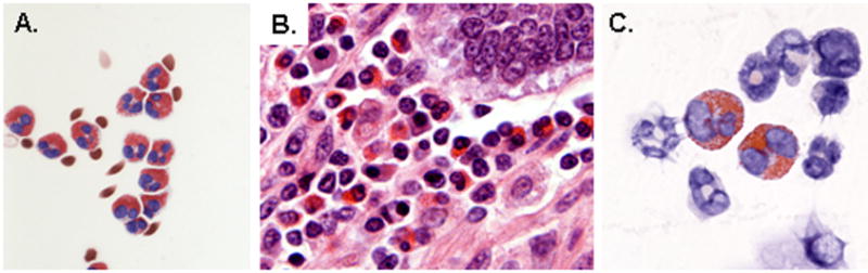
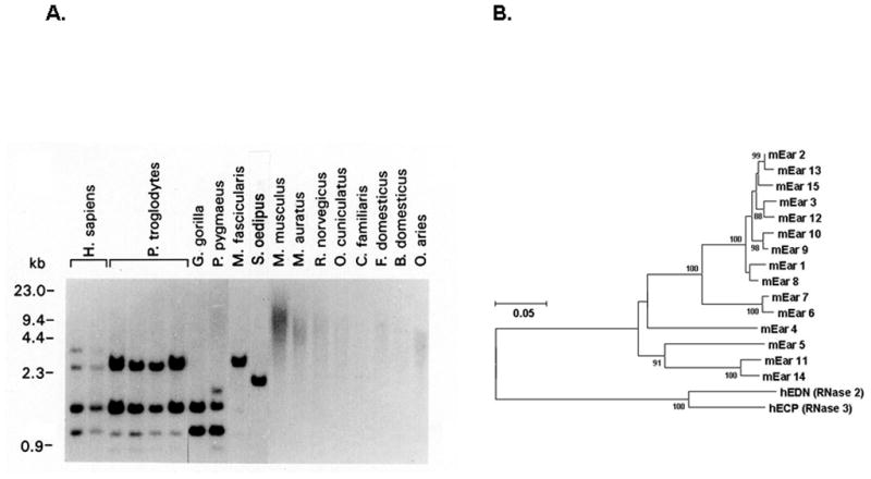
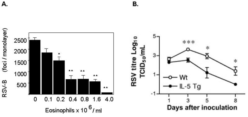
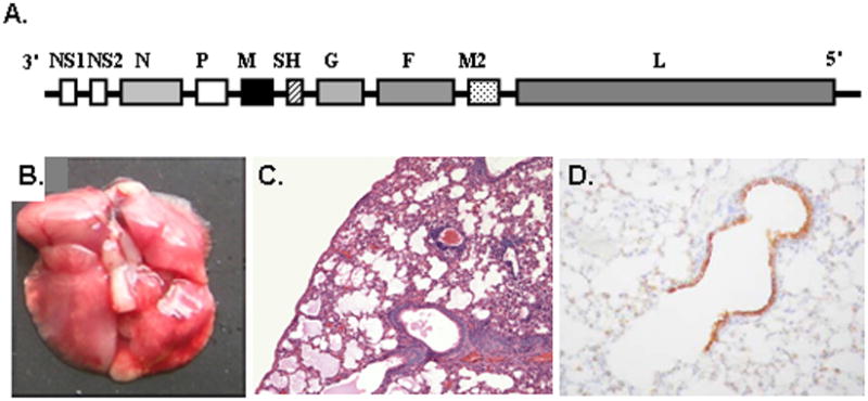
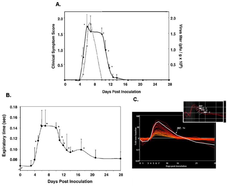
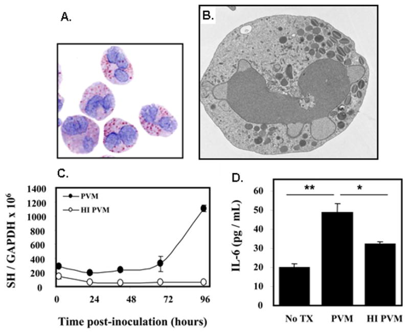
Eosinophils are implicated in the pathophysiology of respiratory virus infection, most typically in negative roles, such as promoting wheezing and bronchoconstriction in conjunction with virus-induced exacerbations of reactive airways disease and in association with aberrant hypersensitivity responses to viral vaccines. However, experiments carried out in vitro and in vivo suggest positive roles for eosinophils, as they have been shown to reduce virus infectivity in tissue culture and promote clearance of the human pathogen, respiratory syncytial virus in a mouse challenge model. The related natural rodent pathogen, pneumonia virus of mice (PVM), is highly virulent in mice, and is not readily cleared by eosinophils in vivo. Interestingly, PVM replicates in eosinophils and promotes cytokine release. The molecular basis of virus infection in eosinophils and its relationship to disease outcome is currently under study.


Figures

Figure 1
Figure 1. Eosinophils
(A) Human eosinophils isolated from peripheral blood by negative selection (B) Eosinophilic infiltrates in the intestinal wall of an Owl monkey, A. trivirgatus (image courtesy of Dr. Alfonso Gozalo, Comparative Medicine Branch, NIAID) (C) Eosinophils in mouse bone marrow cytospin. Eosinophils can be found in all mammalian species, and are easily recognized by their large, prominently stained cytoplasmic granules. Panels (A) and (C) reprinted with permission from reference [36].
Figure 2
Figure 2. Evolution of eosinophil secretory ribonucleases
(A) Genomic DNA probed with the coding sequence of the eosinophil-derived neurotoxin (EDN/RNase 2), demonstrating variant genomic structure among closely-related primates, and no detectable hybridizing sequence among non-primate mammals. EDN and eosinophil cationic protein (ECP) are among the most rapidly evolving functional coding sequences identified among primate species. (B) Neighbor-joining tree documenting relationships among mouse eosinophil-associated ribonucleases and human EDN and ECP. Panel (A) reprinted with permission from reference [20].
Figure 3
Figure 3. Eosinophils and antiviral interactions with respiratory syncytial virus (RSV)
(A) Eosinophils reduce the infectivity of respiratory syncytial virus (RSV) in tissue culture. Increasing concentrations of eosinophils function in a dose dependent fashion to reduce the number of infectious virions as shown. (B) Eosinophils promote clearance of RSV in a mouse challenge model. Virus is cleared more rapidly from the lungs of eosinophil-enriched interleukin-5 (IL-5) transgenic mice when compared to wild type. Panels (A) and (B) reprinted with permission from references [24] and [26], respectively.
Figure 4
Figure 4. Pneumonia virus of mice (PVM) is a natural pathogen of rodent species
(A) PVM is a negative sense non-segmented single stranded RNA virus, family Paramyxoviridae, genus Pneumovirus, gene order as shown. (B) Challenge with a minimal inoculum (<100 pfu) results in a severe respiratory infection, with macroscopic evidence of hemorrhage and (C) microscopic pathology including profound granulocytic inflammation and edema. (D) Immunoreactive virus can be detected in bronchiolar epithelial cells. Panels (A), (B), (C), and (D) reprinted with permission from references [29], [37], [38], and [33], respectively.
Figure 5
Figure 5. Virus replication and prolonged respiratory dysfunction
(A) As shown, virus replication (dotted line) peaks at day 7 in response to a minimal inoculum, and infectious virus can no longer be detected after day 10. In contrast, clinical symptom scores (full line; based on 6-point objective criteria) remain elevated through day 14, and (B) respiratory dysfunction, documented here as expiratory time, remains statistically above baseline through day 16. (C) Prolonged respiratory dysfunction correlates directly with persistent production of proinflammatory chemokines, despite resolution of the infection per se. Reprinted with permission from reference [33].
Figure 6
Figure 6. Eosinophils are targets of virus infection
(A) Mouse eosinophils grown in culture from unselected normal bone marrow, (B) Electron micrograph of cultured mouse eosinophil shown in (A) depicting normal nuclear, cytoplasmic and granule morphology; image courtesy of Dr. Elizabeth Fischer, Research Technologies Section, Rocky Mountain Laboratories, NIAID; (C) Replication of PVM in mouse eosinophils as determined by quantitative RT-PCR detection of the virus SH gene; no replication of heat-inactivated (HI) virus is observed. (D) Active replication is accompanied by release of interleukin-6. Panels (A), (C) and (D) are reprinted with permission from reference [35].

References

    1. Rothenberg ME, Hogan SP. The eosinophil. Annu Rev Immunol. 2006;24:147–174. - PubMed
    1. Jacobsen EA, Taranova AG, Lee NA, Lee JJ. Eosinophils: singularly destructive effector cells or purveyors of immunoregulation? J Allergy Clin Immunol. 2007;119:1313–1320. - PubMed
    1. Klion AD, Nutman TB. The role of eosinophils in host defense against helminth parasites. J Allergy Clin Immunol. 2004;113:30–37. - PubMed
    1. Menzies-Gow A, Robinson DS. Eosinophils, eosinophilic cytokines (interleukin-5), and antieosinophilic therapy in asthma. Curr Opin Pulm Med. 2002;8:33–38. - PubMed
    1. Leckie MJ. Anti-interleukin-5 monoclonal antibodies: preclinical and clinical evidence in asthma models. Am J Respir Med. 2003;2:245–259. - PubMed

Publication types

MeSH terms