. 2009 Jul;30(7):2157-72.
doi: 10.1002/hbm.20658.

Brain activity during visual versus kinesthetic imagery: an fMRI study

Affiliations

Brain activity during visual versus kinesthetic imagery: an fMRI study

Aymeric Guillot et al. Hum Brain Mapp. 2009 Jul.

Abstract

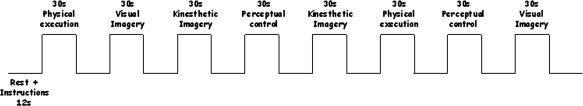
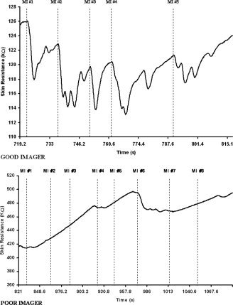
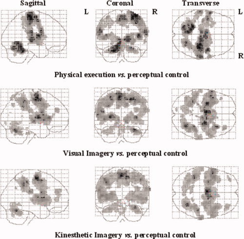
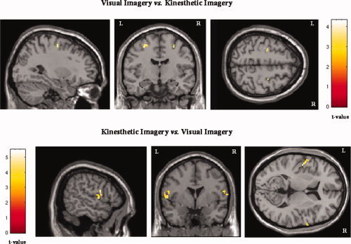
Although there is ample evidence that motor imagery activates similar cerebral regions to those solicited during actual movements, it is still unknown whether visual (VI) and kinesthetic imagery (KI) recruit comparable or distinct neural networks. The present study was thus designed to identify, through functional magnetic resonance imaging at 3.0 Tesla in 13 skilled imagers, the cerebral structures implicated in VI and KI. Participants were scanned in a perceptual control condition and while physically executing or focusing during motor imagery on either the visual or kinesthetic components of an explicitly known sequence of finger movements. Subjects' imagery abilities were assessed using well-established psychological, chronometric, and new physiological measures from the autonomic nervous system. Compared with the perceptual condition, physical executing, VI, and KI resulted in overlapping (albeit non-identical) brain activations, including motor-related regions and the inferior and superior parietal lobules. By contrast, a divergent pattern of increased activity was observed when VI and KI were compared directly: VI activated predominantly the occipital regions and the superior parietal lobules, whereas KI yielded more activity in motor-associated structures and the inferior parietal lobule. These results suggest that VI and KI are mediated through separate neural systems, which contribute differently during processes of motor learning and neurological rehabilitation.


Figures

Figure 1
Figure 1
Schematic representation of the experimental block‐design. After reading the instructions, the participants were subjected to a 30 s‐period during which they either physically performed the movement, mentally rehearsed the sequence using visual or kinesthetic imagery, or remained motionless while closing their eyes. Similar “start” and “stop” sound signals were used in each condition.
Figure 2
Figure 2
Skin resistance responses during motor imagery. In the good imager group, a response (indicated by the dotted line) was recorded during each motor imagery trial, hence attesting to mental work. Conversely, in the poor imagers, the lack of skin resistance responses attests the difficulty to form an accurate representation of action. The strong increase in skin resistance throughout the session also indicated that the subject becomes too relaxed and was not able to keep an adequate arousal level. MI, motor imagery.
Figure 3
Figure 3
SPM activation maps comparing the different experimental conditions (physical execution, VI, and KI) with the perceptual control condition. Voxels are displayed on a progressive gray scale. The maps are displayed on Talairach space as a maximum intensity projection (all voxels activated are visible as if viewed in transparency through the brain) viewed from the right side (sagittal), the back (coronal), and the top (transverse) of the brain. [Color figure can be viewed in the online issue, which is available at www.interscience.wiley.com.]
Figure 4
Figure 4
Activation maps of the lateral premotor area during the visual and kinesthetic imagery conditions. When the visual imagery condition was contrasted with the kinesthetic imagery condition (upper row, x = −29; y = −9; z = 53), the activation was located more posteriorly and superiorly than when the kinesthetic imagery condition was compared to the visual imagery condition (lower row, x = −59; y = 8; z = 7).
Figure 5
Figure 5
Activation maps of the exclusive brain activations during both visual and kinesthetic imagery conditions. When the visual imagery condition was contrasted to the kinesthetic imagery condition (left side), selective activation was found in the superior parietal lobule (BA 5–7; X = −1) and occipital cortex (BA 17–19, Z = 12 and Z = 40). When the visual imagery condition was subtracted from the kinesthetic imagery condition (right side), exclusive activation was located in the putamen (Z = 1), the cerebellar cortex (Z = −23), and the inferior prefrontal area (BA 44, Z = 13).

References

    1. Amador N,Fried I ( 2004): Single‐neuron activity in the human supplementary motor area underlying preparation for action. J Neurosurg 100: 250–259. - PubMed
    1. Andersen RA,Asanuma C,Cowan WM ( 1985): Callosal and prefrontal associational projecting cell populations in area 7A of the macaque monkey: A study using retrogradely transported fluorescent dyes. J Comp Neurol 232: 443–455. - PubMed
    1. Binkofski F,Amunts K,Stephan KM,Posse S,Schormann T,Freund HJ,Zilles K,Seitz RJ ( 2000): Broca's region subserves imagery of motion: A combined cytoarchitectonic and fMRI study. Hum Brain Mapp 11: 273–285. - PMC - PubMed
    1. Boucsein W ( 1993): Methodological issues in electrodermal measurement In: Roy JC,Boucsein W, Fowles DC,Gruzelier JH, editors. Progress in Electrodermal Research. London: Plenum; pp. 31–41.
    1. Buccino G,Binkofski F,Fink GR,Fadiga L,Fogassi L,Gallese V,Seitz RJ,Zilles K,Rizzolatti G,Freund HJ ( 2001): Action observation activates premotor and parietal areas in a somatotopic manner: An fMRI study. Eur J Neurosci 13: 400–404. - PubMed

Publication types