. 2008 Nov;131(Pt 11):3019-33.
doi: 10.1093/brain/awn230. Epub 2008 Sep 26.

Role of microglial IKKbeta in kainic acid-induced hippocampal neuronal cell death

Affiliations

Role of microglial IKKbeta in kainic acid-induced hippocampal neuronal cell death

Ik-Hyun Cho et al. Brain. 2008 Nov.

Abstract

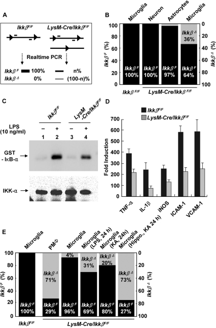
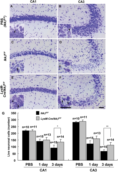
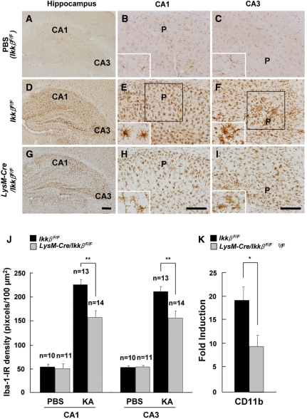
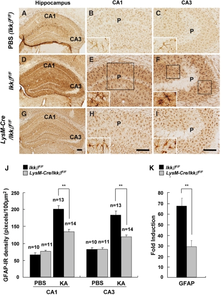
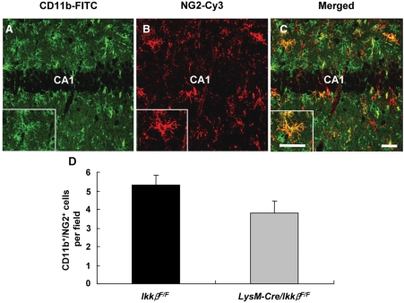
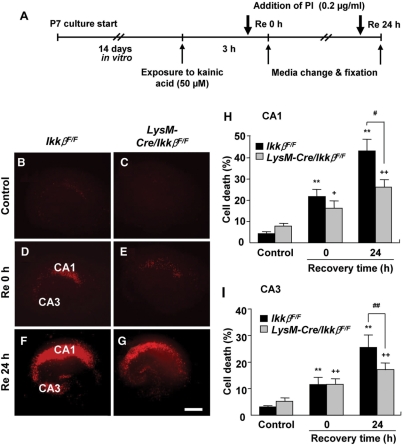
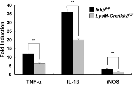
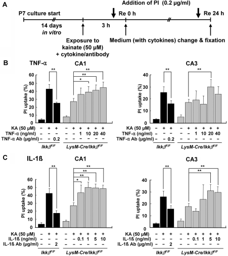
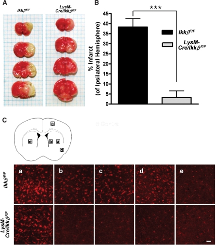
Microglial cells are activated during excitotoxin-induced neurodegeneration. However, the in vivo role of microglia activation in neurodegeneration has not yet been fully elucidated. To this end, we used Ikkbeta conditional knockout mice (LysM-Cre/Ikkbeta(F/F)) in which the Ikkbeta gene is specifically deleted in cells of myeloid lineage, including microglia, in the CNS. This deletion reduced IkappaB kinase (IKK) activity in cultured primary microglia by up to 40% compared with wild-type (Ikkbeta(F/F)), and lipopolysaccharide-induced proinflammatory gene expression was also compromised. Kainic acid (KA)-induced hippocampal neuronal cell death was reduced by 30% in LysM-Cre/Ikkbeta(F/F) mice compared with wild-type mice. Reduced neuronal cell death was accompanied by decreased KA-induced glial cell activation and subsequent expression of proinflammatory genes such as tumour necrosis factor (TNF)-alpha and interleukin (IL)-1beta. Similarly, neurons in organotypic hippocampal slice cultures (OHSCs) from LysM-Cre/Ikkbeta(F/F) mouse brain were less susceptible to KA-induced excitotoxicity compared with wild-type OHSCs, due in part to decreased TNF-alpha and IL-1beta expression. Based on these data, we concluded that IKK/nuclear factor-kappaB dependent microglia activation contributes to KA-induced hippocampal neuronal cell death in vivo through induction of inflammatory mediators.


Figures

Fig. 1
Fig. 1
Microglia-specific Ikkβ gene deletion in the LysM-Cre/IkkβF/F mice. (A and B) Primary microglia and astrocytes were cultured from neonatal wild-type (IkkβF/F) or LysM-Cre/IkkβF/F mice, and cortical neurons were isolated and cultured from E17 embryos of LysM-Cre/IkkβF/F mice. Genomic DNA from each primary cell culture was analysed by real-time PCR to determine the rate of Ikkβ deletion (A). Percentages of undeleted and deleted IkkβF alleles are shown in black and grey bars, respectively (B). (C) Primary microglia from wild-type and LysM-Cre/IkkβF/F mice were stimulated with LPS (10 ng/ml) for 30 min, and cell lysates were used to measure IKK activity. Samples were resolved on SDS–PAGE, and radiolabelled substrates were exposed using a PhosphorImager (upper panel). The same membrane was probed with anti-IKKα antibody to normalize IKK loading (lower panel). (D) Primary microglia from either wild-type (black bars) or LysM-Cre/IkkβF/F mice (grey bars) were stimulated with LPS (10 ng/ml) for 3 h. Total RNA was prepared and used to determine the mRNA expression levels of TNF-α, IL-1β, iNOS, ICAM-1 and VCAM-1, which are represented as fold-induction. (E) Microglia and PMΦ from adult wild-type or LysM-Cre/IkkβF/F mice were used to prepare genomic DNA to determine the Ikkβ gene deletion rate. Percentages of undeleted and deleted IkkβF alleles are shown in black and grey bars, respectively.
Fig. 2
Fig. 2
KA-induced hippocampal neuronal cell death is decreased in LysM-Cre/IkkβF/F mice. Wild-type (IkkβF/F) (A–D) and LysM-Cre/IkkβF/F (E and F) mice were i.c.v. injected with either PBS (A and B) or KA (0.2 µg in 4 µl PBS) (C–F). Cryosections (30 µm thick) were stained with cresyl violet. Arrows indicate dying neurons and arrow heads show live cells. Scale bars: 50 µm. (G) The rate of neuronal loss in the CA1 or CA3 area of ipsilateral side hippocampus was evaluated by counting live cells. Data are presented as mean ± SEM. (Student's t-test, *P < 0.05, **P < 0.01; versus wild-type mice.)
Fig. 3
Fig. 3
KA-induced microglia activation is reduced in LysM-Cre/IkkβF/F mice. Wild-type (IkkβF/F) (A–F) and LysM-Cre/IkkβF/F (G–I) mice were i.c.v. injected with either PBS (A–C) or KA (D–I). After 3 days, mice were sacrificed and cryosections were immunostained with anti-Iba-1 (A–I) antibodies. P: pyramidal cell layer. Scale bars: 100 µm. (J) Expression levels of Iba-1 in the CA1 and CA3 subfields of wild-type and LysM-Cre/IkkβF/F mice were quantified. (K) The mRNA levels of CD11b in the ipsilateral hippocampus were examined 36 h after KA injection. Data are presented as mean ± SEM. (Student's t-test, *P < 0.05, **P < 0.01; versus wild-type mice.)
Fig. 4
Fig. 4
KA-induced astrocyte activation is reduced in LysM-Cre/IkkβF/F mice. Wild-type (IkkβF/F) (A–F) and LysM-Cre/IkkβF/F (G–I) mice were i.c.v. injected with either PBS (A–C) or KA (D–I). After 3 days, mice were sacrificed and cryosections were immunostained with anti-GFAP (A–I) antibodies. P: pyramidal cell layer. Scale bars: 100 µm. (J) Expression levels of GFAP in the CA1 and CA3 subfields of wild-type and LysM-Cre/IkkβF/F mice were quantified. (K) The mRNA levels of GFAP in the ipsilateral hippocampus were examined 36 h after KA injection. Data are presented as mean ± SEM. (Student's t-test, *P < 0.05, **P < 0.01; versus wild-type mice.)
Fig. 5
Fig. 5
Microglial IKKβ deletion does not block immune cell recruitment from the periphery. (A–C) Infiltration of peripheral monocytes in the ipsilateral hippocampus was examined by immunostaining with anti-CD11b and anti-NG2 antibodies 3 days after KA injection in wild-type and LysM-Cre/IkkβF/F mice. Scale bars: 50 µm. (D) CD11b+/NG2+ cells were counted in 27 optical fields (350 µm × 350 µm) per animal group (two fields per section, two sections per animal in the seven animals per group). Data are expressed as mean ± SEM. (Student's t-test, P = 0.09; versus wild-type mice.)
Fig. 6
Fig. 6
Neurons in OHSCs from LysM-Cre/IkkβF/F mice are less susceptible to KA-induced excitotoxicity. (A) Schematic representation of the protocols used to study KA excitotoxicity in OHSCs. OHSCs were maintained in slice culture medium for 2 weeks, then exposed to KA (50 µM) for 3 h. After a media change, OHSCs were either directly fixed (D and E), or allowed to recover in fresh media for 24 h before fixation (F and G). One hour prior to fixation, PI (0.2 μg/ml) was added to the media. Neuronal cell death in OHSCs was determined by PI uptake in the CA1 and CA3 pyramidal cell layers. The extent of neuronal degeneration was quantified by PI fluorescence intensity (H and I), and representative images are shown (B–G). Scale bars: 200 µm. Data are presented as mean ± SEM. (ANOVA test with a Fisher's post hoc test, **P < 0.01, versus control wild-type OHSCs;+P < 0.05, ++P < 0.01, versus control LysM-Cre/IkkβF/F OHSCs; #P < 0.05, ##P < 0.01, versus KA-treated wild-type OHSCs.)
Fig. 7
Fig. 7
KA-induced proinflammatory gene expression is reduced in LysM-Cre/IkkβF/F mice. Wild-type and LysM-Cre/IkkβF/F mice were i.c.v. injected with KA or PBS. After 36 h, total RNA was prepared from ipsilateral hippocampus and used for quantitative real-time PCR to measure TNF-α, IL-1β and iNOS mRNA levels. The relative mRNA expression levels in KA-injected mice compared with those in PBS-injected mice are presented as fold induction (**P < 0.01 by Student's t-test; versus wild-type mice; n > 5).
Fig. 8
Fig. 8
Treatment with TNF-α and IL-1β potentiates KA-induced excitotoxicity in OHSCs from LysM-Cre/IkkβF/F mice. (A) Schematic representation of the protocols used to study the effects of TNF-α or IL-1β on KA-induced neuronal cell death in OHSCs. LysM-Cre/IkkβF/F OHSCs were co-treated with KA (50 µM) and cytokines for 3 h, then incubated in recovery medium with or without various concentrations of TNF-α (B) or IL-1β (C). In addition, wild-type OHSCs were treated with KA, in the presence or absence of either anti-TNF-α (B) or anti-IL-1β (C) blocking antibodies. After 24 h of recovery, OHSCs were fixed and cellular PI uptake was measured in the CA1 and CA3 pyramidal cell layers. The extent of neuronal degeneration was quantified by fluorescence intensity of PI (B and C). Data are expressed as mean ± SEM. (ANOVA test with a Fisher's post hoc test; *P < 0.05, **P < 0.01; versus KA-treated LysM-Cre/IkkβF/F OHSCs.)
Fig. 9
Fig. 9
Infarct size and microglial activation following MCAO are reduced in LysM-Cre/IkkβF/F mice. Wild-type and LysM-Cre/IkkβF/F mice were subjected to transient MCAO for 1 h and reperfused. (A) After 71 h, the brains were removed, cut into 2-mm thick blocks and stained with triphenyl tetrazolium chloride. (B) The infarct area was measured and expressed as the percentage of the ipsilateral hemisphere. Data are presented as mean ± SEM. (***P < 0.001 by Student's t-test; versus wild-type mice; n = 4). (C) Cryosections of the second blocks were stained with anti-Iba-1 antibody. Representative images of five different regions were captured and are presented (ipsilateral: a–d, contralateral: e). Scale bars: 50 μm.

Similar articles

Cited by

References

    1. Acarin L, Gonzalez B, Castellano B. STAT3 and NFkappaB activation precedes glial reactivity in the excitotoxically injured young cortex but not in the corresponding distal thalamic nuclei. J Neuropathol Exp Neurol. 2000;59:151–63. - PubMed
    1. Andersson PB, Perry VH, Gordon S. The CNS acute inflammatory response to excitotoxic neuronal cell death. Immunol Lett. 1991a;30:177–81. - PubMed
    1. Andersson PB, Perry VH, Gordon S. The kinetics and morphological characteristics of the macrophage-microglial response to kainic acid-induced neuronal degeneration. Neuroscience. 1991b;42:201–14. - PubMed
    1. Barone FC, Feuerstein GZ. Inflammatory mediators and stroke: new opportunities for novel therapeutics. J Cereb Blood Flow Metab. 1999;19:819–34. - PubMed
    1. Block ML, Zecca L, Hong JS. Microglia-mediated neurotoxicity: uncovering the molecular mechanisms. Nat Rev Neurosci. 2007;8:57–69. - PubMed

Publication types

MeSH terms