. 2008 Dec 15;112(13):4981-90.
doi: 10.1182/blood-2008-05-160077. Epub 2008 Sep 26.

Intestinal double-positive CD4+CD8+ T cells of neonatal rhesus macaques are proliferating, activated memory cells and primary targets for SIVMAC251 infection

Affiliations

Intestinal double-positive CD4+CD8+ T cells of neonatal rhesus macaques are proliferating, activated memory cells and primary targets for SIVMAC251 infection

Xiaolei Wang et al. Blood. .

Abstract

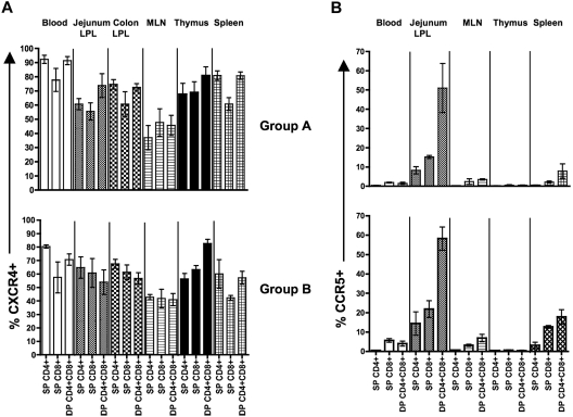
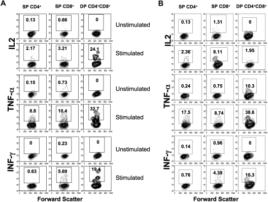
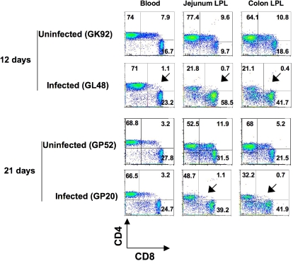
Peripheral blood and thymic double-positive (DP) CD4(+)CD8(+) T cells from neonates have been described earlier, but the function and immunophenotypic characteristics of other tissue-derived DP T cells are not clearly understood. Here, we demonstrate the functional and immunophenotypic characteristics of DP cells in 6 different tissues, including thymus from normal neonatal rhesus macaques (Macaca mulatta) between 0 and 21 days of age. In general, intestinal DP T cells of neonates have higher percentages of memory markers (CD28(+)CD95(+)CD45RA(low)CD62L(low)) and proliferation compared with single-positive (SP) CD4(+) and CD8(+) T cells. In addition, percentages of DP T cells increase and CD62L expression decreases as animals mature, suggesting that DP cells mature and proliferate with maturity and/or antigen exposure. Consistent with this, intestinal DP T cells in neonates express higher levels of CCR5 and are the primary targets in simian immunodeficiency virus (SIV) infection. Finally, DP T cells produce higher levels of cytokine in response to mitogen stimulation compared with SP CD4(+) or CD8(+) T cells. Collectively, these findings demonstrate that intestinal DP T cells of neonates are proliferating, activated memory cells and are likely involved in regulating immune responses, in contrast to immature DP T cells in the thymus.


Figures

Figure 1
Figure 1
Distribution of SP CD4+, CD8+ and DP T cells in various tissues and their phenotype from normal uninfected neonates of 2 age groups. (A) Mean percentages of SP and DP CD4+/CD8+ T-cell populations of normal uninfected neonates are shown. Each bar represents the mean plus or minus SEM percentage of T-cell subsets in newborn to 3-day-old (group A) versus 12- to 21-day-old (group B) neonates. Note that DP T cells increase in tissues, particularly the intestine with age. (B) Representative flow cytograms of SP CD4+, CD8+, and DP T cells showing the percentages of naive (CD28+CD95), central memory (CD28+CD95+), and effector memory (CD28CD95+) T-cell populations in each polygonal gate as well as other naive/memory markers (CD45RA, CCR7), L-selectin (CD62L), and the early activation marker (CD69) in jejunum LPLs in each quadrant from a normal uninfected 21-day-old neonate examined by 9-color flow cytometry. Note, that in contrast to SP T cells, essentially all DP cells in the jejunum have a “transitional memory” (CD28+CD95+CCR7+) phenotype. Plots were generated by gating first through lymphocytes using forward scatter (FSC) and side scatter (SSC) parameters and then through CD3+ T cells. (C) Mean frequencies of naive and memory cell populations are shown for different tissues from neonates of different ages. Each bar represents mean plus or minus SEM. * indicates significant differences between group A and B neonates for the specified T-cell subsets.
Figure 2
Figure 2
Relative expression of activation, trafficking, and naive/memory markers expression in different subpopulations of CD3+ T cells. Mean percentages of CD69, CD62L, CD45RA, and CCR7 are compared between different T-cell subsets (SP, DP) and between age groups. Bars represent mean values of 3 to 4 macaques plus or minus SEM. * indicates significant differences between group A (0- to 3-day-old) and B (12- to 21-day-old) neonates for the specified T-cell subsets.
Figure 3
Figure 3
Increased CCR5 expression by DP CD4+CD8+ T cells subsets in neonates. Mean percentages of cells expressing CXCR4 (A) and CCR5 (B) are shown for all subpopulation of T cells as indicated. Percentages represent means of 3 to 4 macaques plus or minus SEM.
Figure 4
Figure 4
Increased cytokine production by DP CD4+CD8+ T-cell subsets in neonates. Cytokine production by mitogen-stimulated or unstimulated jejunum lamina propria (A) and spleen (B) T-cell subsets in a normal, uninfected neonate at 12 days old. Cells were unstimulated or stimulated with PMA/ionomycin, and cytokine production was detected by intracellular cytokine staining. The percentage of cytokine-producing cells is indicated in the top box of each panel. Note that DP T cells had higher IL-2, TNF-α, and IFN-γ responses compared with SP CD4+/CD8+ T cells in jejunum LPLs, whereas SP CD8+ T cells had higher IL-2 responses in spleen lymphocytes.
Figure 5
Figure 5
Increased proliferative capacity in DP CD4+CD8+ T cells. (A) Bar charts showing the mean BrdU+ proliferative responses in different T-cell subsets from different tissues. (B) Mean BrdU expression in naive (CD95) and memory (CD95+) T-cell subsets are shown from different tissues in normal, uninfected neonates 12 to 21 days old. Each bar represents mean plus or minus SEM. Representative flow cytograms show increased proliferative capacity in DP T cells in whole blood (C) and jejunum LPLs (D). Note that memory (CD95+) cells have higher proliferative responses compared with naive (CD95) cells. Percentages of CD95+ and BrdU+ cells are shown in each quadrant (C and D).
Figure 6
Figure 6
Early loss of DP T cells in intestinal tissues in SIV infection. Dot plots comparing T-cell subsets from intestinal (jejunum and colon) tissues and peripheral blood of 2 normal, uninfected neonatal macaques (GK92 and GP52) to age-matched SIVmac251-infected (GL48 and GP20) neonates. Plots were generated by gating on lymphocytes and then through CD3+ T cells. Note that a more dramatic loss of DP CD4+CD8+ T cells was detected in intestinal tissues in infected macaques very early in infection (as indicated by the arrows) compared with other tissues. The percentages of CD4+ and CD8+ cells are shown in each quadrant.

References

    1. Veazey RS, Mansfield KG, Tham IC, et al. Dynamics of CCR5 expression by CD4(+) T cells in lymphoid tissues during simian immunodeficiency virus infection. J Virol. 2000;74:11001–11007. - PMC - PubMed
    1. Pahar B, Lackner AA, Veazey RS. Intestinal double-positive CD4+CD8+ T cells are highly activated memory cells with an increased capacity to produce cytokines. Eur J Immunol. 2006;36:583–592. - PubMed
    1. Sala P, Tonutti E, Feruglio C, Florian F, Colombatti A. Persistent expansions of CD4+ CD8+ peripheral blood T cells. Blood. 1993;82:1546–1552. - PubMed
    1. Ortolani C, Forti E, Radin E, Cibin R, Cossarizza A. Cytofluorimetric identification of two populations of double positive (CD4+,CD8+) T lymphocytes in human peripheral blood. Biochem Biophys Res Commun. 1993;191:601–609. - PubMed
    1. Weiss L, Roux A, Garcia S, et al. Persistent expansion, in a human immunodeficiency virus-infected person, of V beta-restricted CD4+CD8+ T lymphocytes that express cytotoxicity-associated molecules and are committed to produce interferon-gamma and tumor necrosis factor-alpha. J Infect Dis. 1998;178:1158–1162. - PubMed

Publication types

MeSH terms