. 2008 Nov 21;283(47):32704-14.
doi: 10.1074/jbc.M802066200. Epub 2008 Sep 26.

Distinct roles of beta1 metal ion-dependent adhesion site (MIDAS), adjacent to MIDAS (ADMIDAS), and ligand-associated metal-binding site (LIMBS) cation-binding sites in ligand recognition by integrin alpha2beta1

Affiliations

Distinct roles of beta1 metal ion-dependent adhesion site (MIDAS), adjacent to MIDAS (ADMIDAS), and ligand-associated metal-binding site (LIMBS) cation-binding sites in ligand recognition by integrin alpha2beta1

Dimitra Valdramidou et al. J Biol Chem. .

Abstract

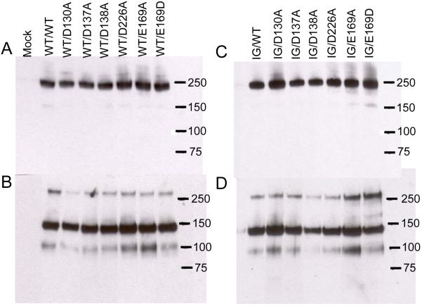
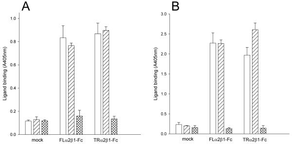
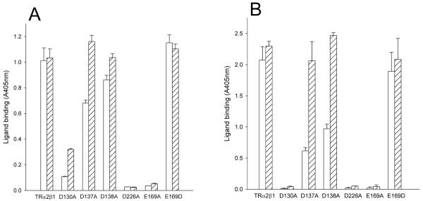
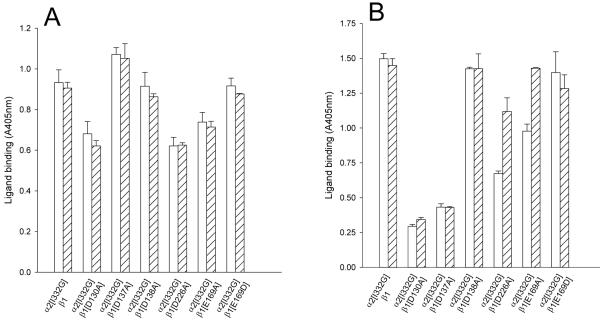
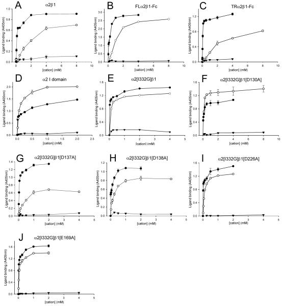
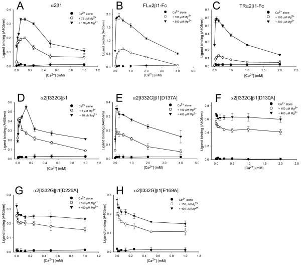
Integrin-ligand interactions are regulated in a complex manner by divalent cations, and previous studies have identified ligand-competent, stimulatory, and inhibitory cation-binding sites. In collagen-binding integrins, such as alpha2beta1, ligand recognition takes place exclusively at the alpha subunit I domain. However, activation of the alphaI domain depends on its interaction with a structurally similar domain in the beta subunit known as the I-like or betaI domain. The top face of the betaI domain contains three cation-binding sites: the metal-ion dependent adhesion site (MIDAS), the ADMIDAS (adjacent to MIDAS), and LIMBS (ligand-associated metal-binding site). The role of these sites in controlling ligand binding to the alphaI domain has yet to be elucidated. Mutation of the MIDAS or LIMBS completely blocked collagen binding to alpha2beta1; in contrast mutation of the ADMIDAS reduced ligand recognition but this effect could be overcome by the activating monoclonal antibody TS2/16. Hence, the MIDAS and LIMBS appear to be essential for the interaction between alphaI and betaI, whereas occupancy of the ADMIDAS has an allosteric effect on the conformation of betaI. An activating mutation in the alpha2 I domain partially restored ligand binding to the MIDAS and LIMBS mutants. Analysis of the effects of Ca(2+), Mg(2+), and Mn(2+) on ligand binding to these mutants showed that the MIDAS is a ligand-competent site through which Mn(2+) stimulates ligand binding, whereas the LIMBS is a stimulatory Ca(2+)-binding site, occupancy of which increases the affinity of Mg(2+) for the MIDAS.


Figures

FIGURE 1
FIGURE 1. Western blotting of recombinant TRα2β1-Fc integrins
Recombinant integrins were partially purified from cell culture supernatants using protein A Sepharose. Aliquots of purified proteins were run on 3-8 % SDS-PAGE gels under non-reducing (panels A, B) or reducing conditions (panels C, D), transferred to nitrocellulose and blotted with anti-human Fc conjugated with horseradish peroxidase. Panels A and C, culture supernatants from cells transfected with wild-type (WT) TRα2 and WT or cation-binding site mutant TRβ1. Panels B and D, culture supernatants from cells transfected with I332G mutant TRα2 (IG) and WT or cation-binding site mutant TRβ1. Migration positions of molecular weight markers are indicated (kDa). Under non-reducing conditions a predominant band at ~ 240 kDa is observed, close to the expected size for the TRα2β1-Fc heterodimer. Under reducing conditions disulfide bonds between the Fc hinge regions are broken and two predominant bands of ~ 140 kDa and ~ 100 kDa are observed, corresponding to the expected sizes of TRα2 and TRβ1 proteins, respectively. For unknown reasons the β subunit band reacts much more weakly with the anti-human Fc antibody than the α subunit band (31). The 240 and 100 kDa bands are reactive with the anti-β1 mAb TS2/16; the 240 and 140 kDa bands are reactive with anti-α2 mAb JA212 (not shown). These bands are not observed in proteins purified from the supernatants of mock-transfected cells.
FIGURE 2
FIGURE 2. Collagen binding to recombinant α2β1-Fc integrins
Culture supernatants from CHO L761h cells. transfected with FLα2 and FLβ1 constructs (FLα2β1-Fc), TRα2 and TRβ1 constructs (TRα2β1-Fc) or no DNA (mock) were analysed by Fc-capture assays. Binding of collagen I (1 μg/ml) to captured proteins was measured in 5 mM Mg2+ (A) or 1 mM Mn2+ (B) in the absence (open bars) or presence of the activating anti-β1 mAb TS2/16 (diagonally-hatched bars) or 5 mM EDTA (cross-hatched bars). Supernatants from cells transfected with either α2 or β1 constructs alone did not show any collagen binding above that of supernatants from mock-transfected cells (data not shown). Absorbance values and error bars represent the mean and standard deviation of three replicate wells from one experiment, representative of four separate experiments.
FIGURE 3
FIGURE 3. Collagen binding to TRα2β1-Fc cation-binding site mutants
Culture supernatants from transfected CHO L761h cells with TRα2 and wild-type TRβ1 or TRβ1 mutated at the MIDAS (D130A), ADMIDAS (D137A, D138A) or LIMBS (D226A, E169A, E169D) were analysed in Fc-capture assays, performed in the presence of 5 mM Mg2+ (A) or 1 mM Mn2+ (B) in the absence (open bars) or presence of the activating mAb TS2/16 (diagonally-hatched bars). Absorbance values and error bars represent the mean and standard deviation of three replicate wells from one experiment, representative of three separate experiments. Non-specific collagen binding (as estimated from the binding to wells treated with supernatant from mock-transfected cells) has been subtracted in this and all subsequent figures.
FIGURE 4
FIGURE 4. Collagen binding to TRα2[I332G]β1-Fc cation-binding site mutants
Culture supernatants from transfected CHO L761h cells with TRα2 and wild-type TRβ1 or TRβ1 mutated at the MIDAS (D130A), ADMIDAS (D137A, D138A) or LIMBS (D226A, E169A, E169D) were analysed in Fc-capture assays, performed in the presence of 5mM Mg2+ (A) or 1 mM Mn2+ (B). The ligand-binding capacity of the α2β1-Fc mutants was detected with 0.5 μg/ml collagen I in the absence (open bars) or the presence of anti-β1 antibody TS2/16 (diagonally hatched bars). Absorbance values and error bars represent the mean and standard deviation from one experiment, representative of three separate experiments.
FIGURE 5
FIGURE 5. Effect of divalent cations on the binding of collagen to purified α2β1 and recombinant proteins
Binding of collagen I to purified α2β1 and to recombinant proteins was measured in the presence of varying concentrations of Mn2+ (●), Mg2+ (○), or Ca2+ (▲). Purified α2β1 (A), FLα2β1-Fc (B), TRα2β1-Fc (C), α2 I domain (D), TRα2[I332G]β1-Fc (E), TRα2[I332G]β1[D130A]-Fc (F), TRα2[I332G]β1[D137A]-Fc (G), TRα2[I332G]β1[D138A]-Fc (H), TRα2[I332G]β1[D226A]-Fc (I), and TRα2[I332G]β1[E169A]-Fc (J). Each experiment shown is representative of at least three separate experiments.
FIGURE 6
FIGURE 6. Effect of Ca2+ on Mg2+-dependent collagen binding to purified α2β 1 and recombinant proteins
Purified α2β1 (A), FLα2β1-Fc (B), TRα2β1-Fc (C), TRα2[I332G]β1-Fc (D), TRα2[I332G]β1[D137A]-Fc (E), TRα2[I332G]β1[D130A]-Fc (F), TRα2[I332G]β1[D226A]-Fc (G), TRα2[I332G]β1[E169A]-Fc( H). Binding of collagen I was measured in the presence of two constant concentrations of Mg2+ (as indicated in the panel legend) with varying concentrations of Ca2+. The level of ligand binding supported by Ca2+ alone is also shown. Each experiment shown is representative of at least three separate experiments.

References

    1. Hynes RO. Cell. 2002;110:673–687. - PubMed
    1. Takada Y, Ye X, Simon S. Genome Biol. 2007;8:215. - PMC - PubMed
    1. Luo BH, Carman CV, Springer TA. Annu. Rev. Immunol. 2007;25:619–647. - PMC - PubMed
    1. Mould AP, Humphries MJ. Curr. Opin. Cell Biol. 2004;16:544–551. - PubMed
    1. Takagi J, Petre BM, Walz T, Springer TA. Cell. 2002;110:599–611. - PubMed

Publication types