. 2009 Feb;24(2):404-12.
doi: 10.1093/ndt/gfn528. Epub 2008 Sep 27.

Rosuvastatin protects against podocyte apoptosis in vitro

Affiliations

Rosuvastatin protects against podocyte apoptosis in vitro

Fionnuala C Cormack-Aboud et al. Nephrol Dial Transplant. 2009 Feb.

Abstract

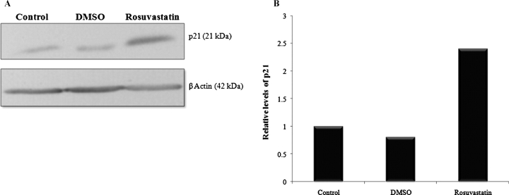
Background: Clinical studies suggest that statins reduce proteinuria and slow the decline in kidney function in chronic kidney disease. Given a rich literature identifying podocyte apoptosis as an early step in the pathophysiological progression to proteinuria and glomerulosclerosis, we hypothesized that rosuvastatin protects podocytes from undergoing apoptosis. Regarding a potential mechanism, our lab has shown that the cell cycle protein, p21, has a prosurvial role in podocytes and there is literature showing statins upregulate p21 in other renal cells. Therefore, we queried whether rosuvastatin is prosurvival in podocytes through a p21-dependent pathway.

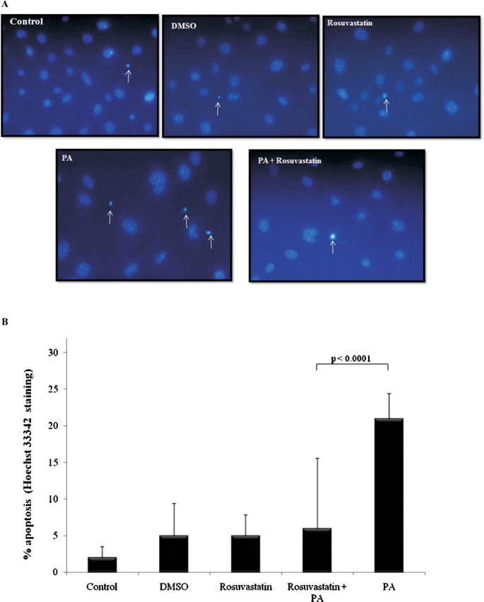
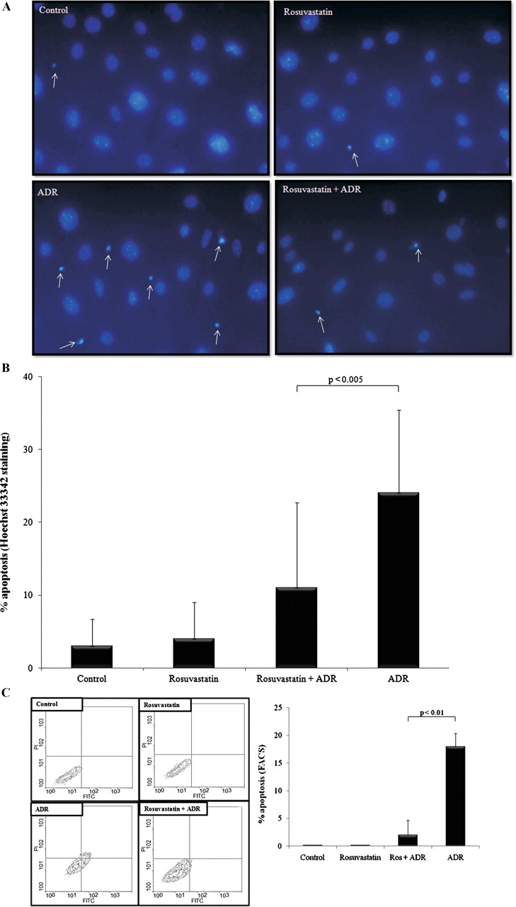
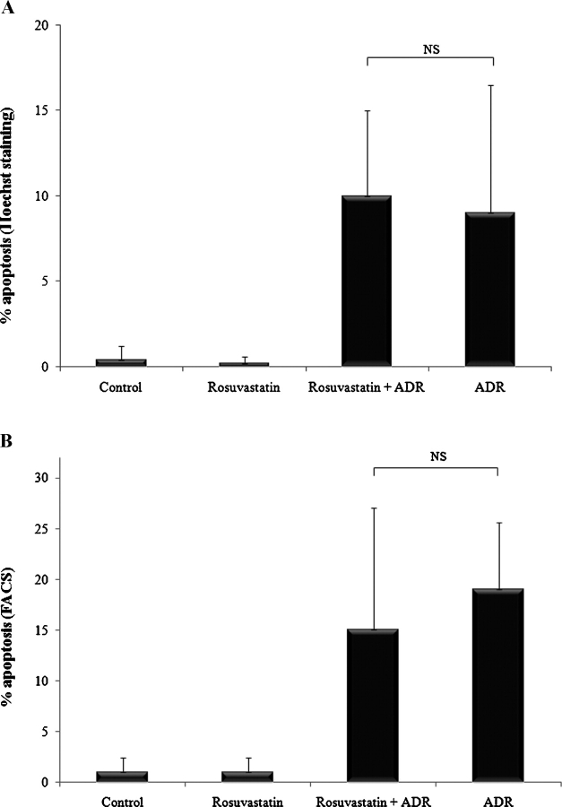
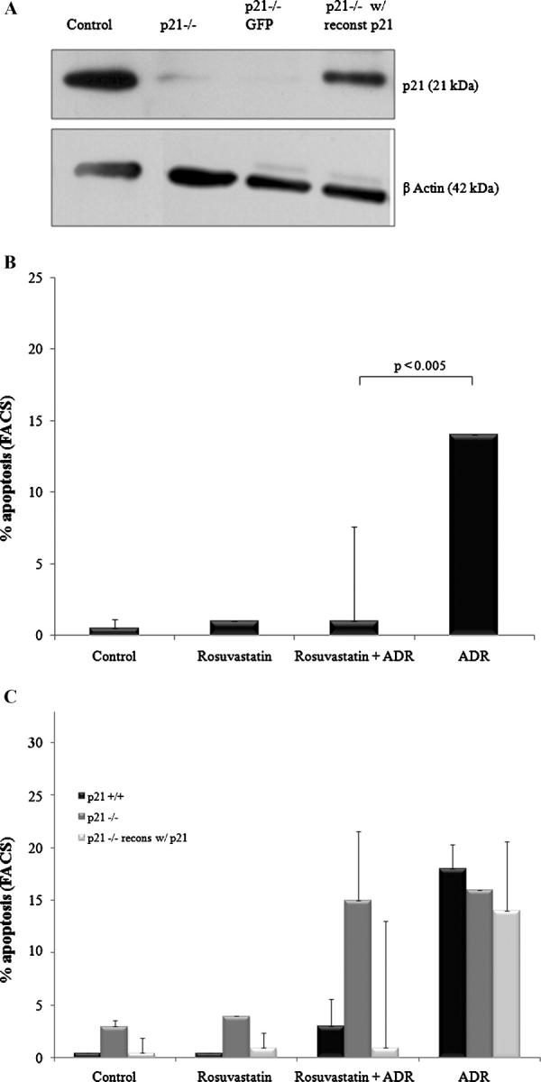
Methods: Two independent apoptotic triggers, puromycin aminonucleoside (PA) and adriamycin (ADR), were used to induce apoptosis in p21 +/+ and p21 -/- conditionally immortalized mouse podocytes with or without pre-exposure to rosuvastatin. Apoptosis was measured by two methods: Hoechst 33342 staining and fluorescence-activated cell sorting (FACS). To establish a role for p21, p21 levels were measured by western blotting following rosuvastatin exposure and p21 was stably transduced into p21 -/- mouse podocytes.

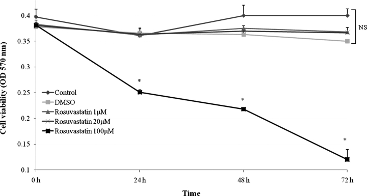
Results: Rosuvastatin protects against ADR- and PA-induced apoptosis in podocytes. Further, exposure to rosuvastatin increases p21 levels in podocytes in vitro. ADR induces apoptosis in p21 -/- mouse podocytes, but rosuvastatin's protective effect is not seen in the absence of p21. Reconstituting p21 in p21 -/- podocytes restores rosuvastatin's prosurvival effect.

Conclusion: Rosuvastatin is prosurvival in injured podocytes. Rosuvastatin exerts its protective effect through a p21-dependent antiapoptotic pathway. These findings suggest that statins decrease proteinuria by protecting against podocyte apoptosis and subsequent podocyte depopulation.


Figures

Fig. 1
Fig. 1
Low-dose rosuvastatin does not affect podocyte viability in vitro. Growth-restricted conditionally immortalized mouse podocytes were incubated with escalating doses of rosuvastatin or DMSO as a vehicle control. Cell viability was determined by MTT assay at 24, 48 and 72 h time points (*P < 0.05).
Fig. 2
Fig. 2
Rosuvastatin protects podocytes from puromycin aminonucleoside-induced apoptosis. Growth-restricted mouse podocytes pre-incubated with rosuvastatin (20 μM) or DMSO (vehicle) for 24 h were exposed to puromycin aminonucleoside (PA, 30 μg/ml) for 48 h. Hoechst staining was performed (A) and the percentage of apoptosis was measured (B).
Fig. 3
Fig. 3
Rosuvastatin protects podocytes from adriamycin-induced apoptosis. Podocytes pre-incubated for 24 h with rosuvastatin or DMSO were exposed to adriamycin (ADR, 0.25 μg/ml) for 48 h. Hoechst staining was performed (A) and the percentage of apoptosis was measured (B). Additionally, cells were dual-labeled with FITC-Annexin V and propidium iodide and fluorescent-activated cell sorting (FACS) performed. Representative contour plots (C) show virtually all of the cells in control and rosuvastatin-treated groups were viable, as detected in the lower-left quadrant (FITC, PI). ADR exposure significantly increased the percentage of apoptotic (FITC+, PI) cells, as detected in the lower-right quadrant. Pre-incubation with rosuvastatin largely abrogated the rightward shift induced by ADR, consistent with a prosurvival effect. The results of three separate FACS experiments were pooled and averaged results are shown graphically.
Fig. 4
Fig. 4
Rosuvastatin increases protein levels of p21 in podocytes in vitro. Growth-restricted mouse podocytes were pre-incubated for 24 h with rosuvastatin or DMSO and protein levels of p21WAF1/CIP1 +/+ measured by Western blot analysis. A representative blot is shown (A). β-actin was used as a housekeeping protein to ensure equal protein loading and densitometric analysis performed (B). Values are averages of three separate experiments and normalized for differences in protein loading.
Fig. 5
Fig. 5
The prosurvival effect of rosuvastatin is negated in the absence of p21. p21 −/− growth-restricted conditionally immortalized mouse podocytes were pre-incubated with rosuvastatin or DMSO. Cells were exposed to ADR and apoptosis measured 48 h later by Hoechst staining (A) and FACS analysis (B).
Fig. 6
Fig. 6
Reconstituting p21 expression in p21 −/− podocytes restores the prosurvival effect of rosuvastatin. Growth-restricted p21 −/− mouse podocytes were stably transfected with a human p21 vector or GFP vector as a control. Western blotting for p21WAF1/CIP1 +/+ showed successful re-expression of p21 in the p21 −/− podocytes (A). Following stable transfection, cells were pre-incubated with rosuvastatin or DMSO and ADR-induced apoptosis was measured by FACS analysis (B). The prosurvival effects of rosuvastatin in podocytes as a function of p21 status are summarized in (C).

References

    1. Mundel P, Shankland SJ. Podocyte biology and response to injury. J Am Soc Nephrol. 2002;13:3005–3015. - PubMed
    1. Kriz W, Elger M, Nagata M, et al. The role of podocytes in the development of glomerular sclerosis. Kidney Int Suppl. 1994;45:S64–S72. - PubMed
    1. Kriz W, Gretz N, Lemley KV. Progression of glomerular diseases: is the podocyte the culprit? Kidney Int. 1998;54:687–697. - PubMed
    1. Kriz W, Hosser H, Hahnel B, et al. From segmental glomerulosclerosis to total nephron degeneration and interstitial fibrosis: a histopathological study in rat models and human glomerulopathies. Nephrol Dial Transplant. 1998;13:2781–2798. - PubMed
    1. Pagtalunan ME, Miller PL, Jumping-Eagle S, et al. Podocyte loss and progressive glomerular injury in type II diabetes. J Clin Invest. 1997;99:342–348. - PMC - PubMed

Publication types

MeSH terms