Review
. 2008;26(2-3):97-115.

Mechanisms of CNS myelin inhibition: evidence for distinct and neuronal cell type specific receptor systems

Affiliations
Review

Mechanisms of CNS myelin inhibition: evidence for distinct and neuronal cell type specific receptor systems

Roman J Giger et al. Restor Neurol Neurosci. 2008.

Abstract

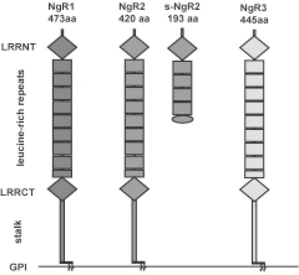
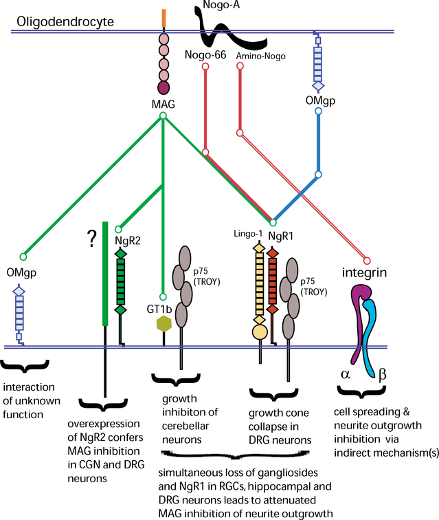
Following injury to the adult mammalian central nervous system, regenerative growth of severed axons is very limited. The lack of neuronal repair is often associated with significant functional deficits, and depending on the severity of injury, may result in permanent paralysis distal to the site of injury. A detailed understanding of the molecular mechanisms that limit neuronal growth in the injured spinal cord is an important step toward the development of specific strategies aimed at restoring functional connectivity lost as a consequence of injury. While rapid progress is being made in defining the molecular identity of CNS growth inhibitory constituents, comparatively little is known about their receptors and downstream signaling mechanisms. Emerging new evidence suggests that the mechanisms for myelin inhibition are likely to be complex, involving multiple and distinct receptor systems that may operate in a redundant manner. Furthermore, the relative contribution of a specific ligand-receptor system to bring about growth inhibition may greatly vary among different neuronal cell types. Myelin-associated glycoprotein (MAG), for example, employs different mechanisms to inhibit neurite outgrowth of cerebellar, sensory, and retinal ganglion neurons in vitro. Nogo-A harbors distinct growth inhibitory regions, which employ different signaling mechanisms. The Nogo-66 receptor 1 (NgR1), a shared ligand binding component in a receptor complex for Nogo-66, MAG, and OMgp, participates in neuronal growth cone collapse to acutely presented myelin inhibitors, but is dispensable for longitudinal neurite outgrowth inhibition on substrate-bound Nogo-66, MAG, OMgp, or crude CNS myelin in vitro. Consistent with the idea of cell-type specific mechanisms for myelin inhibition, different types of CNS neurons possess very different regenerative capacities and respond differently to experimental treatment strategies in vivo. We speculate that differences in regenerative axonal growth among different fiber systems are a reflection of their intrinsic ability to elongate axons and their distinct cell surface receptor profiles to respond to the growth inhibitory extracellular milieu. The existence of cell type specific mechanisms to impair regenerative axonal growth in the CNS may have important implications for the development of treatment strategies. Depending on the fiber tract injured, different ligand-receptor systems may need to be targeted in order to elicit robust and long-distance regenerative axonal growth.


Figures

Fig. 1.
Fig. 1.
The Nogo receptor family. The Nogo receptors NgR1, NgR2, and NgR3 comprise a small subfamily of leucine-rich repeat (LRR) proteins. The three receptors share an identical overall domain organization composed of a cluster of 8.5 canonical LRRs flanked N-terminally by a LRRNT capping domain and C-terminally by a LRRCT capping domain. The LRRNT-LRR-LRRCT domains are connected via a stalk region to a glycosylphosphatidyl inositol (GPI) anchor. We identified s-NgR2, a soluble form of NgR2 that is the product of alternative splicing of exon III of the NgR2 gene. The first 171 amino acid residues of s-NgR2 are identical with full-length NgR2 and are followed by 22 amino acids unique to s-NgR2. NgR1 is a 65-kDa glycoprotein that supports binding of Nogo, MAG, and OMgp. NgR2 is a 65-kDa glycoprotein and supports binding of MAG but not Nogo or OMgp. Thus far no binding partners for NgR3 have been identified.
Fig. 2.
Fig. 2.
Molecular players of neurite outgrowth inhibition in the CNS. “Metro map” of ligand-receptor interactions of the prototypic myelin inhibitors Nogo-A, MAG, and OMgp. The “Green line” depicts the associations of MAG with its receptors, the “Red line” depicts the associations of Nogo-A (Amino-Nogo and Nogo-66) with its receptors, and the “Blue line” depicts the association of OMgp with its receptor. Nogo-66, MAG, and OMgp associate with the NgR1/p75/Lingo-1 complex. MAG also binds to gangliosides (including GT1b), NgR2, and axonal OMgp. NgR2 does not associate with p75, TROY, or Lingo-1 and the signal-transducing component (green box) in the NgR2 complex is not known. Amino-Nogo interacts with neuronal integrins, however, this interaction is thought to be indirect. See main text for more details on the functional significance of these interactions.
Fig. 3.
Fig. 3.
NgR1 expression in brain and spinal cord. β-galactosidase reporter gene expression analysis in NgR1tauLacZ mice (Zheng et al., 2005), as assessed by X-gal histochemistry. A. In brain tissue sections of one-month-old mice, strongest β-galactosidase activity is observed in the cerebral cortex, the corpus callosum (CC), anterior commissure (AC), and optic chiasm (OC). Weaker β-galactosidase activity is found in the medial septum and the striatum/caudate putamen (CPu). B. In the spinal cord of one-month-old mice, β-galactosidase activity is most robust in the corticospinal tract (CST). Weaker labeling is detected in the dorso-lateral spinal while matter including presumptive fiber tracts such as the rubrospinal or raphespinal tract. Also weak labeling is detected in the dorsal gray matter of the spinal cord, and may arise from DRG neuron afferent projections. No reporter gene expression was detected in motoneuron pools or the ventral gray and white matter of the spinal cord. This suggests that robust NgR1 expression in the spinal cord is restricted to a small number of fiber tracts.

References

    1. Barrette B, Vallieres N, Dube M, & Lacroix S (2007). Expression profile of receptors for myelin-associated inhibitors of axonal regeneration in the intact and injured mouse central nervous system. Mol Cell Neurosci, 34, 519–538. - PubMed
    1. Barton WA, Liu BP, Tzvetkova D, Jeffrey PD, Fournier AE, Sah D, et al. (2003). Structure and axon outgrowth inhibitor binding of the Nogo-66 receptor and related proteins. EMBO J, 22, 3291–3302. - PMC - PubMed
    1. Bartsch U, Bandtlow CE, Schnell L, Bartsch S, Spillmann AA, Rubin BP, et al. (1995). Lack of evidence that myelin-associated glycoprotein is a major inhibitor of axonal regeneration in the CNS. Neuron, 15, 1375–1381. - PubMed
    1. Benson MD, Romero MI, Lush ME, Lu QR, Henkemeyer M, & Parada LF (2005). Ephrin-B3 is a myelin-based inhibitor of neurite outgrowth. Proc Natl Acad Sci USA, 102, 10694–10699. - PMC - PubMed
    1. Bonilla IE, Tanabe K,& Strittmatter SM(2002). Small prolinerich repeat protein 1A is expressed by axotomized neurons and promotes axonal outgrowth. J Neurosci, 22, 1303–1315. - PMC - PubMed

Publication types