. 2008 Nov 6;456(7218):116-20.
doi: 10.1038/nature07338. Epub 2008 Sep 28.

The CRAC channel consists of a tetramer formed by Stim-induced dimerization of Orai dimers

Affiliations

The CRAC channel consists of a tetramer formed by Stim-induced dimerization of Orai dimers

Aubin Penna et al. Nature. .

Abstract

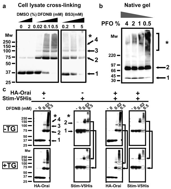
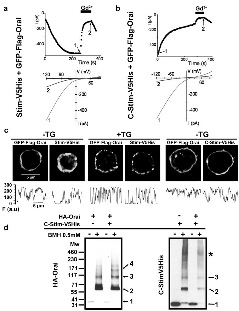
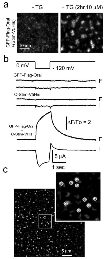
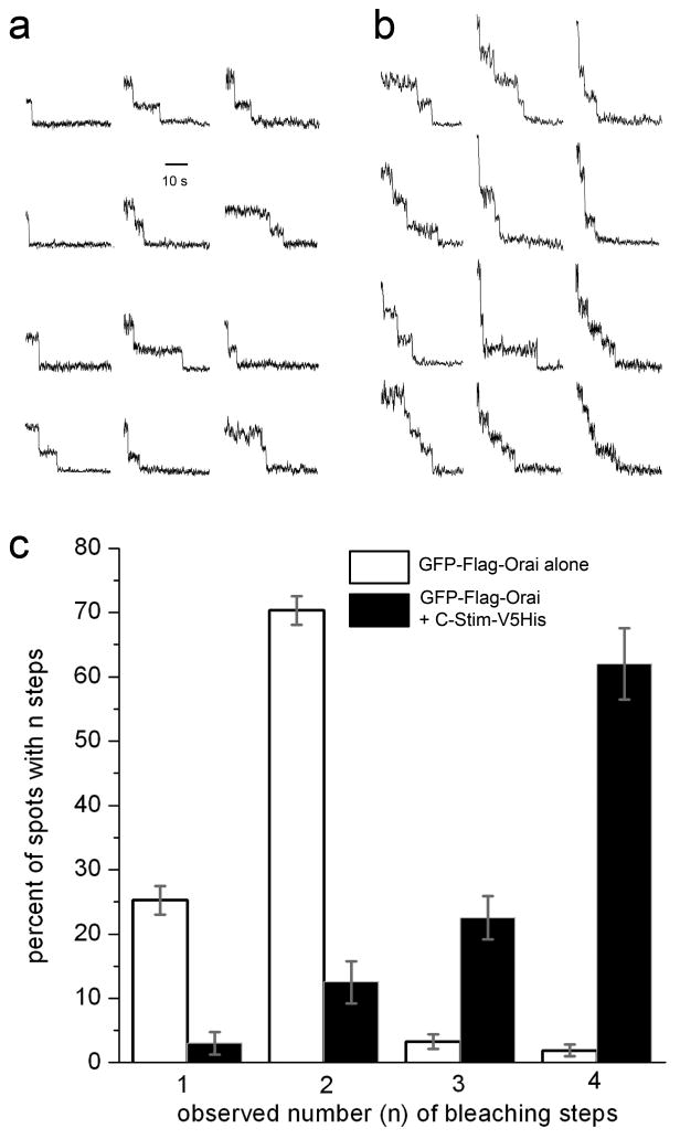
Ca(2+)-release-activated Ca(2+) (CRAC) channels underlie sustained Ca(2+) signalling in lymphocytes and numerous other cells after Ca(2+) liberation from the endoplasmic reticulum (ER). RNA interference screening approaches identified two proteins, Stim and Orai, that together form the molecular basis for CRAC channel activity. Stim senses depletion of the ER Ca(2+) store and physically relays this information by translocating from the ER to junctions adjacent to the plasma membrane, and Orai embodies the pore of the plasma membrane calcium channel. A close interaction between Stim and Orai, identified by co-immunoprecipitation and by Förster resonance energy transfer, is involved in the opening of the Ca(2+) channel formed by Orai subunits. Most ion channels are multimers of pore-forming subunits surrounding a central channel, which are preassembled in the ER and transported in their final stoichiometry to the plasma membrane. Here we show, by biochemical analysis after cross-linking in cell lysates and intact cells and by using non-denaturing gel electrophoresis without cross-linking, that Orai is predominantly a dimer in the plasma membrane under resting conditions. Moreover, single-molecule imaging of green fluorescent protein (GFP)-tagged Orai expressed in Xenopus oocytes showed predominantly two-step photobleaching, again consistent with a dimeric basal state. In contrast, co-expression of GFP-tagged Orai with the carboxy terminus of Stim as a cytosolic protein to activate the Orai channel without inducing Ca(2+) store depletion or clustering of Orai into punctae yielded mostly four-step photobleaching, consistent with a tetrameric stoichiometry of the active Orai channel. Interaction with the C terminus of Stim thus induces Orai dimers to dimerize, forming tetramers that constitute the Ca(2+)-selective pore. This represents a new mechanism in which assembly and activation of the functional ion channel are mediated by the same triggering molecule.


Conflict of interest statement

The authors declare no competing financial interests.

Figures

Figure 1
Figure 1. Orai is present mainly as homodimers in resting S2 cells
Each panel is representative of at least 3 independent experiments. Numbers represent assigned state of oligomerization: 1, monomer; 2, dimer; 3, trimer; 4, tetramer; * high-order aggregates. a, Determination of Orai oligomeric structure using chemical cross-linking. DFDNB (1,5-difluoro-2,4-dinitrobenzene, membrane-permeant) and BS3 (bis(sulfosuccimidyl)suberate, membrane-impermeant) were incubated with HA-Orai transfected S2 cells lysates and the sizes of the cross-linked products were analyzed by SDS-PAGE on 4-12% gradient gels. Orai oligomers, from dimer to tetramer, were observed with dimer always being the predominant population. Similar results were obtained in intact cells using DSP (dithiobis succinimidylpropionate, membrane-permeant, data not shown), DFDNB (see Supplementary Fig. 2, 3), and a cysteine-reactive cross-linker BMH (maleimide 1,6-bismaleimidohexane, membrane permeable). b, Confirmation of Orai dimerization using PFO-PAGE. HA-Orai transfected S2 cell lysates were incubated with sample buffer containing different PFO concentration for 30 minutes at room temperature before electrophoresis. c, DFDNB cross-linking of total cell lysates of S2 cells tranfected with HA-Orai only, Stim-V5His only or co-transfected. Labels indicate untreated cells (-), DMSO vehicle control (0) and concentrations of DFDNB. Arrows show the reduction in Stim and Orai low-order oligomers upon Ca2+ store depletion by TG (1.5 μM TG for 15 min).
Figure 2
Figure 2. The C-terminus of Stim (C-Stim) constitutively activates Orai without forming punctae
a, b Time course (upper) and I-V curves (lower) comparing CRAC current in representative cells co-transfected with Stim + GFP-Orai (a) or with C-Stim + GFP-Orai (b). Application of 5 nM Gd3+ reversibly blocked most of the current in cells transfected with either Stim + GFP-Orai or C-Stim + GFP-Orai. c, Subcellular localization of GFP-Orai together with Stim-V5His or C-Stim-V5His in resting (StimV5His and C-StimV5His) or store-depleted (2 μM TG for 15 min, Stim-V5His only) co-transfected S2 cells. Puncta formation was only observed upon store-depletion in cells expressing StimV5His and GFP-Orai. The panels underneath each picture display fluorescence intensity profiles (F) for Orai and Stim obtained from the same regions of interest, tracing the perimeter of each cell clockwise from top. To quantify the extent to which GFP-Orai was inhomogeneously distributed we calculated the ratio of fluorescence variance/mean fluorescence from profiles like those illustrated. Mean ratio values for GFP-Orai (±SEM; n=6 cells for each condition) were: StimV5His, -TG, 29.03±2.9; StimV5His, +TG, 62.9±4.5 (p = 0.00002); C-Stim-V5His, -TG, 22.4±3.7 (not significantly different from StimV5His). d, BMH cross-linking in intact S2 cells transfected with HA-Orai only, C-Stim-V5His only or co-transfected. Numbers represent inferred state of oligomerization: 1, monomer; 2, dimer; 3, trimer; 4, tetramer; *, higher order aggregates. The cross-linking pattern of Orai showed a clear tetrameric population when co-expressed with C-Stim, but not in the absence of C-Stim. The cross-linking profile of C-Stim was not affected by the presence of Orai, and revealed a majority of dimers and trimers.
Figure 3
Figure 3. Single-molecule photo-bleaching of GFP-Orai in intact oocytes
a, Store depletion induces formation of Orai punctae in Xenopus oocytes. Images were obtained by TIRF microscopy of oocytes expressing GFP-Orai together with StimV5His and show 40 × 40 μm regions in the animal hemisphere before (left) and 2 hr after (right) bath application of 10 μM TG in zero-calcium Ringer's solution. b, Oocytes expressing GFP-Orai together with C-Stim showed strong Ca2+ influx, whereas this was absent with expression of GFP-Orai or C-Stim alone. Pairs of traces show cytosolic [Ca2+] as reported by normalized fluorescence pseudoratio changes of Fluo-4 (F) and whole-cell voltage-clamp measurements of Ca2+-activated Cl- current (I) in response to a hyperpolarizing step from 0 mV to -120 mV (top trace). c, Representative TIRFM image, acquired before photobleaching, showing fluorescent spots (circled) sparsely distributed in the membrane of an oocyte expressing GFP-Orai together with C-Stim. Inset shows a magnified view of a small region (white box), with circular regions of interest used to measure bleaching steps overlaid on the image.
Figure 4
Figure 4. GFP-Orai forms dimers in the basal state and predominantly tetramers when co-expressed with C-Stim
a, b Representative examples of single-molecule bleaching records obtained from oocytes expressing, respectively, GFP-Orai alone and GFP-Orai together with C-Stim. c, Histogram shows percentages of spots that showed 1, 2, 3, and 4 bleaching steps in oocytes expressing GFP-Orai alone (open bars) and GFP-Orai plus C-Stim (filled bars). Errors bars indicate ± 1 SEM. Data for GFP-Orai were obtained from 400 spots, 11 imaging records, 6 oocytes; and data for GFP-Orai + C-Stim from 278 spots, 5 imaging records, 3 oocytes. Comparison of bleaching step distributions with and without C-Stim yielded a Chi square value of 590; p < 0.001. This cannot be attributed to an increased likelihood of two GFP-Orai dimers happening to lie indistinguishably close to each other because of increased expression level or C-Stim-induced clustering because fluorescence spots in both conditions showed similar random distributions and densities (respectively, 37±6 and 41±4 spots in a 40×40 μm2 region), and we did not observe spots with >4 bleaching steps as might be expected for a macro-molecular clustering.

References

    1. Liou J, et al. STIM is a Ca2+ sensor essential for Ca2+-store-depletion-triggered Ca2+ influx. Curr Biol. 2005;15:1235–41. - PMC - PubMed
    1. Roos J, et al. STIM1, an essential and conserved component of store-operated Ca2+ channel function. J Cell Biol. 2005;169:435–45. - PMC - PubMed
    1. Feske S, et al. A mutation in Orai1 causes immune deficiency by abrogating CRAC channel function. Nature. 2006;441:179–85. - PubMed
    1. Zhang SL, et al. Genome-wide RNAi screen of Ca2+ influx identifies genes that regulate Ca2+ release-activated Ca2+ channel activity. Proc Natl Acad Sci U S A. 2006;103:9357–62. - PMC - PubMed
    1. Vig M, et al. CRACM1 is a plasma membrane protein essential for store-operated Ca2+ entry. Science. 2006;312:1220–3. - PMC - PubMed

Publication types

MeSH terms