. 2008 Oct 23;51(20):6334-47.
doi: 10.1021/jm800389v. Epub 2008 Sep 27.

The importance of micelle-bound states for the bioactivities of bifunctional peptide derivatives for delta/mu opioid receptor agonists and neurokinin 1 receptor antagonists

Affiliations

The importance of micelle-bound states for the bioactivities of bifunctional peptide derivatives for delta/mu opioid receptor agonists and neurokinin 1 receptor antagonists

Takashi Yamamoto et al. J Med Chem. .

Abstract

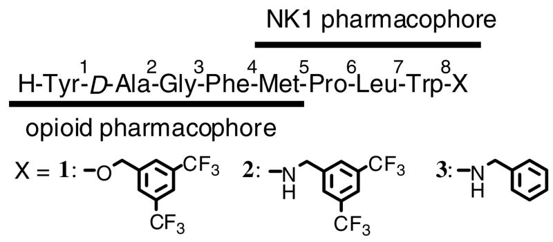
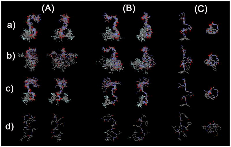
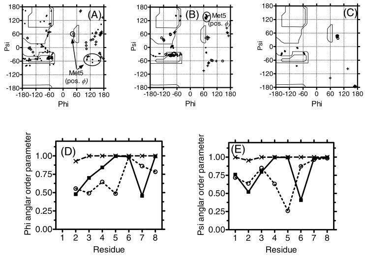
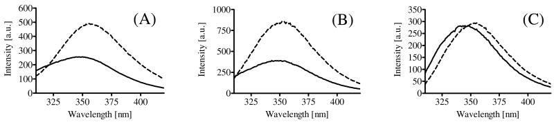
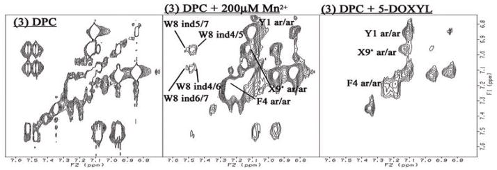
To provide new insight into the determining factors of membrane-bound peptide conformation that might play an important role in peptide-receptor docking and further biological behaviors, the dodecylphosphocholine (DPC) micelle-bound conformations of bifunctional peptide derivatives of delta-preferring opioid agonists and NK1 antagonists (1: Tyr-D-Ala-Gly-Phe-Met-Pro-Leu-Trp-O-3,5-Bzl(CF 3) 2; 2: Tyr-D-Ala-Gly-Phe-Met-Pro-Leu-Trp-NH-3,5-Bzl(CF 3) 2; 3: Tyr-D-Ala-Gly-Phe-Met-Pro-Leu-Trp-NH-Bzl) were determined based on 2D NMR studies. Although the differences in the primary sequence were limited to the C-terminus, the obtained NMR conformations were unexpectedly different for each compound. Moreover, their biological activities showed different trends in direct relation to the compound-specific conformations in DPC micelles. The important result is that not only were the NK1 antagonist activities different (the pharmacophore located at the C-terminus)but the opioid agonist activities (this pharmacophore was at the structurally preserved N-terminus) also were shifted, suggesting that a general conformational change in the bioactive state was induced due to relatively small and limited structural modifications.


Figures

Figure 1
Figure 1
Sequence of the bifunctional ligands synthesized and examined.
Figure 2
Figure 2
Fingerprint (HN-Hα) region of the NOESY spectrum of (A) 1, (B) 2 and (C) 3 in DPC micelles. Intraresidue HN-HαNOE cross-peaks are labeled with residue number, and arrows indicate the connectivity path from N-terminal to C-terminal. X9 represents the cross-peaks derived from the corresponding C-terminal HN and benzyl protons.
Figure 2
Figure 2
Fingerprint (HN-Hα) region of the NOESY spectrum of (A) 1, (B) 2 and (C) 3 in DPC micelles. Intraresidue HN-HαNOE cross-peaks are labeled with residue number, and arrows indicate the connectivity path from N-terminal to C-terminal. X9 represents the cross-peaks derived from the corresponding C-terminal HN and benzyl protons.
Figure 3
Figure 3
Diagram of HN-Hα coupling constants, NOE connectivities, and Hα chemical shift index (CSI) for (A) 1, (B) 2 and (C) 3. The Hα CSI was calculated using the random-coil values reported by Andersen et al., The residual interresidue NOE distance restraints of 1 (left), 2 (middle) and 3 (right) (D). Each column shows the sequential (i, i + 1; open), medium-range (i, i + 2–4; hatched) and long-range restraints (i, i + >4; filled), respectively. The residue Bzl or 9 stands for the respective C-terminal moieties.
Figure 4
Figure 4
Ensembles of the best 20 calculated structures in 40-fold DPC micelle/pH 4.5 buffer for (A) 1, (B) 2 and (C) 3 with the lowest restraint energy, aligned on backbone atoms of residues (a) 1-8, (b) 1-4 and (c) 5-8, from N-terminal (up in the left image) to C-terminal (down). Only backbone atoms were illustrated in (a) and (b) for easier comparison, and the most stable conformers (c) are shown with all non-hydrogen atoms.
Figure 5
Figure 5
The D-Ala2 (crosses), Gly3 (open circle) and Met5 with positive ϕ angles (circled) were indicated in the Ramachandran ϕ, ψ plots for (A) 1, (B) 2 and (C) 3 for residues 2-7 of 20 final structures. Angular order parameters for ϕ (D) and ψ (E) angles calculated from the 20 final structures for 1 (open circles), 2 (filled squares) and 3 (crosses). For calculating the ψ angles of Trp8, Non-carbonyl oxygen atoms of the C-terminal ester (1) and nitrogen atoms of C-terminal amide (2 and 3) were used instead of N (i + 3), respectively.
Figure 6
Figure 6
Fluorescence spectra of (A) 1 (B) 2 (C) 3: the spectra were recorded at 500 μg/mL at 290 nm excitation in 40-fold DPC micelle/pH 7.4 HEPES buffer (solid line) or EtOH : pH 7.4 HEPES buffer = 1 : 1 solution (broken line).
Figure 7
Figure 7
Typical example of the paramagnetic effects on TOCSY Spectra. The aromatic region of 3 with DPC micelles (left column), with 200 μM Mn2+ (middle) and 5-DOXYL stearic acid (right). Preserved resonances (labeled) are in a phase not missed by the phase-specific radical probe (Mn2+ or DOXYL). Spectra were compared from the same noise level. The full spectra for 1-3 are available in the Supporting Information.

References

    1. Dobson CM. Protein folding and misfolding. Nature. 2003;426:884–890. - PubMed
    1. Stefani M, Dobson CM. Protein aggregation and aggregate toxicity: new insights into protein folding, misfolding diseases and biological evolution. J Mol Med. 2003;81:678–699. - PubMed
    1. Selkoe DJ. Folding proteins in fatal ways. Nature. 2003;426:900–904. - PubMed
    1. Barrow CJ, Zagorski MG. Solution structures of beta peptide and its constituent fragments: relation to amyloid deposition. Science. 1991;253:179–182. - PubMed
    1. Fraser PE, Nguyen JT, Surewicz WK, Kirschner DA. pH-dependent structural transitions of Alzheimer amyloid peptides. Biophys J. 1991;60:1190–1201. - PMC - PubMed

Publication types

MeSH terms