

Identification of cell surface markers to differentiate rat endothelial and fibroblast cells using lectin arrays and LC-ESI-MS/MS
- PMID: 18821777
- PMCID: PMC2950091
- DOI: 10.1021/ac801390b
Identification of cell surface markers to differentiate rat endothelial and fibroblast cells using lectin arrays and LC-ESI-MS/MS
Abstract
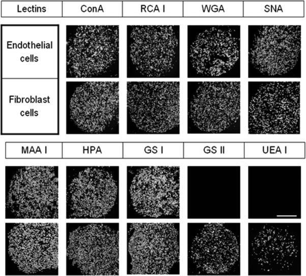
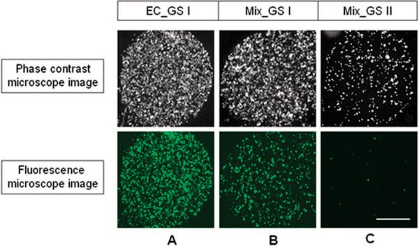
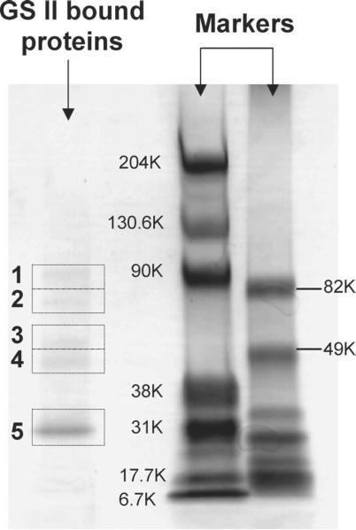
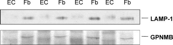
Vascular endothelial cells located at the inner surface of blood vessels are a key component in angiogenesis and are employed as a primary cell type in the study of angiogenesis. These endothelial cells are, however, easily contaminated with fibroblast cells, which are located in proximity to the endothelial cells, during their isolation from tissue. It is thus important to find markers to distinguish the two cell types. In the present work, lectin arrays were prepared using aldehyde-terminated self-assembled monolayers (SAMs) and utilized to explore cell surface carbohydrate expression patterns on endothelial and fibroblast cells. It was found that the lectins Griffonia simplicifolia II (GS II) and Ulex europaeus agglutinin I (UEA I) selectively bind to rat fibroblast cells and not to rat endothelial cells. GS II-binding glycoproteins on fibroblast cells, which are potential cell surface markers to differentiate endothelial and fibroblast cells, were captured on a GS II lectin column and analyzed by LC-ESI-MS/MS. Six candidate cell surface glycoproteins were identified. Differential expression was confirmed by Western blot analysis for two of these proteins, lysosome-associated membrane glycoprotein-1 and transmembrane glycoprotein NMB.
Figures
Similar articles
-
Identification of cell surface glycoprotein markers for glioblastoma-derived stem-like cells using a lectin microarray and LC-MS/MS approach.J Proteome Res. 2010 May 7;9(5):2565-72. doi: 10.1021/pr100012p. J Proteome Res. 2010. PMID: 20235609 Free PMC article.
-
Identification of Plasma Membrane Glycoproteins Specific to Human Glioblastoma Multiforme Cells Using Lectin Arrays and LC-MS/MS.Proteomics. 2018 Jan;18(1). doi: 10.1002/pmic.201700302. Epub 2017 Dec 19. Proteomics. 2018. PMID: 29136334
-
Changes in lectin binding by differentiating cutaneous keratinocytes from the newborn rat.J Invest Dermatol. 1987 Jun;88(6):719-26. doi: 10.1111/1523-1747.ep12470390. J Invest Dermatol. 1987. PMID: 2438357
-
Derivatization in liquid chromatography for mass spectrometric detection.Drug Discov Ther. 2013 Feb;7(1):9-17. doi: 10.5582/ddt.2013.v7.1.9. Drug Discov Ther. 2013. PMID: 23524938 Review.
-
Isotope-coded ESI-enhancing derivatization reagents for differential analysis, quantification and profiling of metabolites in biological samples by LC/MS: A review.J Pharm Biomed Anal. 2016 Oct 25;130:181-193. doi: 10.1016/j.jpba.2016.04.033. Epub 2016 Apr 30. J Pharm Biomed Anal. 2016. PMID: 27178301 Review.
Cited by
-
Glycoprotein analysis using protein microarrays and mass spectrometry.Mass Spectrom Rev. 2010 Sep-Oct;29(5):830-44. doi: 10.1002/mas.20269. Mass Spectrom Rev. 2010. PMID: 20077480 Free PMC article. Review.
-
Cell surface lectin array: parameters affecting cell glycan signature.Glycoconj J. 2013 Apr;30(3):195-203. doi: 10.1007/s10719-012-9433-y. Epub 2012 Aug 17. Glycoconj J. 2013. PMID: 22899543
-
Mitochondrial proteomic analysis reveals deficiencies in oxygen utilization in medullary thick ascending limb of Henle in the Dahl salt-sensitive rat.Physiol Genomics. 2012 Sep 1;44(17):829-42. doi: 10.1152/physiolgenomics.00060.2012. Epub 2012 Jul 17. Physiol Genomics. 2012. PMID: 22805345 Free PMC article.
-
Identification of cell surface glycoprotein markers for glioblastoma-derived stem-like cells using a lectin microarray and LC-MS/MS approach.J Proteome Res. 2010 May 7;9(5):2565-72. doi: 10.1021/pr100012p. J Proteome Res. 2010. PMID: 20235609 Free PMC article.
-
A novel lectin from Agrocybe aegerita shows high binding selectivity for terminal N-acetylglucosamine.Biochem J. 2012 Apr 15;443(2):369-78. doi: 10.1042/BJ20112061. Biochem J. 2012. PMID: 22268569 Free PMC article.
References
-
- Folkman J, Shing Y. J. Biol. Chem. 1992;267:10931–10934. - PubMed
-
- Conway E, Collen D, Carmeliet P. Cardiovasc. Res. 2001;49:507–521. - PubMed
-
- Dzau VJ, Gnecchi M, Pachori AS, Morello F, Melo LG. Hypertension. 2005;46:7–18. - PubMed
-
- Gulati R, Lerman A, Simari RD. Clin. Sci. 2005;109:27–37. - PubMed
-
- Folkman J, Haudenschild C. Nature. 1980;288:551–556. - PubMed
Publication types
MeSH terms
Substances
Grants and funding
LinkOut - more resources
Full Text Sources
