. 2008 Dec 5;384(1):73-86.
doi: 10.1016/j.jmb.2008.08.089. Epub 2008 Sep 16.

Electrostatics in the ribosomal tunnel modulate chain elongation rates

Affiliations

Electrostatics in the ribosomal tunnel modulate chain elongation rates

Jianli Lu et al. J Mol Biol. .

Abstract

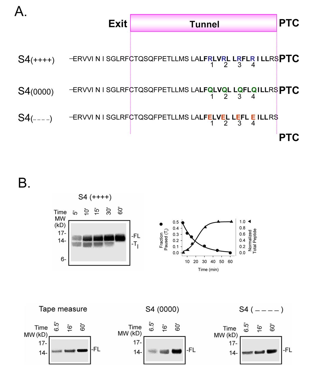
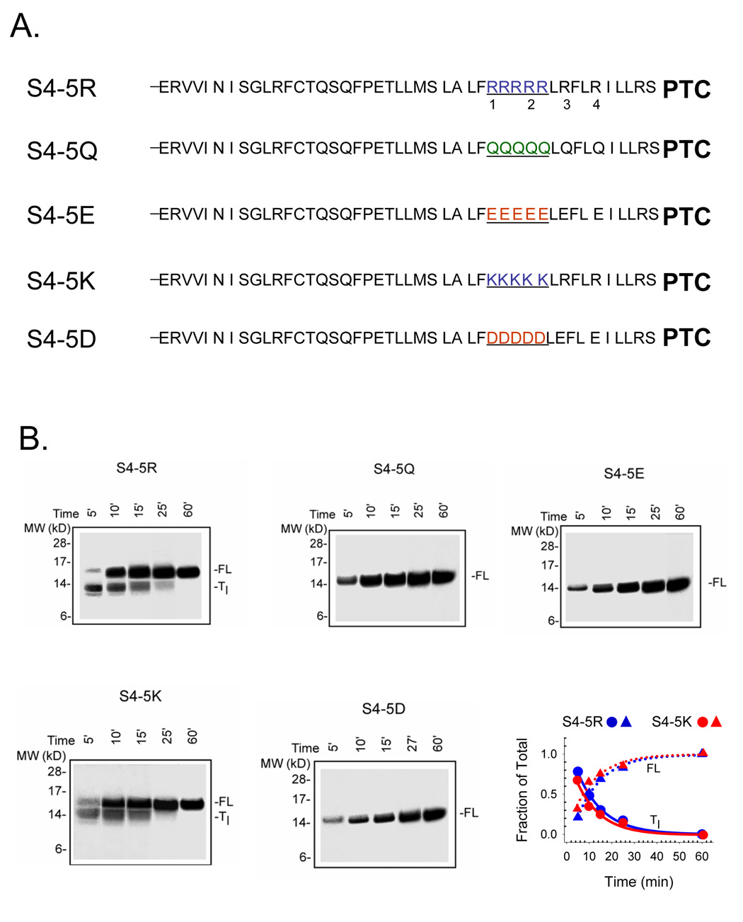
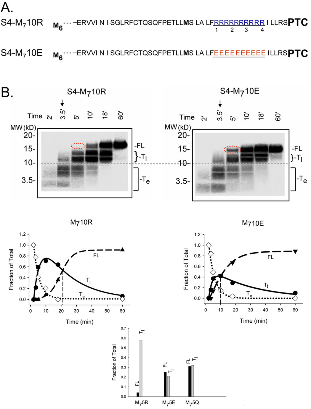
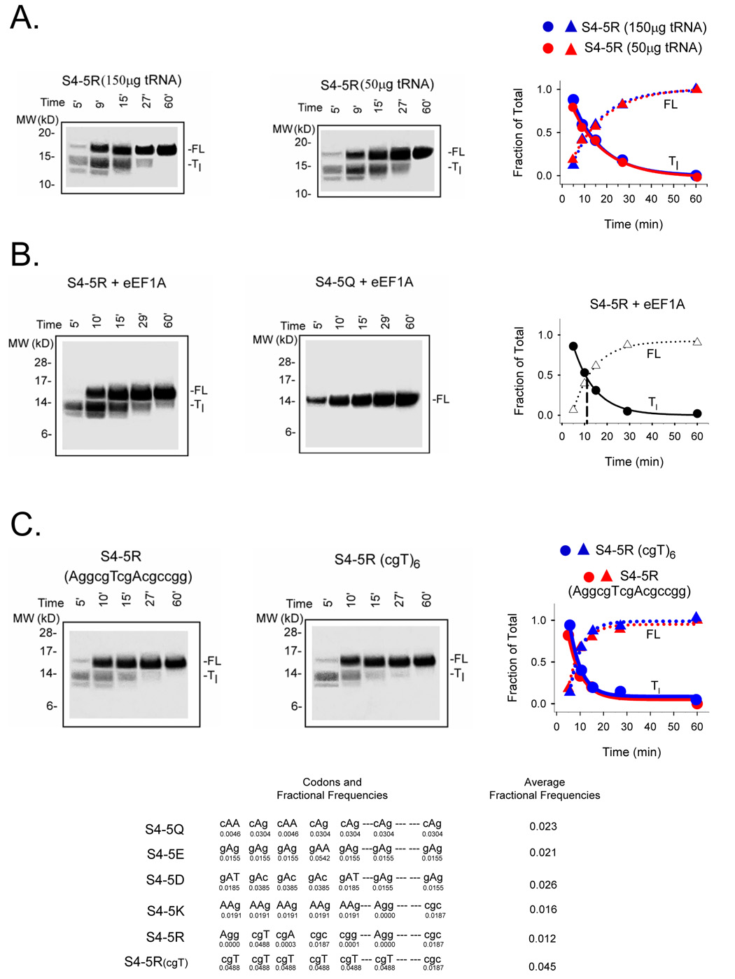
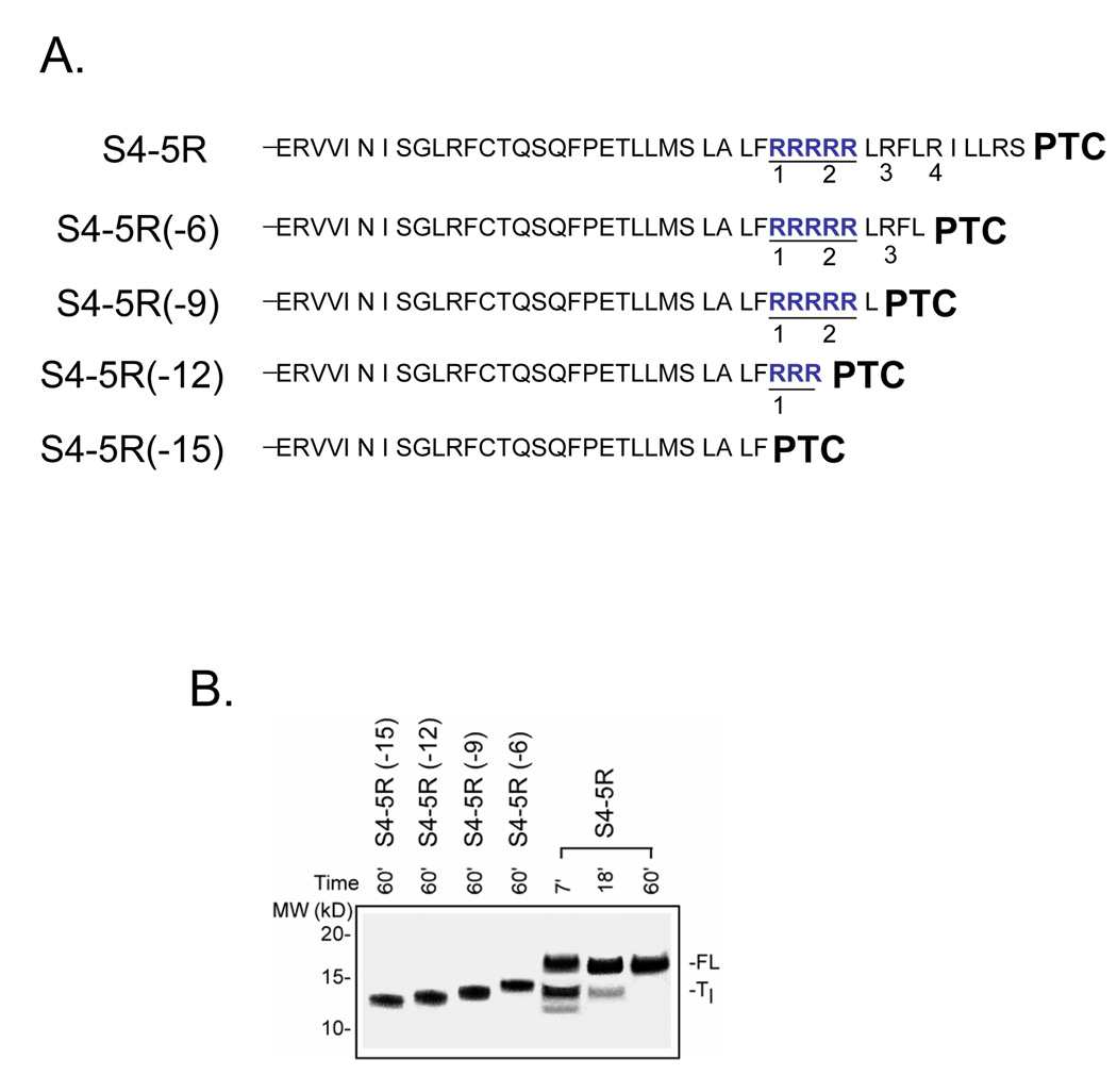
Electrostatic potentials along the ribosomal exit tunnel are nonuniform and negative. The significance of electrostatics in the tunnel remains relatively uninvestigated, yet they are likely to play a role in translation and secondary folding of nascent peptides. To probe the role of nascent peptide charges in ribosome function, we used a molecular tape measure that was engineered to contain different numbers of charged amino acids localized to known regions of the tunnel and measured chain elongation rates. Positively charged arginine or lysine sequences produce transient arrest (pausing) before the nascent peptide is fully elongated. The rate of conversion from transiently arrested to full-length nascent peptide is faster for peptides containing neutral or negatively charged residues than for those containing positively charged residues. We provide experimental evidence that extraribosomal mechanisms do not account for this charge-specific pausing. We conclude that pausing is due to charge-specific interactions between the tunnel and the nascent peptide.


Figures

Figure 1
Figure 1. Time course of translation of S4-substituted tape measure transcripts
A. Schematic sequence of S4 (++++), S4(0000), and S4(−−−−) constructs. The substituted tape measure is a portion of the indicated sequence that resides inside the ribosomal exit tunnel. S4(++++) is the designation for a substituted tape measure in which 19 residues have been replaced with a 19-residue sequence from the fourth transmembrane segment (S4) of Kv channels. The bolded sequence is the proximal portion of the S4 from KvAP, a bacterial Kv channel. The four arginines that translocate across the membrane electric field in the function of Kv channels are labeled 1–4. S4(0000) designates an S4-substituted tape measure in which R1–R4 have been mutated to glutamines (Q1–Q4). S4(−−−−) designates an S4-substituted tape measure in which R1–R4 have been mutated to glutamates (E1–E4). All sequences extend an additional 51 residues (not shown) to the N-terminus. B. Translation rates. Translation was carried out by standard means and aliquots quenched at the indicated times (min), treated with RNase, and fractionated on NuPAGE protein gels (Bis-Tris 12%; top gel, lanes 1–5 loaded with 10, 6, 3, 2, and 1.5 µl, respectively; bottom gels, lanes 1–3 loaded with 6, 3, and 1.5 µl, respectively) for the constructs shown in A. The higher molecular weight band in the S4(++++) gel is the full-length nascent peptide (FL). The lower band in this gel is a transient species (Tl). The fraction of Tl peptide, calculated as the intensity of the lower band divided by the sum of the intensities of the lower and higher bands in a given lane, is plotted as a function of translation time (top row, right), along with normalized total peptide, calculated as the sum of both bands divided by the sum at 60 min. The 45-min time point was omitted from the S4(++++) gel display. The data were fit (solid curve) with a single exponential function and a sigmoidal function, respectively. The three gels (bottom row) for the unmodified tape measure, S4(0000), and S4(−−−−) show only one band, the full-length peptide.
Figure 2
Figure 2. Effect of additional consecutive charges
A. Schematic sequences of S4-substituted tape measures. S4-5R designates a construct with 5 consecutive arginines (highlighted in blue), including R1 and R2, as indicated. S4-5Q, S4- 5E, S4-5K, and S4-5D designate constructs with 5 consecutive glutamines (green), glutamates (red), lysines (blue), and aspartates (red), respectively, each including replacement of R1 and R2. B. Translation time course for the constructs shown in A. as described in Figure 1B (Bis-Tris 12%; all gels, lanes 1–5 loaded with 20, 10, 5, 3, and 1.5 µl, respectively). For S4-5R and S4-5K, the fraction of Tl transient peptide, calculated as the intensity of the lower band divided by the sum of the intensities of the lower and higher bands in a given lane, is plotted (lower right) as a function of translation time, along with fraction of full-length peptide (1-fraction Tl). S4-5R (blue) and S4-5K (red) are plotted as circles and triangles, respectively, to give Tc values of 10 and 8 min. S4-5Q, S4-5E, and S4-5D displayed only one band at the full-length molecular weight and therefore were not plotted.
Figure 3
Figure 3. Pulse-chase studies of S4-substituted tape measures
A. Schematic sequences of methionine-enriched S4-substituted tape measures with 10 consecutive charges. S4-M710R and S4-M710E designate constructs with a total of 7 methionines (bolded) and either 10 consecutive arginines (blue) or 10 consecutive glutamates (red), which replace R1–R4 and the intervening residues in the original S4-substituted tape measure. B. Translation time course for the constructs shown in A. Translation was carried out as described in the Materials and Methods. At the time indicated by the arrow (3.5 min), 1 mM non-radioactive methionine (103-fold dilution of radioactive methionine) was added and additional time points sampled. Samples were fractionated on NuPAGE gels (Bis-Tris 12%, top row; lanes 1–6 loaded with 8, 8, 6, 3, 3, and 3 µl, respectively). The dashed red oval highlights the absence and presence, respectively, of a FL band for S4-M710R and S4-M710E. The data are plotted as fraction of total peptide at the indicated times (below gels). The fraction of a specified population (full-length (FL), early transient (Te), late transient (Tl)) peptide was calculated as the intensity of the specified band divided by the sum of the intensities of all the bands in a given lane. The intensity of Te was multiplied by 1.167 to normalize peptide species containing 6 methionines to the FL containing 7 methionines. The FL data were fit with a sigmoidal function, the Te data with an exponential decay function, and the Tl data with a double exponential (4-parameter) function to give the corresponding solid curves. The vertical line indicates the Tc value, the time at which the fraction of FL equals the fraction of Tl. The bar graph shows results obtained at 5 min from similar experiments using M75R, M75E, and M75Q, which were analyzed as described above.
Figure 4
Figure 4. Potential factors in charge-dependent pausing
A. Addition of supplemental tRNAs. Translation reactions were set up exactly as described in the Materials and Methods and Figure 2, but calf liver tRNAs (100 µg/ml; Novagen) was added to give a total of 150 µg/ml tRNAs in the translation reaction. Samples were fractionated on NuPAGE gels (Bis-Tris 12%; lanes 1–5 loaded with 22, 15, 5, 3, and 3 µl, respectively). Data are plotted as fraction of Tl and FL for the indicated times to give a Tc value of ~12 min. B. Addition of supplemental eEF1A. Translations were carried out as described in the Materials and Methods and Figure 2, but in the presence of additional purified eEF1A as described in the text. Samples were fractionated on NuPAGE gels (Bis-Tris 12%; lanes 1–5 loaded with 20, 10, 5, 3, and 1.5 µl, respectively). The data are plotted as fraction of Tl and FL for the indicated times to give a Tc value of ~12 min. C. Rare vs common codons. An S4-5R construct containing 5 consecutive arginines was translated as described in Figure 1. Samples were fractionated on NuPAGE gels (Bis-Tris 12%; lanes 1–5 loaded with 25, 15, 5, 3, and 2.5 µl, respectively). The gel on the left derives from a peptide encoded by the 5 consecutive arginine codons AggcgTcgAcgccgg, some of which are rare codons. The gel on the right derives from a peptide encoded by 5 consecutive cgT codons plus a sixth cgT (R3), which are common. A plot of the fraction of Tl and FL for the indicated times gives Tc values of 10.2 ± 1.8 (n = 2) and 10.4 min, respectively. Below are the average fractional frequencies of occurrence for the 7 codons in each construct (see Figure 2), based on the study of Thanaraj and Argos .
Figure 5
Figure 5. Identity of Tl intermediate
A. Schematic of truncated peptides used as calibration standards. The number in parenthesis indicates the number of amino acids deleted from the C-terminus of the S4-5R substituted tape measure. B. Migration of standards. The constructs shown in A. were translated for 60 min (produces final full-length peptide) as described in Figure 1 and run on a NuPAGE gel (Bis-Tris 12%; lanes 1–4 loaded with 2, 2, 2, 2 µl, respectively) along with samples from a parallel translation of S4-5R (lanes 5–7 loaded with 20, 10, and 2.5 µl, respectively) using the same batch of reticulocyte lysate and identical reagents and conditions. The gel was run for a longer time to improve resolution of the bands and more accurate assessment of the mass of the paused peptide. The standards (lanes 1–4) exhibit an ascending ladder pattern with increasing mass and charge of the peptide.
Figure 6
Figure 6. Energy barrier model
This is a schematic representation of the energetics of transit of S4-substituted tape measures along the region of the tunnel that is 10–20 Å from the PTC. The trajectory for 5R is shown in blue, for 5Q in green, and 5E in red. The height of the first barrier is ΔG*1 and the second is ΔG*2.

References

    1. Kosolapov A, Tu L, Wang J, Deutsch C. Structure acquisition of the T1 domain of Kv1.3 during biogenesis. Neuron. 2004;44:295–307. - PubMed
    1. Lu J, Deutsch C. Secondary structure formation of a transmembrane segment in Kv channels. Biochemistry. 2005;44:8230–8243. - PubMed
    1. Tu L, Wang J, Deutsch C. Biogenesis of the T1-S1 linker of voltage-gated K+ channels. Biochemistry. 2007;46:8075–8084. - PubMed
    1. Woolhead CA, McCormick PJ, Johnson AE. Nascent membrane and secretory proteins differ in FRET-detected folding far inside the ribosome and in their exposure to ribosomal proteins. Cell. 2004;116:725–736. - PubMed
    1. Mingarro I, Nilsson I, Whitley P, von Heijne G. Different conformations of nascent polypeptides during translocation across the ER membrane. BMC Cell Biology. 2000;1:3. - PMC - PubMed

Publication types

LinkOut - more resources