. 2008 Dec 1;45(11):1510-9.
doi: 10.1016/j.freeradbiomed.2008.08.025. Epub 2008 Sep 9.

Protective effect of Pycnogenol in human neuroblastoma SH-SY5Y cells following acrolein-induced cytotoxicity

Affiliations

Protective effect of Pycnogenol in human neuroblastoma SH-SY5Y cells following acrolein-induced cytotoxicity

Mubeen A Ansari et al. Free Radic Biol Med. .

Abstract

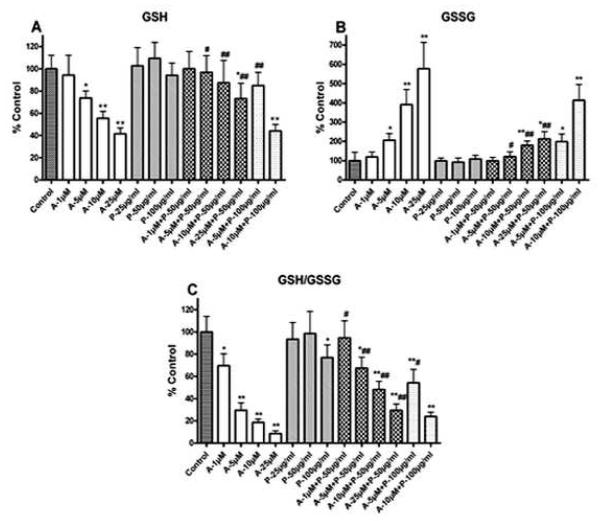
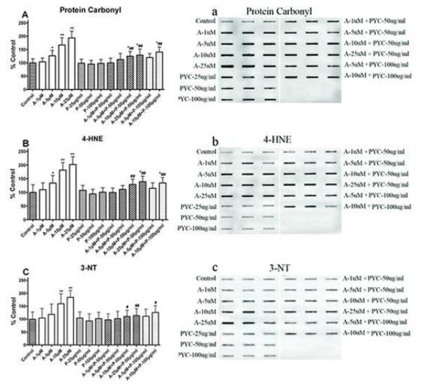
Oxidative stress is one of the hypotheses involved in the etiology of Alzheimer's disease (AD). Considerable attention has been focused on increasing the intracellular glutathione (GSH) levels in many neurodegenerative diseases, including AD. Pycnogenol (PYC) has antioxidant properties and stabilizes intracellular antioxidant defense systems including glutathione levels. The present study investigated the protective effects of PYC on acrolein-induced oxidative cell toxicity in cultured SH-SY5Y neuroblastoma cells. Decreased cell survival in SH-SY5Y cultures treated with acrolein correlated with oxidative stress, increased NADPH oxidase activity, free radical production, protein oxidation/nitration (protein carbonyl, 3-nitrotyrosine), and lipid peroxidation (4-hydroxy-2-nonenal). Pretreatment with PYC significantly attenuated acrolein-induced cytotoxicity, protein damage, lipid peroxidation, and cell death. A dose-response study suggested that PYC showed protective effects against acrolein toxicity by modulating oxidative stress and increasing GSH. These findings provide support that PYC may provide a promising approach for the treatment of oxidative stress-related neurodegenerative diseases such as AD.


Figures

Fig. 1
Fig. 1
PYC prevented loss of SH-SY5Y cell viability. The viability was measured by the reduction of MTT. Acrolein caused loss of cell viability compared with control. 1 h pretreatment with PYC (50 and 100 μg/ml) and incubation for 24 h protected SH-SY5Y cells from acrolein cytotoxicity compared with group acrolein only. Each bar represents the group mean ± SD of six cultures/group. *p < 0.05 and **p < 0.001 versus control, #p < 0.05 and ##p < 0.001 versus acrolein only.
Fig. 2
Fig. 2
Phase contrast microphotographs of SH-SY5Y cells treated with acrolein with and without PYC. Acrolein treatment resulted in significant cell death with some membrane blobbing, but no other morphological changes in surviving SH-SY5Y cells. PYC (50 and 100μg/ml) significantly has reduced cell death, even at high acrolein levels. (A) Control; (B) treated with acrolein 1 μM; (C) acrolein 5 μM; (D) acrolein 10 μM; (E) acrolein 25 μM; (F) treated with PYC 50 μg/ml; (G) acrolein 1 μM + PYC 50 μg/ml; (H) acrolein 5 μM + PYC 50 μg/ml; (I) acrolein 10 μM + PYC 50 μg/ml; (J) acrolein 25 μM + PYC 50 μg/ml; (K) treated with PYC 100 μg/ml; (L) acrolein 1 μM + PYC 100 μg/ml; (M) acrolein 5 μM + PYC 100 μg/ml; (N) acrolein 10 μM + PYC 100 μg/ml; (O) acrolein 25 μM + PYC 100 μg/ml. Microphotographs were taken at 40 X and calibration bar was 10μ.
Fig. 3
Fig. 3
(A) PYC reduces ROS production in SH-SY5Y cells. ROS level was measured by the oxidation of DCFH-DA. Acrolein treatment increased ROS production in SH-SY5Y cells compared with control. 1 h pretreatment with PYC (50 and 100 μg/ml) and incubation for 24 h reduced ROS production by acrolein treatment in SH-SY5Y cell compared the culture treated with acrolein only. Each bar represents the group mean ± SD of six cultures/group. *p < 0.05 and **p < 0.001 versus control, #p < 0.05 and ##p < 0.001 versus acrolein only. PYC depletes O2·− production in SH-SY5Y cells. (B) O2·− level and (C) O2·− producing enzyme NADPH-oxidase activity was increased with acrolein treatment in SH-SY5Y cells compared with control. 1 h pretreatment with PYC (50 and 100 μg/ml) and incubation for 24 h reduced O2·− production by acrolein treatment in cell cultures compared with acrolein only. Each bar represents the group mean ± SD of six cultures/group. *p < 0.05 and **p < 0.001 versus control, #p <0.05 and ##p < 0.001 versus acrolein only.
Fig. 4
Fig. 4
Level of GSH system was alleviated with PYC treatment in SH-SY5Y cells. Acrolein caused loss of GSH (A) increased GSSG (B) and discrepancy of GSH/GSSG (C) in SH-SY5Y cells compared with control. 1 h pretreatment with PYC (50 and 100 μg/ml) and incubation for 24 h protected cells from acrolein induced adverse effect on GSH system compared with group acrolein only. Each bar represents the group mean ± SD of six cultures/group. *p < 0.05 and **p < 0.001 versus control, #p < 0.05 and ##p < 0.001 versus acrolein only.
Fig. 5
Fig. 5
PYC treatment protects SH-SY5Y cells from protein and lipid damages. Acrolein increased protein oxidation (protein carbonyl formation A & a), lipid peroxidation (4-HNE formation B & b), and protein nitration (3-NT formation C & c) in SH-SY5Y cells compared from acrolein induced protein damages compared with group acrolein only. Blot represents one sample of each group and each bar represents the group mean ± SD of six cultures/group. *p < 0.05 and **p < 0.001 versus control, #p < 0.05 and ##p < 0.001 versus acrolein only.
Fig. 6
Fig. 6
NADPH-oxidase subunit proteins in SH-SY5Y cells were significantly affected with acrolein exposure. Cells were processed for immunobloting followed by Western-blot. The levls of p67Phox (B), p47Phox (C), and p40Phox (D) demonstrated increase in membrane and p67Phox decreased in cytosol (G). The effect of acrolein exposure was attenuated with PYC (50 μg/ml ). The gp91Phox (A) and p22Phox (E) failed to demonstrate significant changes. Na+/K+-ATPase (F) and GAPDH (G) show equal proteins.
Fig. 7
Fig. 7
Cytosolic protein subunits of NADPH-oxidase p67Phox (A & B), levels in the SH-SY5Y cells increased in membrane and declined in cytosol with acrolein exposure were significantly attenuated by PYC (50 μg/ml). In same fashion p47Phox (C), and p407Phox (D) also show increase with acrolein exposre and significantly decreased with PYC treatment. Each bar represents the group mean ± SD of six cultures/group. *p < 0.05 and **p < 0.001 versus control, #p < 0.05 and ##p < 0.001 versus acrolein only.
Fig. 8
Fig. 8
Membrane protein subunits of NADPH-oxidase gp91Phox (A) and p22Phox (B) show non significant elevation with acrolien exposure. PYC attenuates the levels of both subunits in membrane.
Fig. 9
Fig. 9
Schematic presentation of proposed mechanism of protective effects of pycnogenal (PYC) on acrolein-induced cytotoxicity in SH-SY5Y cells. The thiol (-SH) – reactive property of acrolein activates NADPH-oxidase complex, leading to the production of superoxide (O2−.). Normally superoxide dismutase (SOD) converts O2−. into hydrogen peroxide (H2O2). O2−. activates nitric oxide synthase (iNOS) leading to the production of nitric oxide (NO) and the formation of peroxynitrites (ONOO), which ultimately causes oxidative/nitrosative stress. Increased oxidative/nitrosative stress causes mitochondrial dysfunction, lipid peroxidation, protein and DNA damage, which can result in cell death. Lipid peroxidation further produces toxic aldehyde products including elevated acrolein that further aggravates degenerative process. ONOO is known to activate poly(ADP-ribrosyl) polymerase (PARP) leading to ATP depletion and cell death. PYC prevents assembling of NADPH-oxidase subunits (both in cytosol and the membrane), and has protective effects on the beginning of acrolein-induced deleterious cascade. PYC prevents O2−. production from NADPH-oxidase activation and NO from iNOS. PYC scavenges O2−., NO, ONOO, H2O2 and hydroxyl radicals (OH.) to prevent oxidative/nitosative stress, and elevates internal antioxidant glutathione (GSH) defenses that can enhance cell survival.

References

    1. Kondo M, Oya-Ito T, Kumagai T, Osawa T, Uchida K. Cyclopentenone prostaglandins as potential inducers of intracellular oxidative stress. The Journal of biological chemistry. 2001;276:12076–12083. - PubMed
    1. Naoi M, Maruyama W, Shamoto-Nagai M, Yi H, Akao Y, Tanaka M. Oxidative stress in mitochondria: decision to survival and death of neurons in neurodegenerative disorders. Molecular neurobiology. 2005;31:81–93. - PubMed
    1. Sundaresan M, Yu ZX, Ferrans VJ, Irani K, Finkel T. Requirement for generation of H2O2 for platelet-derived growth factor signal transduction. Science (New York, N.Y. 1995;270:296–299. - PubMed
    1. Ansari MA, Joshi G, Huang Q, Opii WO, Abdul HM, Sultana R, Butterfield DA. In vivo administration of D609 leads to protection of subsequently isolated gerbil brain mitochondria subjected to in vitro oxidative stress induced by amyloid beta-peptide and other oxidative stressors: relevance to Alzheimer’s disease and other oxidative stress-related neurodegenerative disorders. Free radical biology & medicine. 2006;41:1694–1703. - PMC - PubMed
    1. Greenamyre JT, Betarbet R, Sherer TB. The rotenone model of Parkinson’s disease: genes, environment and mitochondria. Parkinsonism & related disorders. 2003;9(Suppl 2):S59–64. - PubMed

Publication types

MeSH terms