. 2008 Dec 1;233(2):169-78.
doi: 10.1016/j.taap.2008.08.016. Epub 2008 Sep 11.

Regulation of cytochrome P4501A1 expression by hyperoxia in human lung cell lines: Implications for hyperoxic lung injury

Affiliations

Regulation of cytochrome P4501A1 expression by hyperoxia in human lung cell lines: Implications for hyperoxic lung injury

Kushal Y Bhakta et al. Toxicol Appl Pharmacol. .

Abstract

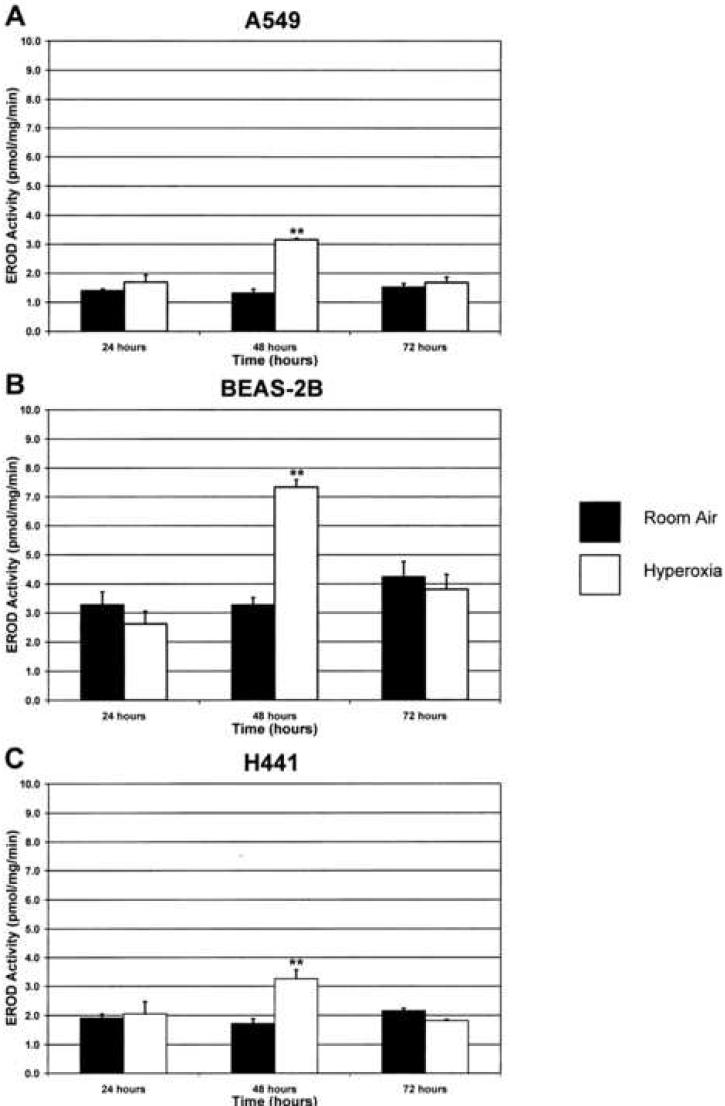
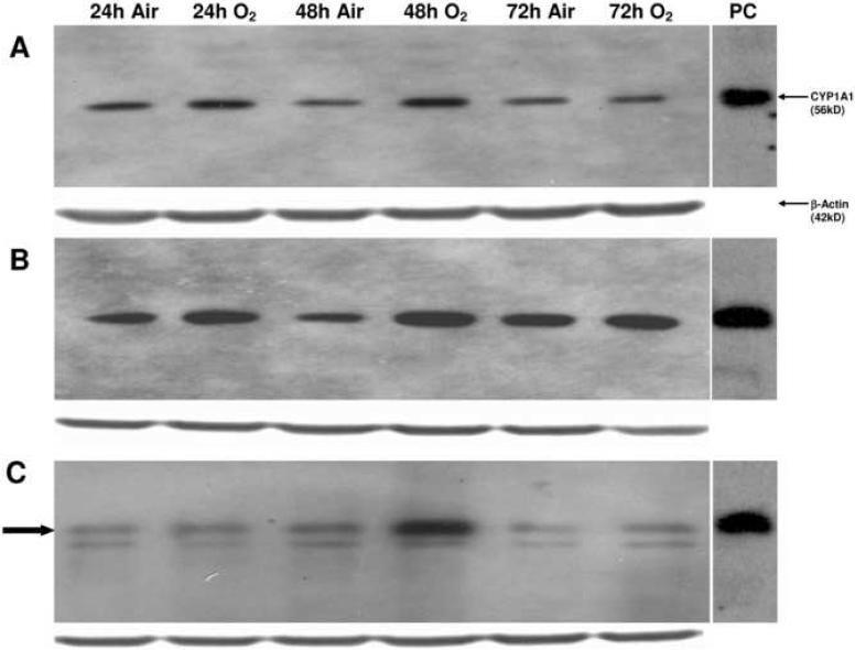
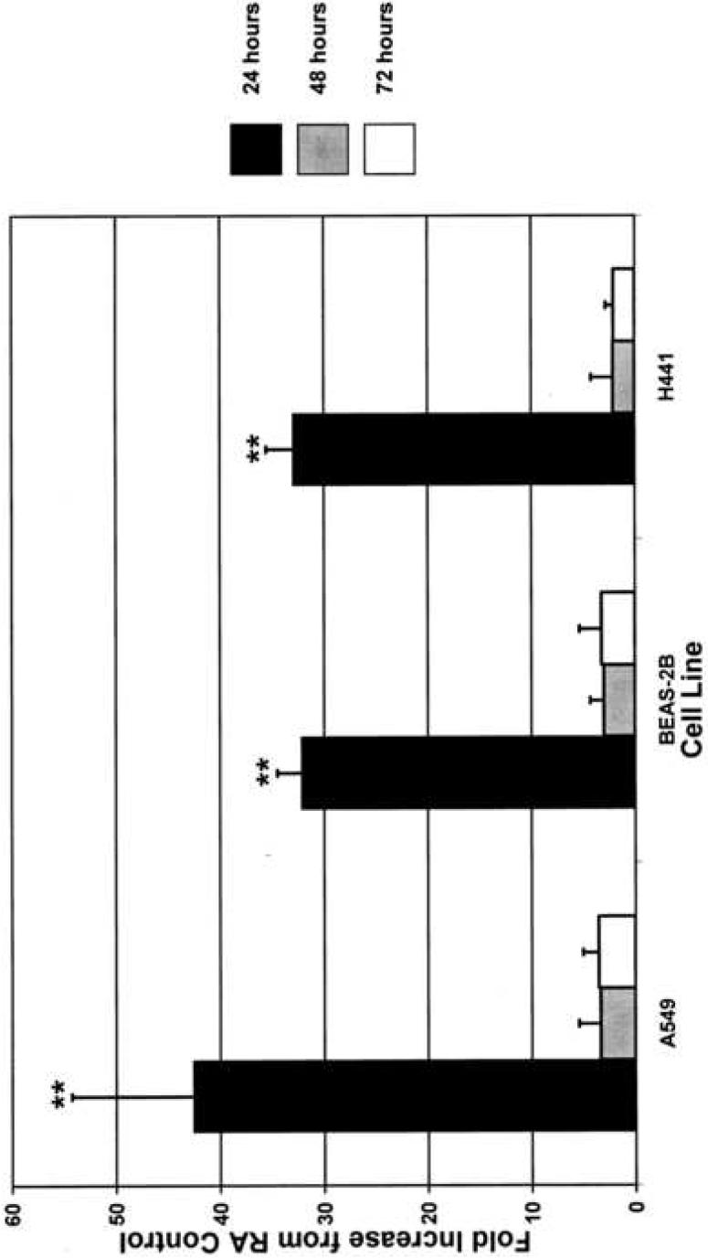
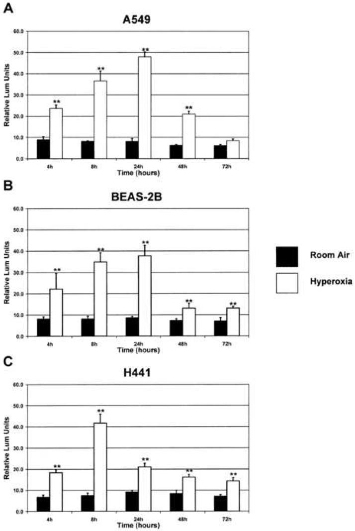
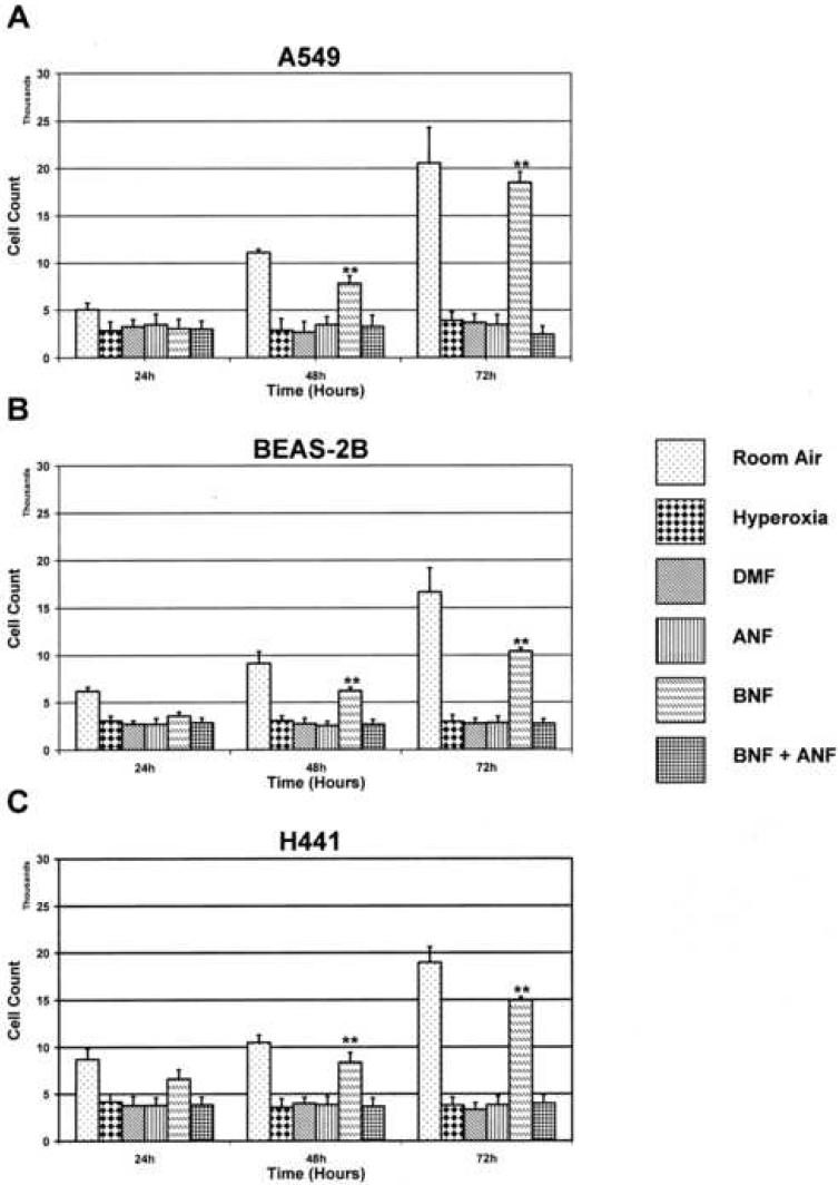
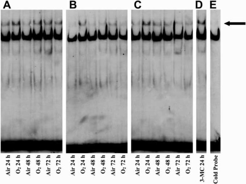
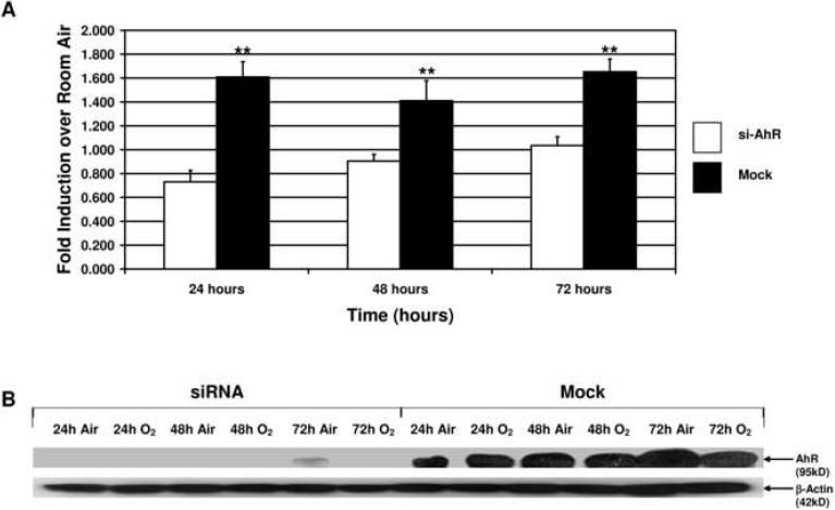
Supplemental oxygen, used to treat pulmonary insufficiency in newborns, contributes to the development of bronchopulmonary dysplasia (BPD). Cytochrome P4501A enzymes are induced by hyperoxia in animal models, but their role in human systems is unknown. Here we investigated the molecular mechanisms of induction of CYP1A1 by hyperoxia in human lung cell lines. Three human lung cell lines were exposed to hyperoxia (95% O2) for 0-72 h, and CYP1A1 activities, apoprotein contents, and mRNA levels were determined. Hyperoxia significantly induced CYP1A1 activity and protein contents (2-4 fold), and mRNA levels (30-40 fold) over control in each cell line. Transfection of a CYP1A1 promoter/luciferase reporter construct, followed by hyperoxia (4-72 h), showed marked (2-6 fold) induction of luciferase expression. EMSA and siRNA experiments strongly suggest that the Ah receptor (AHR) is involved in the hyperoxic induction of CYP1A1. MTT reduction assays showed attenuation of cell injury with the CYP1A1 inducer beta-naphthoflavone (BNF). Our results strongly suggest that hyperoxia transcriptionally activates CYP1A1 expression in human lung cell lines by AHR-dependent mechanisms, and that CYP1A1 induction is associated with decreased toxicity. This novel finding of induction of CYP1A1 in the absence of exogenous AHR ligands could lead to novel interventions in the treatment of BPD.


Figures

Figure 1
Figure 1. Hyperoxia induces CYP1A1 activity
Each cell line was maintained in room air or exposed to hyperoxia as outlined in “Materials and Methods.” CYP1A1 activity was then measured using the ethoxyresorufin-O-deethylase (EROD) assay. **p<0.01 compared to room air at 48 hours or hyperoxia at 24 hours. Values represent means ± SD (n=16).
Figure 2
Figure 2. Hyperoxia increases apoprotein expression
The whole cell suspensions (100 μg protein) were subjected to Western blotting using monoclonal antibodies raised against CYP1A1, as described in “Materials and Methods.” Panel A represents samples from the A549 cell line, panel B represents samples from the BEAS-2B cell line, and panel C represents samples from the H441 cell line. In the H441 cell line, the CYP1A1 protein is highlighted by the closed arrow. Under each sample lane is the corresponding β-actin blot to assess for protein loading. The positive control (labeled “PC”) was 0.5 μg of liver microsomes from mice treated with 3-methylcholanthrene.
Figure 3
Figure 3. Real time RT-PCR analysis of CYP1A1 mRNA shows significant increase in expression at 24 hours
Each cell line was exposed to hyperoxia or maintained in room air as described in “Materials and Methods,” and CYP1A1 mRNA expression was analyzed by RT-PCR. 18S primers were used as an internal control. **p<0.01 compared to hyperoxia at 48 and 72 hours and room air at 24 hours. Values represent means ± SD (n=8).
Figure 4
Figure 4. Hyperoxia increases expression of the luciferase reporter gene driven by the CYP1A1 gene promoter
A 1.6kb fragment of the human CYP1A1 gene promoter was cloned upstream of the luciferase reporter gene, and this construct was then transfected into each cell line. A plasmid containing the Renilla luciferase gene was co-transfected as an internal control. The cells were then exposed to hyperoxia or maintained in room air, and luciferase activity was measured as described in “Materials and Methods.” Results were normalized to Renilla luciferase activity. **p<0.01 as compared to respective room air controls (pGL3-1A1-RA). Values represent means ± SD (n=18).
Figure 5
Figure 5. CYP-1A1 expression modulates cell toxicity during hyperoxia exposure
In order to determine the relative role of CYP-1A1 in modulating cell injury, each cell line was treated with either of the chemical regimens shown. Following chemical treatment, each line was exposed to hyperoxia or maintained in room air as described in “Materials and Methods.” MTT reduction assays were performed to assess cell count and viability at each time point. **p<0.01 compared to alternative treatments. Values represent means ± SD (n=16).
Figure 6
Figure 6. Interaction of nuclear proteins with AhREs in hyperoxia exposed cells
Nuclear extracts from cells maintained in room air or exposed to hyperoxia for 24, 48, or 72 hours were subjected to EMSA, as described in “Materials and Methods.” Panel A represents samples from the A549 cell line, Panel B represents samples from the BEAS-2B cell line, Panel C represents samples from the H441 cell line, Panel D represents nuclear extract from treatment of the A549 cell line with the prototypical CYP1A1 inducer 3-methylcholanthrene (3-MC) for 24 hours, and Panel E represents the nuclear extract from the BEAS-2B cell line exposed to hyperoxia for 24 hours incubated with a 25-fold excess of cold probe before being subjected to EMSA. The closed arrow indicates the shifted band.
Figure 7
Figure 7. siRNA directed against AHR mRNA abolishes expression of the luciferase reporter gene driven by the CYP1A1 gene promoter
siAhR or control siRNA was co-transfected the CYP1A1 promoter-luciferase construct as described in “Materials and Methods.” Panel A depicts fold induction over room air control of the luciferase reporter in the control siRNA (Mock) transfected cells and in the siAhR transfected cells. Results were normalized to Renilla luciferase activity. **p<0.03 as compared to siAhR. Values represent means ± SD. Panel B depicts the representative Western blot for the AhR protein in the Mock and siAhR samples. There is significant decrease in the intensity of the AhR band in the siAhR transfected cells as compared to the Mock transfections.

References

    1. Bland RD, Coalson JJ. Chronic lung disease in early infancy. M. Dekker; New York: 2000.
    1. Bonzo JA, Belanger A, Tukey RH. The role of chrysin and the ah receptor in induction of the human UGT1A1 gene in vitro and in transgenic UGT1 mice. Hepatology. 2007;45:349–360. - PubMed
    1. Chomczynski P, Sacchi N. Single-step method of RNA isolation by acid guanidinium thiocyanate-phenol-chloroform extraction. Anal Biochem. 1987;162:156–159. - PubMed
    1. Cotton RB, Hazinski TA, Morrow JD, Roberts LJ, Zeldin DC, Lindstrom DP, Lappalainen U, Law AB, Steele S. Cimetidine does not prevent lung injury in newborn premature infants. Pediatr Res. 2006;59:795–800. - PMC - PubMed
    1. Couroucli XI, Liang YW, Jiang W, Barrios R, Moorthy B. Attenuation of oxygen-induced abnormal lung maturation in rats by retinoic acid: possible role of cytochrome P4501A enzymes. J Pharmacol Exp Ther. 2006;317:946–954. - PubMed

Publication types

MeSH terms