Review
. 2009 Feb 6;158(3):972-82.
doi: 10.1016/j.neuroscience.2008.08.028. Epub 2008 Aug 27.

Inflammation and the neurovascular unit in the setting of focal cerebral ischemia

Affiliations
Review

Inflammation and the neurovascular unit in the setting of focal cerebral ischemia

G J del Zoppo. Neuroscience. .

Abstract

Responses to focal cerebral ischemia by neurons and adjacent microvessels are rapid, simultaneous, and topographically related. Recent observations indicate the simultaneous appearance of proteases by components of nearby microvessels that are also expressed by neurons in the ischemic territory, implying that the events could be coordinated. The structural relationship of neurons to their microvascular supply, the direct functional participation of glial cells, and the observation of a highly ordered microvessel-neuron response to ischemia suggest that these elements are arranged in and behave in a unitary fashion, the neurovascular unit. Their roles as a unit in the stimulation of cellular inflammation and the generation of inflammatory mediators during focal cerebral ischemia have not been explored yet. However, components of the neurovascular unit both generate and respond to these influences under the conditions of ischemia. Here we briefly explore the potential inter-relationships of the components of the neurovascular unit with respect to their potential roles in ischemia-induced inflammatory responses.
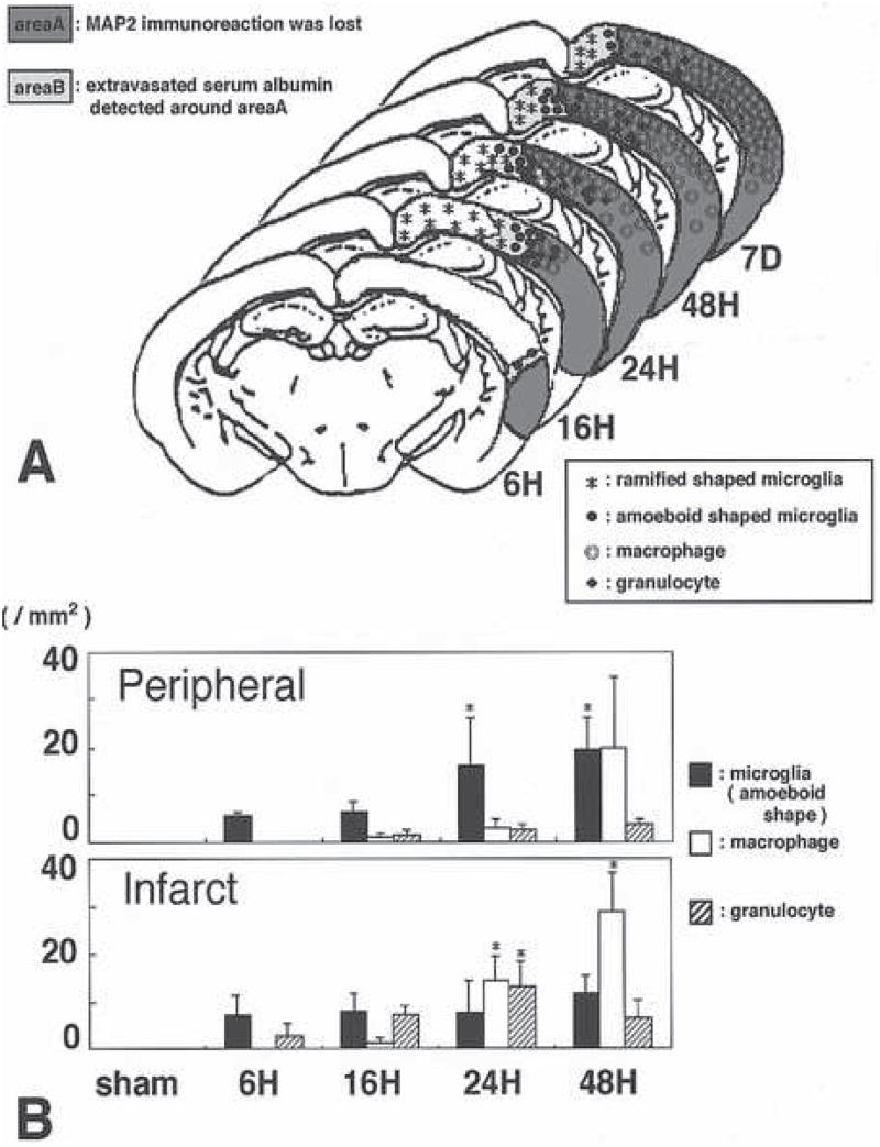

Figures

Figure 1
Figure 1
Microvessel-neuron (m–n) inter-relationships based upon [m–n] distance distributions. A, the neurovascular unit and definition of [m–n] distance. B, indication of positions of microvessels (m) and neurons with evidence of dUTP incorporation (n*) and neurons without evidence of DNA scission (n) at 2 hours following middle cerebral occlusion (MCA:O) in the striatum of the non-human primate. C, untransformed data of [m–n] distances in the non-ischemic striatum of the non-human primate (in μm). D, neurons with evidence of injury (n*) at 2 hours MCA:O are at significantly greater distance from their nearest neighboring microvessel ([m–n*]) than those without evidence of injury ([m–n]). Data and figures from Mabuchi et al.(Mabuchi et al., 2005)
Figure 2
Figure 2
Schema indicating the potential interactions of pro-MMP-2 within 2 hours MCA:O in the ischemic striatum of the non-human primate, the appearance of pro-MMP-9, and their respective activations systems. Proteases identified within this system are noted in bold and in boxes. The known matrix substrates of possibly active proteases are indicated below each system. From data presented in (Heo et al., 1999; Hosomi et al., 2001; Chang et al., 2003).
Figure 3
Figure 3
Complete inhibition of the loss of β-dystroglycan immunoreactivity from primary murine astrocytes cultured on laminin under conditions of experimental ischemia (hatched bars) compared to normoxia (solid bars) by blockade of MMP-like activity (with GM 6001 and with 1, 10-phenanthroline, *). Inhibition of serine proteases (aprotonin) and some cathepsins (E64c and E64d) had no effect on the loss of β-dystroglycan immunoreactivity during experimental ischemia.(Milner et al., 2008b)
Figure 4
Figure 4
Separation of the end-feet of astrocytes from the abluminal surface of the basal lamina matrix of select microvessels very early following MCA:O in the rat (B) compared to normoxia (A). Note swelling of the astrocytes (G), but no swelling of the endothelium. Used with permission of the principal author.(Heo et al., 2005)
Figure 5
Figure 5
Distribution of activated microglial cells compared with peripheral blood macrophages in the evolution of focal cerebral ischemia in the rat. A, topographical distribution of activated microglia in the peripheral region of the ischemic lesion and the incursion of macrophages within the region of ischemic injury identified by MAP2 immunoreactivity at various times after MCA:O. B, relative distribution of microglia, macrophages, and PMN leukocytes identified in the peripheral region and the central region of the evolving infarction. Figure 4 from Mabuchi et al used with the permission of the principal author.(Mabuchi et al., 2000)

References

    1. Abumiya T, Fitridge R, Mazur C, Copeland BR, Koziol JA, Tschopp JF, Pierschbacher MD, del Zoppo GJ. Integrin alpha(IIb)beta(3) inhibitor preserves microvascular patency in experimental acute focal cerebral ischemia. Stroke. 2000;31:1402–1410. - PubMed
    1. Abumiya T, Lucero J, Heo JH, Tagaya M, Koziol JA, Copeland BR, del Zoppo GJ. Activated microvessels express vascular endothelial growth factor and integrin alpha(v)beta3 during focal cerebral ischemia. J Cereb Blood Flow Metab. 1999;19:1038–1050. - PubMed
    1. Aloisi F. Immune function of microglia. GLIA. 2001;36:165–179. - PubMed
    1. Balabanov R, Washington R, Wagnerova J, Dore-Duffy P. CNS microvascular pericytes express macrophage-like function, cell surface integrin alpha M, and macrophage marker ED-2. Microvasc Res. 1996;52:127–142. - PubMed
    1. Bär T. Morphometric evaluation of capillaries in different laminae of rat cerebral cortex by automatic image analysis: Changes during development and aging. In: Cervos-Navarro J, editor. Advances in Neurology. New York: Raven Press; 1978. pp. 1–9. - PubMed

MeSH terms