. 2008 Nov;14(11):2379-93.
doi: 10.1261/rna.1227808. Epub 2008 Sep 29.

The 3' proximal translational enhancer of Turnip crinkle virus binds to 60S ribosomal subunits

Affiliations

The 3' proximal translational enhancer of Turnip crinkle virus binds to 60S ribosomal subunits

Vera A Stupina et al. RNA. 2008 Nov.

Abstract

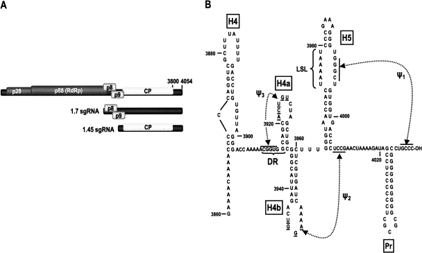
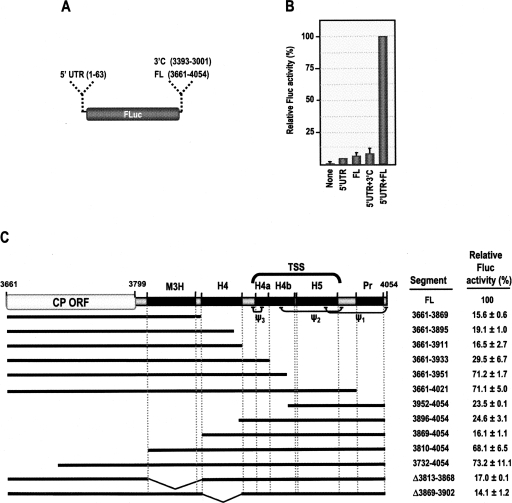
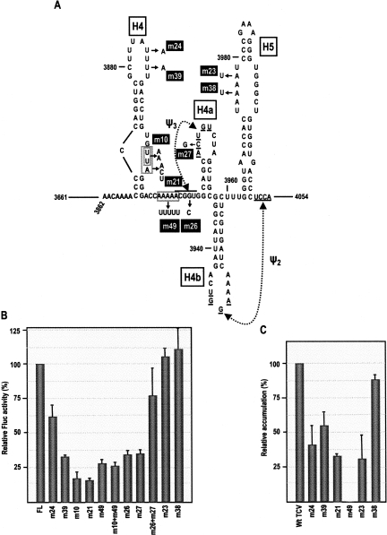
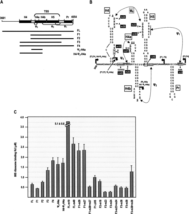
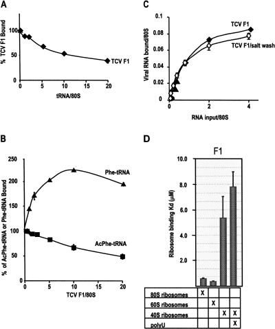
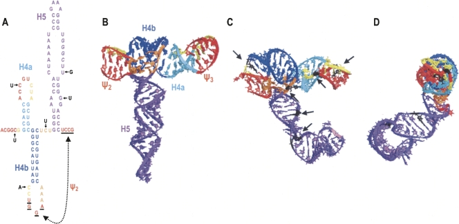
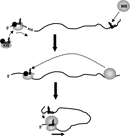
During cap-dependent translation of eukaryotic mRNAs, initiation factors interact with the 5' cap to attract ribosomes. When animal viruses translate in a cap-independent fashion, ribosomes assemble upstream of initiation codons at internal ribosome entry sites (IRES). In contrast, many plant viral genomes do not contain 5' ends with substantial IRES activity but instead have 3' translational enhancers that function by an unknown mechanism. A 393-nucleotide (nt) region that includes the entire 3' UTR of the Turnip crinkle virus (TCV) synergistically enhances translation of a reporter gene when associated with the TCV 5' UTR. The major enhancer activity was mapped to an internal region of approximately 140 nt that partially overlaps with a 100-nt structural domain previously predicted to adopt a form with some resemblance to a tRNA, according to a recent study by J.C. McCormack and colleagues. The T-shaped structure binds to 80S ribosomes and 60S ribosomal subunits, and binding is more efficient in the absence of surrounding sequences and in the presence of a pseudoknot that mimics the tRNA-acceptor stem. Untranslated TCV satellite RNA satC, which contains the TCV 3' end and 6-nt differences in the region corresponding to the T-shaped element, does not detectably bind to 80S ribosomes and is not predicted to form a comparable structure. Binding of the TCV T-shaped element by 80S ribosomes was unaffected by salt-washing, reduced in the presence of AcPhe-tRNA, which binds to the P-site, and enhanced binding of Phe-tRNA to the ribosome A site. Mutations that reduced translation in vivo had similar effects on ribosome binding in vitro. This strong correlation suggests that ribosome entry in the 3' UTR is a key function of the 3' translational enhancer of TCV and that the T-shaped element contains some tRNA-like properties.


Figures

FIGURE 1.
FIGURE 1.
Genome organization and structure of the 3′ terminal region of TCV. (A) Genomic organization of TCV genomic and subgenomic RNAs. The RdRp (p88) is expressed as a ribosomal readthrough product of p28. The larger subgenomic RNA is the bi-cistronic mRNA for p8 and p9 movement proteins (Li et al. 1998), and the smaller subgenomic RNA encodes the viral capsid protein (Hacker et al. 1992). (B) Structure of the 3′ region of the TCV 3′ UTR. All secondary and tertiary structures have been previously confirmed using single and compensatory mutations (Zhang et al. 2006c; McCormack et al. 2008). (LSL) large symmetrical loop. Residues involved in pseudoknots are underlined or overlined. Names of the hairpins are boxed and pseudoknots designations are shown.
FIGURE 2.
FIGURE 2.
In vivo translation of a luciferase reporter construct in the presence and absence of viral 5′ and 3′ sequences. (A) Firefly luciferase reporter construct used for in vivo translation in protoplasts. The TCV 5′ UTR is 63 nt. The full-length TCV 3′ fragment (FL) includes the entire 3′ UTR and 139 nt from the CP ORF (positions 3661–4054). The 3′C control fragment is an internal segment of TCV (positions 3001–3393), inserted in the minus-sense orientation. (B) Relative translation in the presence and absence of the 5′ UTR and FL. Arabidopsis protoplasts were co-inoculated with 30 μg of RNA from experimental Fluc constructs and 10 μg of RNA synthesized from an internal control construct containing RLuc. Luciferase activity was determined at 18 hpi. All values are averages from at least three independent experiments, and standard deviation bars are shown. Transfected Fluc RNAs contained either no added TCV sequences (none), only the TCV 5′ UTR (5′UTR), only the 3′ region (FL), or both 5′ and 3′ sequences (5′ UTR+FL). (C) Effect of deletions within the FL fragment on translation from the 5′ UTR–Fluc construct. Fragments included in the constructs are denoted by a thick line. The positions of known TCV elements (hairpins and pseudoknots) and the TSS predicted to form from the Ψ3 → Ψ2 domain (McCormack et al. 2008) are shown. Percent luciferase activity (averages of at least three independent experiments) with standard deviations is given.
FIGURE 3.
FIGURE 3.
Mutations in H4 and Ψ3 region repress translation in vivo. (A) Location of mutations generated in 5′UTR-Fluc-FL. Mutation designations are boxed. m10 replaces the consecutive two uridylates in the H4 asymmetric loop with adenylates; m21 replaces “UUA” in the same location with “ACU.” (B) RNA transcribed from 5′UTR-Fluc-FL (FL) or 5′UTR-Fluc-FL containing mutations described in A along with RNA from the control Rluc construct were inoculated into protoplasts and luciferase activity measured at 18 hpi. Values are averages of at least three independent experiments. Bars reflect standard deviation. (C) Relative accumulation of TCV viral genomic RNA in protoplasts containing mutations in the 3′ region. Mutations were incorporated into full-length TCV cDNA and transcribed RNA inoculated into protoplasts. Viral RNA levels were determined by Northern analyses of total extracted RNA using a TCV-specific probe and normalized to the levels of ribosomal RNA. Values are from three independent assays. Bars reflect standard deviation. Accumulation of TCV containing m26, m27,m26+m27, and m10 were previously assayed (see text; Sun and Simon 2006; McCormack et al. 2008).
FIGURE 4.
FIGURE 4.
TCV 3′ sequences bind to yeast 80S ribosomes. (A) Location of fragments used for ribosome binding. Precise locations of the end points are indicated in B. Names of the fragments are given to the right. Location of 3′ elements is shown above fragments. (B) Mutations introduced into fragments are shown. Mutation designations are boxed. Thick bent arrows denote location of fragment end points, with identity of fragments using particular end points shown in parentheses. Ψ4 forms between H4 and H5 in fragment F3 (see the text). (C) Wt and mutant fragments were subjected to filter binding assays using yeast 80S ribosomes. Columns 1–7 (left to right) are wt fragments. Remaining columns reflect fragments (FL, F1, F2, or F3) containing various mutations described in B. K d were calculated from three independent experiments. Standard error bars are shown.
FIGURE 5.
FIGURE 5.
Features of ribosome binding to fragment F1. (A) Effect of deacylated tRNA on ribosome binding to F1. Ribosomes (30 pmol) were preincubated with 0–600 pmol of tRNA followed by addition of 30 pmol of [32P] 5′-end labeled F1. (B) Effect of F1 on Phe-tRNA and Ac-Phe-tRNA binding. For P-site specificity, ribosomes (30 pmol) were incubated with 0–600 pmol of F1 followed by addition of 30 pmol of labeled Ac-Phe-tRNA. For A site competition, ribosomes were preincubated with 0–600 pmol of F1, followed by addition of labeled Phe-tRNA. Data are expressed as percentage of initial binding (without competing RNA) at given competing RNA/ribosomes molar ratios. (C) F1 binding to 80S ribosomes. Two to 100 pmol of labeled F1 were combined with 25 pmol of yeast 80S ribosomes that were and were not salt washed, and bound RNA was detected following filter binding. The fraction of ribosomes active in F1 binding is comparable with yeast Phe-tRNA binding with similarly prepared yeast ribosomes from the same yeast strain (Petrov et al. 2004). (D) Binding of 80S ribosomes and 60S and 40S ribosomal subunits to labeled F1 in the presence and absence of poly(U). For all assays, K d were calculated from three independent experiments. Standard error bars are shown.
FIGURE 6.
FIGURE 6.
SatC structure predicted by RNA2D3D and molecular modeling. (A) SatC sequence in the region corresponding to the TCV Ψ3 → Ψ2 domain. The 6-nt differences with the TCV sequence (in black) are shown. Color coding of sequences is used to help identify regions in the 3D structure shown in BD. Underlined residues participate in Ψ2 interaction in satC. Although presented with the hairpins in the figure, this pseudoknot does not coexist in the same satC structure as H5 (Zhang et al. 2006a). (B) TCV TSS predicted by RNA2D3D and molecular modeling (McCormack et al. 2008). (C) Predicted structure of the comparable region of satC. Arrows point to the locations in the structure occupied by the 6-nt differences. Note that Ψ3 and Ψ2 are not stably maintained. (D) Side view of the satC structure.
FIGURE 7.
FIGURE 7.
Model for translational enhancement by 3′ sequences in TCV. Our data suggest that the 5′ UTR and 3′ UTR work synergistically to mediate translation in TCV. We suggest that the large 60S ribosomal subunit binds to the TSS while the 43S preinitiation complex enters at the 5′ UTR. Circularization of the template could be mediated by assembly of the 80S ribosome, which would require that the 60S subunit release the TSS to position the initiator met-tRNA in the P-site. We propose that H4 (not shown) plays a critical role in translation, possibly by interfering with a ribosome-binding repressor. Such a repressor could become active following RdRp binding to the same region, to restrict ribosome binding and thus help mediate the switch between translation and replication. Additional upstream sequences are also important for translation, such as those in the M3H region, possibly to assist in ribosome relocation to the 5′ end.

Similar articles

Cited by

References

    1. Allen G.S., Frank J. Structural insights on the translation initiation complex: Ghosts of a universal initiation complex. Mol. Microbiol. 2007;63:941–950. - PubMed
    1. Baird S.D., Turcotte M., Korneluk R.G., Holcik M. Searching for IRES. RNA. 2006;12:1755–1785. - PMC - PubMed
    1. Barends S., Bink H.H., van den Worm S.H., Pleij C.W., Kraal B. Entrapping ribosomes for viral translation: tRNA mimicry as a molecular Trojan horse. Cell. 2003;112:123–129. - PubMed
    1. Bert A.G., Grépin R., Vadas M.A., Goodall G.J. Assessing IRES activity in the HIF-1α and other cellular 5′ UTRs. RNA. 2006;12:1074–1083. - PMC - PubMed
    1. Bradrick S.S., Walters R.W., Gromeier M. The hepatitis C virus 3′-untranslated region or a poly(A) tract promote efficient translation subsequent to the initiation phase. Nucleic Acids Res. 2006;34:1293–1303. - PMC - PubMed

Publication types

MeSH terms