Review
. 2009 Feb;19(2):102-11.
doi: 10.1093/glycob/cwn096. Epub 2008 Sep 29.

Glycobiology on the fly: developmental and mechanistic insights from Drosophila

Affiliations
Review

Glycobiology on the fly: developmental and mechanistic insights from Drosophila

Kelly G ten Hagen et al. Glycobiology. 2009 Feb.

Abstract

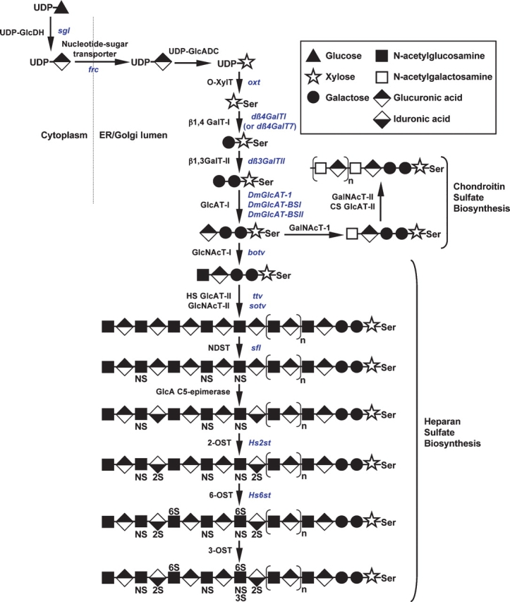
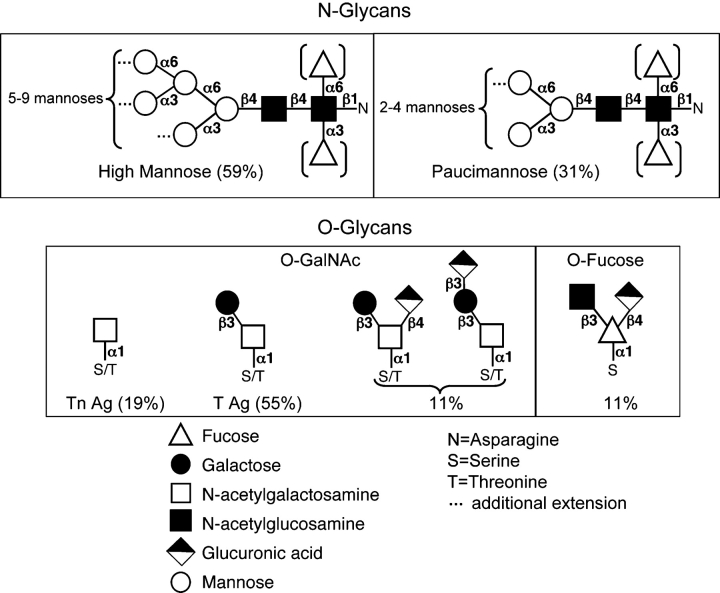
Drosophila melanogaster offers many unique advantages for deciphering the complexities of glycan biosynthesis and function. The completion of the Drosophila genome sequencing project as well as the comprehensive catalogue of existing mutations and phenotypes have lead to a prolific database where many of the genes involved in glycan synthesis, assembly, modification, and recognition have been identified and characterized. Recent biochemical and molecular studies have elucidated the structure of the glycans present in Drosophila. Powerful genetic approaches have uncovered a number of critical biological roles for glycans during development that impact on our understanding of their function during mammalian development. Here, we summarize key recent findings and provide evidence for the usefulness of this model organism in unraveling the complexities of glycobiology across many species.


Figures

Fig. 1
Fig. 1
Biosynthesis of glycosaminoglycans. The initiation of chondroitin sulfate (CS) and the complete synthesis of heparan sulfate (HS) are shown. Enzymes responsible for catalyzing each step are shown in black and the corresponding Drosophila genes are shown in blue. Enzyme abbreviations are as follows: UDP-GlcDH, UDP-glucose dehydrogenase; UDP-GlcADC, UDP-glucuronic acid decarboxylase; O-XylT, polypeptide O-xylosyltransferase; β1,4GalT-I, xylose-β1,4-galactosyltransferase; β1,3GalT-II, galactose-β1,3-galactosyltransferase; GlcAT-I, galactose-β1,3-glucuronyltransferase; GalNAcT-I, glucuronic acid-β1,4-N-acetylgalactosaminyltransferase; CS GlcAT-II, chondroitin sulfate GalNAc-β1,3-glucuronyltransferase; GalNAcT-II, glucuronic acid-β1, 4-N-acetylgalactosaminyltransferase; GlcNAcT-I, glucuronic acid-α1,4-N-acetylglucosaminyltransferase; HS GlcAT-II, heparan sulfate GlcNAc-β1, 4-glucuronyltransferase; GlcNAcT-II, glucuronic acid α1,4-N-acetylglucosaminyltransferase; NDST, N-deacetylase/N-sulfotransferase; 2-OST, 2-O-sulfotransferase; 6-OST, 6-O-sulfotransferase; 3-OST, 3-O-sulfotransferase.
Fig. 2
Fig. 2
N- and O-linked glycan structures present in Drosophila melanogaster. Shown are the major types of N-linked and O-linked glycans found in Drosophila and their relative abundance. N-Glycans consist primarily of high mannose (59%) and paucimannose (31%); hybrid (7%) and complex (3%) structures are much less abundant. Brackets indicate that an α1,3 fucose or α1,6 fucose may also be present in these structures. Mucin-type O-linked glycans are predominantly comprised of the Tn antigen (Tn Ag) and T antigen (T Ag or Core 1) structures. The only detectable protein O-fucose glycan in Drosophila is a glucuronyl trisaccharide (Aoki et al. 2008).
Fig. 3
Fig. 3
Mucin-type O-linked glycan expression is found throughout Drosophila embryogenesis. Tn Ag (GalNAcα1-S/T) was detected by immunofluorescence and confocal imaging using antibodies directed against this glycan (described in Tian and Ten Hagen 2007a). Embryos at various stages of development (shown in the bottom-left corner of each image) are shown across the top row. The bottom and middle rows show enlarged images of developing tubular structures (denoted in the top-right corner of each image). O-Glycans are abundant along the apical and luminal regions of the developing organs shown. Dashed white lines are included to illustrate the outer boundaries of certain organs. fg, foregut; hg, hindgut; mp, malpighian tubules; ps, posterior spiracles; sg, salivary gland; tp, tracheal placodes; ts, tracheal system. Adapted from Glycobiology, 17, 820-827 (2007) by copyright permission of Oxford University Press.

References

    1. Acar M, Jafar-Nejad H, Takeuchi H, Rajan A, Ibrani D, Rana NA, Pan H, Haltiwanger RS, Bellen HJ. Rumi is a CAP10 domain glycosyltransferase that modifies Notch and is required for Notch signaling. Cell. 2008;132:247–258. - PMC - PubMed
    1. Alexander W, Viney EM, Zhang J, Metcalf D, Kauppi M, Hyland CD, Carpinelli MR, Stevenson W, Croker BA, Hilton AA, et al. Thrombocytopenia and kidney disease in mice with a mutation in the C1galt1 gene. Proc Natl Acad Sci USA. 2006;103:16442–16447. - PMC - PubMed
    1. Aoki K, Perlman M, Lim J, Cantu R, Wells L, Tiemeyer M. Dynamic developmental elaboration of N-linked glycan complexity in the Drosophila melanogaster embryo. J Biol Chem. 2007;282:9127–9142. - PubMed
    1. Aoki K, Porterfield M, Lee SS, Dong B, Nguyen K, McGlamry KH, Tiemeyer M. The diversity of O-linked glycans expressed during Drosophila melanogaster development reflects stage- and tissue-specific requirements for cell signaling. J Biol Chem. 2008 In press. PMID: 18725413. - PMC - PubMed
    1. Boquet I, Hitier R, Dumas M, Chaminade M, Préat T. Central brain postembryonic development in Drosophila: Implication of genes expressed at the interhemispheric junction. J Neurobiol. 2000;42:33–48. - PubMed

Publication types