

Rotational position of femoral and tibial components in TKA using the femoral transepicondylar axis
- PMID: 18825470
- PMCID: PMC2565051
- DOI: 10.1007/s11999-008-0452-8
Rotational position of femoral and tibial components in TKA using the femoral transepicondylar axis
Abstract
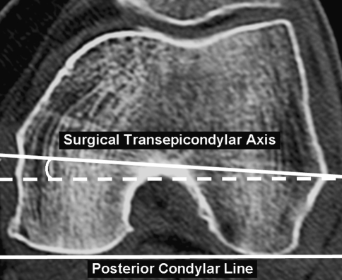
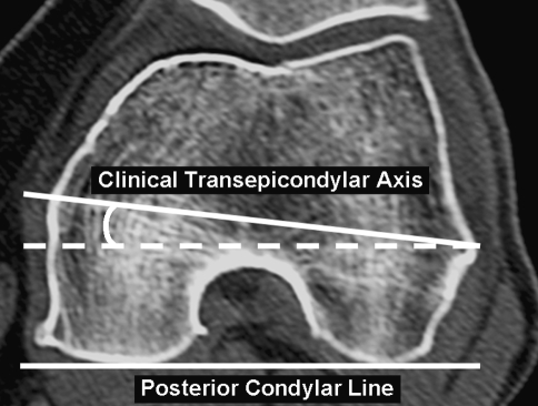
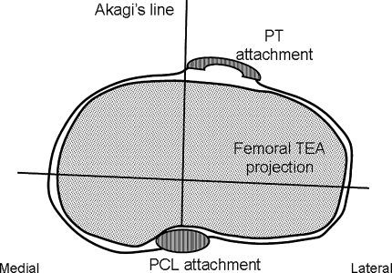
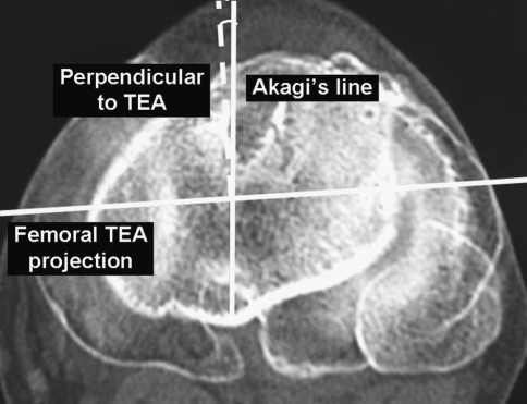
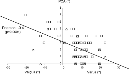
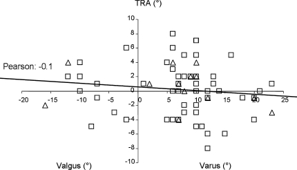
Proper femoral and tibial component rotational positioning in TKA is critical for outcomes. Several rotational landmarks are frequently used with different advantages and limitations. We wondered whether coronal axes in the tibia and femur based on the transepicondylar axis in the femur would correlate with anteroposterior deformity. We obtained computed tomography scans of 100 patients with arthritis before they underwent TKA. We measured the posterior condylar angle on the femoral side and the angle between Akagi's line and perpendicular to the projection of the femoral transepicondylar axis on the tibial side. On the femoral side, we found a linear relationship between the posterior condylar angle and coronal deformity with valgus knees having a larger angle than varus knees, ie, gradual external rotation increased with increased coronal deformity from varus to valgus. On the tibial side, the angle between Akagi's line and the perpendicular line to the femoral transepicondylar axis was on average approximately 0 degrees , but we observed substantial interindividual variability without any relationship to gender or deformity. A preoperative computed tomography scan was a useful, simple, and relatively inexpensive tool to identify relevant anatomy and to adjust rotational positioning. We do not, however, recommend routine use because on the femoral side, we found a relationship between rotational landmarks and coronal deformity.
Figures
References
-
- {'text': '', 'ref_index': 1, 'ids': [{'type': 'DOI', 'value': '10.1097/01.blo.0000160027.52481.32', 'is_inner': False, 'url': 'https://doi.org/10.1097/01.blo.0000160027.52481.32'}, {'type': 'PubMed', 'value': '15995437', 'is_inner': True, 'url': 'https://pubmed.ncbi.nlm.nih.gov/15995437/'}]}
- Akagi M, Mori S, Nishimura S, Nishimura A, Asano T, Hamanishi C. Variability of extraarticular tibial rotation references for total knee arthroplasty. Clin Orthop Relat Res. 2005;436:172–176. - PubMed
-
- {'text': '', 'ref_index': 1, 'ids': [{'type': 'DOI', 'value': '10.1097/00003086-200403000-00030', 'is_inner': False, 'url': 'https://doi.org/10.1097/00003086-200403000-00030'}, {'type': 'PubMed', 'value': '15057100', 'is_inner': True, 'url': 'https://pubmed.ncbi.nlm.nih.gov/15057100/'}]}
- Akagi M, Oh M, Nonaka T, Tsujimoto H, Asano T, Hamanishi C. An anteroposterior axis of the tibia for total knee arthroplasty. Clin Orthop Relat Res. 2004;420:213–219. - PubMed
-
- {'text': '', 'ref_index': 1, 'ids': [{'type': 'DOI', 'value': '10.1097/00003086-200107000-00022', 'is_inner': False, 'url': 'https://doi.org/10.1097/00003086-200107000-00022'}, {'type': 'PubMed', 'value': '11451114', 'is_inner': True, 'url': 'https://pubmed.ncbi.nlm.nih.gov/11451114/'}]}
- Akagi M, Yamashita E, Nakagawa T, Asano T, Nakamura T. Relationship between frontal knee alignment and reference axes in the distal femur. Clin Orthop Relat Res. 2001;388:147–156. - PubMed
-
- {'text': '', 'ref_index': 1, 'ids': [{'type': 'PubMed', 'value': '7673281', 'is_inner': True, 'url': 'https://pubmed.ncbi.nlm.nih.gov/7673281/'}]}
- Arima J, Whiteside LA, McCarthy DS, White SE. Femoral rotational alignment, based on the anteroposterior axis, in total knee arthroplasty in a valgus knee. A technical note. J Bone Joint Surg Am. 1995;77:1331–1334. - PubMed
-
- {'text': '', 'ref_index': 1, 'ids': [{'type': 'PubMed', 'value': '8425366', 'is_inner': True, 'url': 'https://pubmed.ncbi.nlm.nih.gov/8425366/'}]}
- Berger RA, Rubash HE, Seel MJ, Thompson WH, Crossett LS. Determining the rotational alignment of the femoral component in total knee arthroplasty using the epicondylar axis. Clin Orthop Relat Res. 1993;286:40–47. - PubMed
