Review
. 2008 Oct 28;47(43):11174-83.
doi: 10.1021/bi8013938. Epub 2008 Oct 1.

Diverse cell signaling events modulated by perlecan

Affiliations
Review

Diverse cell signaling events modulated by perlecan

John M Whitelock et al. Biochemistry. .

Abstract

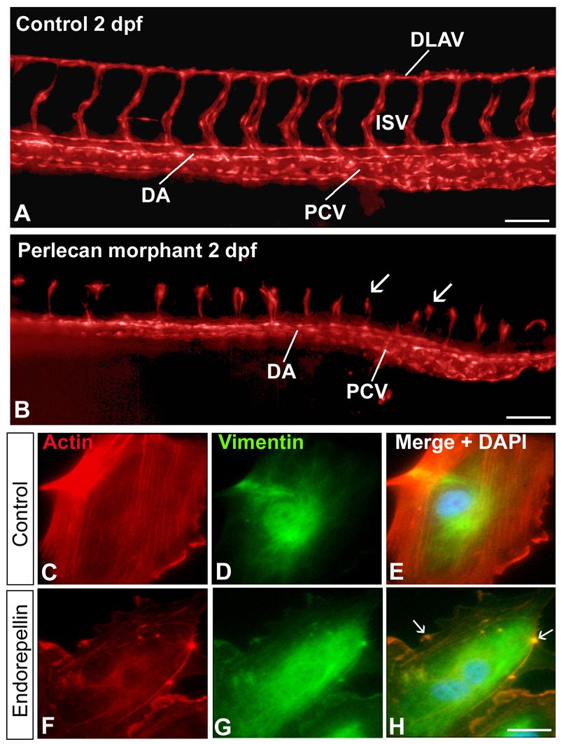
Perlecan is a ubiquitous pericellular proteoglycan ideally placed to mediate cell signaling events controlling migration, proliferation, and differentiation. Its control of growth factor signaling usually involves interactions with the heparan sulfate chains covalently coupled to the protein core's N-terminus. However, this modular protein core also binds with relatively high affinity to a number of growth factors and surface receptors, thereby stabilizing cell-matrix links. This review will focus on perlecan-growth factor interactions and describe recent advances in our understanding of this highly conserved proteoglycan during development, cancer growth, and angiogenesis. The pro-angiogenic capacities of perlecan that involve proliferative and migratory signals in response to bound growth factors will be explored, as well as the anti-angiogenic signals resulting from interactions between the C-terminal domain known as endorepellin and integrins that control adhesion of cells to the extracellular matrix. These two somewhat diametrically opposed roles will be discussed in light of new data emerging from various fields which converge on perlecan as a key regulator of cell growth and angiogenesis.


Figures

FIGURE 1
FIGURE 1
Schematic diagram of human perlecan depicting the various domains (Roman numerals) and the abbreviations for each module (top). The bottom panel shows a list of perlecan interactive partners relative to each protein core domain.
FIGURE 2
FIGURE 2
Pro- and anti-angiogenic activity of perlecan and endorepellin, respectively. (A,B) Effects of perlecan knockdown on the development of zebrafish embryonic vasculature. The vasculature is visualized in red (pseudocolor) and derives from the transgenic expression of GFP driven by the fli1 promoter specific for endothelial cells (A) Epifluorescence microscopy with 3D deconvolution of the trunk vessels from a zebrafish control embryo at 2 days post-fertilization (dpf). Note the correct formation of the dorsal aorta (DA), posterior cardinal vein (PCV), intersegmental vessels (ISV) and dorsal longitudinal anastomotic vessels (DLAV). (B) In contrast, the perlecan morphants exhibit relatively normal axial vessels, but significantly blunted ISVs and no DLAVs (arrows). Bar = 100 µm. (C–H) Double immunofluorescent micrographs of human endothelial cells following a 30 min-exposure to endorepellin using rhodamine-phalloidin to label actin (red) or antibodies against vimentin (green). Notice the endorepellin-evoked disruption of the actin cytoskeleton (F–H) in contrast to the preservation of the actin microfilaments in untreated endothelial cells (C–E). The arrows in panel H show bundles of actin filaments collapsed onto the plasmalemma, Bar = 5 µm.
FIGURE 3
FIGURE 3
Endorepellin is cleaved by the BMP-1/Tolloid proteases, liberating the antiangiogenic LG3 domain. The illustration depicts a graphic illustration of endorepellin and a comparative model of LG3. Highlighted in magenta is a calcium ion shown to be vital for function. To the right are immunofluorescence images of endothelial cells stained for actin (red) and depicting the effects of LG3 (notice the disruption of actin cytoskeleton) and of an LG3 mutant which exhibits reduced affinity for calcium. Nuclei are stained in blue by DAPI.
FIGURE 4
FIGURE 4
Working model showing the potential dual role of perlecan in tumor angiogenesis. Heparanase-mediated release of growth factors, primarily FGF2 and VEGF, bound to the heparan sulfate chains of perlecan would lead to sustained activation of FGF/FGFR and VEGFR2/Akt pathways, with consequent enhanced survival of endothelial cells and increased tumor angiogenesis and tumor progression. On the other hand, limited proteolysis of perlecan protein core could liberate endorepellin and LG3; this evokes a α2β1 integrin-dependent disruption of endothelial cell actin cytoskeleton, inhibition of endothelial cell migration, reduced angiogenesis and reduced tumor growth.

References

    1. Hassell JR, Yamada Y, Arikawa-Hirasawa E. Role of perlecan in skeletal development and diseases. Glycoconj. J. 2003;19:263–267. - PubMed
    1. Iozzo RV, Murdoch AD. Proteoglycans of the extracellular environment: clues from the gene and protein side offer novel perspectives in molecular diversity and function. FASEB J. 1996;10:598–614. - PubMed
    1. Iozzo RV. Basement membrane proteoglycans: from cellar to ceiling. Nature Rev. Mol. Cell Biol. 2005;6:646–656. - PubMed
    1. Marneros AG, Olsen BR. Physiological role of collagen XVIII and endostatin. FASEB J. 2005;19:716–728. - PubMed
    1. Bezakova G, Rüegg MA. New insights into the roles of agrin. Nature Rev. Mol. Cell Biol. 2003;4:295–308. - PubMed

Publication types

MeSH terms

Substances