. 2008 Sep 30:8:274.
doi: 10.1186/1471-2407-8-274.

CXCR4 expression in papillary thyroid carcinoma: induction by nitric oxide and correlation with lymph node metastasis

Affiliations

CXCR4 expression in papillary thyroid carcinoma: induction by nitric oxide and correlation with lymph node metastasis

Hironao Yasuoka et al. BMC Cancer. .

Abstract

Background: Metastasis to regional lymph nodes is a common step in the progression of cancer. Recent evidence suggests that tumor production of CXCR4 promotes lymph node metastasis. Nitric oxide (NO) may also increase metastatic ability in human cancers.

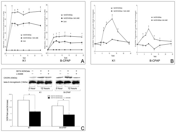
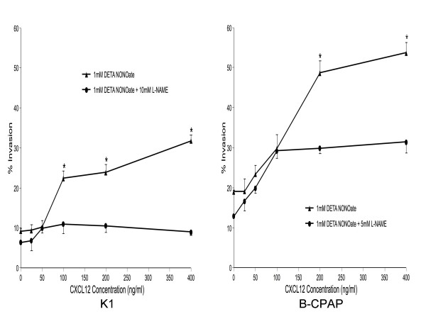
Methods: Nitrite/nitrate levels and functional CXCR4 expression were assessed in K1 and B-CPAP papillary thyroid carcinoma (PTC) cells after induction and/or inhibition of NO synthesis. CXCR4 expression was also analyzed in primary human PTC. The relationship between nitrotyrosine levels, which are a biomarker for peroxynitrate formation from NO in vivo, CXCR4 expression, and lymph node status was also analyzed.

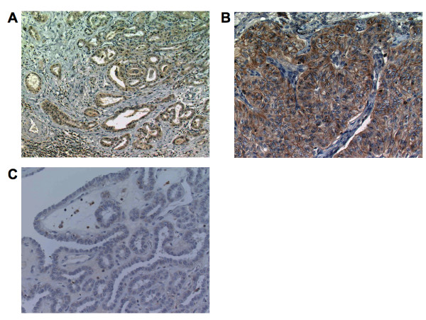
Results: Production of nitrite/nitrate and functional CXCR4 expression in both cell lines was increased by treatment with the NO donor DETA NONOate. The NOS inhibitor L-NAME eliminated this increase. Positive CXCR4 immunostaining was observed in 60.7% (34/56) of PTCs. CXCR4 expression was significantly correlated with nitrotyrosine levels and lymph node metastasis in human PTC.

Conclusion: Our data indicate that NO stimulates CXCR4 expression in vitro. Formation of the NO biomarker nitrotyrosine was also correlated with CXCR4 expression and lymph node metastasis in human PTC. NO may induce lymph node metastasis via CXCR4 induction in papillary thyroid carcinoma.


Figures

Figure 1
Figure 1
Effects of NO on CXCR4 expression. Effects of DETA NONOate in the presence or absence of L-NAME on (A) nitrate/nitrite production, (B) CXCR4 mRNA expression, and (C) CXCR4 protein expression. K1 and B-CPAP cells were treated with 1 mM DETA NONOate in the presence or absence of L-NAME for various time periods and prepared for (A) measurement of nitrate/nitrite production, (B) real-time RT-PCR analysis, and (C) western blot analysis, as described in Materials and Methods. Determinations were performed in triplicate and expressed as mean of three experiments ± SE. (A) Control indicates cells with no treatment. (B-C) Data was expressed as ratio of mRNA or protein levels relative to control (untreated) cells. * indicates significant difference (p < 0.05) from control and/or L-NAME-treated cells.
Figure 2
Figure 2
CXCL12 induced the chemoinvasion of papillary thyroid cancer cells. Cells were seeded into the upper compartments of Matrigel Invasion Chambers as described in Materials and Methods. Uncoated or Matrigel-coated membranes separated the upper compartments from lower compartments containing the indicated concentrations of CXCL12. Cells were treated with DETA NONOate with or without L-NAME. Two days later, cells that had migrated to the bottom of the membrane were stained and counted. The percentage of invasive cells (% Invasion) was calculated as the number of cells penetrating the Matrigel-coated membranes divided by the number penetrating the uncoated membranes. Determinations were performed in triplicate and expressed as mean of three experiments ± SE. * indicates significant difference (p < 0.05) from L-NAME-treated cells.
Figure 3
Figure 3
CXCR4 expression in human papillary thyroid carcinoma tissue. (A) immunohistochemical localization of CXCR4 protein was cytoplasmic. (B) In some cases carcinoma cells showed positive staining in the membrane. (C) In the tumor in which CXCR4 immunodetection are unreactive, staining for CXCR4 occurred in inflammatory cells.

References

    1. Xu W, Liu LZ, Loizidou M, Ahmed M, Charles IG. The role of nitric oxide in cancer. Cell Res. 2002;12:311–320. doi: 10.1038/sj.cr.7290133. - DOI - PubMed
    1. Ambs S, Merriam WG, Ogunfusika MO, Bennett WP, Ishibe N, Hussain SP, Tzeng EE, Geller DA, Billiar TR, Harris CC. p53 and vascular endothelial growth factor regulate tumor growth of NOS2-expressing human carcinoma cells. Nat Med. 1998;4:1371–1376. doi: 10.1038/3957. - DOI - PubMed
    1. Nakamura Y, Yasuoka H, Tsujimoto M, Yoshidome K, Nakahara M, Nakao K, Nakamura M, Kakudo K. Nitric oxide in breast cancer: induction of vascular endothelial growth factor-C and correlation with metastasis and poor prognosis. Clin Cancer Res. 2006;12:1201–1207. doi: 10.1158/1078-0432.CCR-05-1269. - DOI - PubMed
    1. Nakamura Y, Yasuoka H, Zuo H, Takamura Y, Miyauchi A, Nakamura M, Kakudo K. Nitric oxide in papillary thyroid carcinoma: induction of vascular endothelial growth factor d and correlation with lymph node metastasis. J Clin Endocrinol Metab. 2006;91:1582–1585. doi: 10.1210/jc.2005-1790. - DOI - PubMed
    1. Beckman JS. Oxidative damage and tyrosine nitration from peroxynitrite. Chem Res Toxicol. 1996;9:836–844. doi: 10.1021/tx9501445. - DOI - PubMed

Publication types

MeSH terms