. 2008 Oct 15;121(Pt 20):3476-86.
doi: 10.1242/jcs.029454. Epub 2008 Sep 30.

TorsinA binds the KASH domain of nesprins and participates in linkage between nuclear envelope and cytoskeleton

Affiliations

TorsinA binds the KASH domain of nesprins and participates in linkage between nuclear envelope and cytoskeleton

Flávia C Nery et al. J Cell Sci. .

Abstract

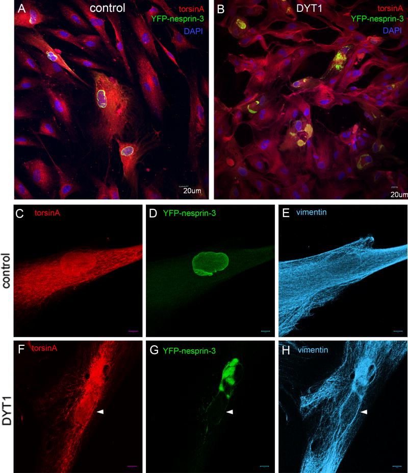
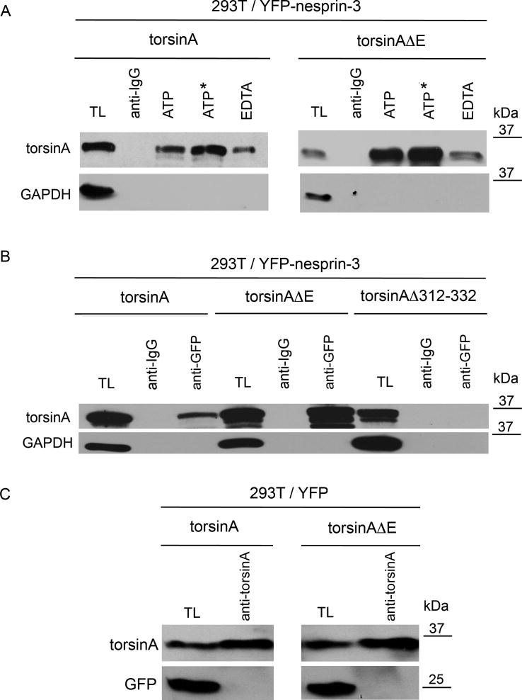
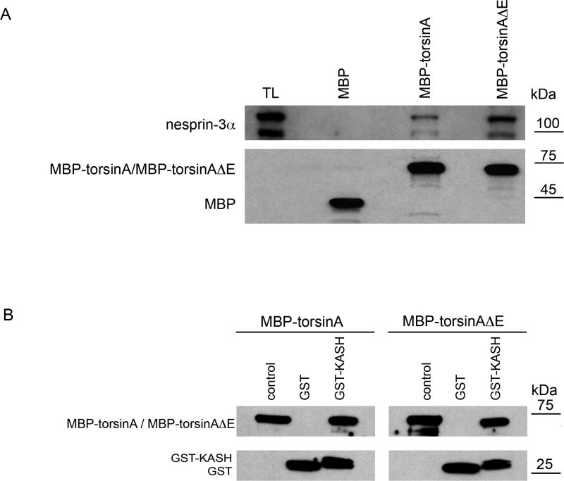
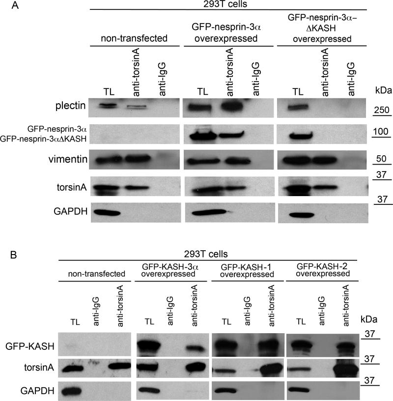
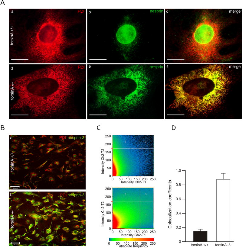
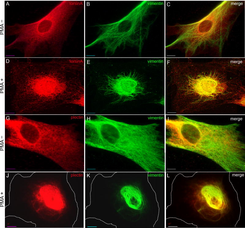
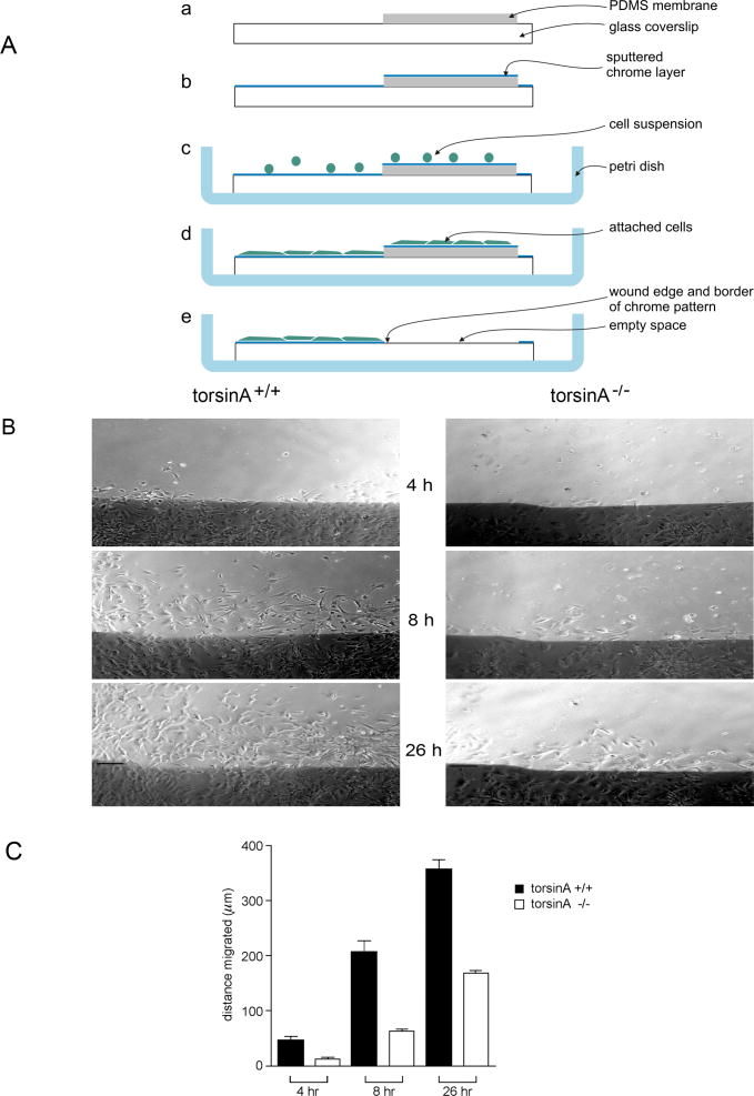
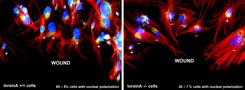
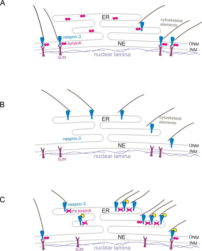
A specific mutation (DeltaE) in torsinA underlies most cases of the dominantly inherited movement disorder, early-onset torsion dystonia (DYT1). TorsinA, a member of the AAA+ ATPase superfamily, is located within the lumen of the nuclear envelope (NE) and endoplasmic reticulum (ER). We investigated an association between torsinA and nesprin-3, which spans the outer nuclear membrane (ONM) of the NE and links it to vimentin via plectin in fibroblasts. Mouse nesprin-3alpha co-immunoprecipitated with torsinA and this involved the C-terminal region of torsinA and the KASH domain of nesprin-3alpha. This association with human nesprin-3 appeared to be stronger for torsinADeltaE than for torsinA. TorsinA also associated with the KASH domains of nesprin-1 and -2 (SYNE1 and 2), which link to actin. In the absence of torsinA, in knockout mouse embryonic fibroblasts (MEFs), nesprin-3alpha was localized predominantly in the ER. Enrichment of yellow fluorescent protein (YFP)-nesprin-3 in the ER was also seen in the fibroblasts of DYT1 patients, with formation of YFP-positive globular structures enriched in torsinA, vimentin and actin. TorsinA-null MEFs had normal NE structure, but nuclear polarization and cell migration were delayed in a wound-healing assay, as compared with wild-type MEFs. These studies support a role for torsinA in dynamic interactions between the KASH domains of nesprins and their protein partners in the lumen of the NE, with torsinA influencing the localization of nesprins and associated cytoskeletal elements and affecting their role in nuclear and cell movement.


Figures

Fig. 1
Fig. 1
Association of endogenous nesprin-3α, plectin and vimentin with torsinA. Lysates from wild-type MFs were immunoprecipitated at 4°C overnight with antibodies to torsinA (D-M2A8 and DMG-10, 1:1) or IgG (anti-mouse IgG) without added ATP. Immunoprecipitates and lysates were resolved on SDS-PAGE gels and immunoblotted with antibodies to plectin, nesprin-3, vimentin, torsinA and GAPDH. TL, total lysates. The position of MW markers is indicated. The experiment was repeated three times and representative blots are shown. The percentage of each protein immunoprecipitated from lysates is: 7±2% for plectin, 17±4% for nesprin-3α, 17±3% for vimentin,12±3% for torsinA (values are mean percentage ± s.e.m., n=3 experiments).
Fig. 2
Fig. 2
Differential distribution of YFP-nesprin-3 in DYT1 and control human fibroblasts. Primary skin fibroblasts from controls and DYT1 subjects were infected with a lentivirus vector expressing YFP-nesprin-3 and fixed 72 hours later. (A,B) Cells were immunostained for torsinA and GFP, with DAPI staining of nuclei. In control cells, YFP-nesprin is primarily localized in the perinuclear region (A), whereas in DYT1 cells it accumulates in globular structures presumably within the ER/NE (B); this was seen in two different cell lines of each type. (C–E) Immunocytochemical staining of infected control cells showing a typical distribution of torsinA (ER/NE) and vimentin (cytoplasm), with YFP-nesprin-3 primarily localized around the nucleus. (F–H) Staining of infected cells from DYT1 patients showing increased concentration of both torsinA and vimentin in the region of YFP-nesprin-3 globular structures. White arrowheads indicate nuclei. Scale bars: 20 μm in A,B; 10 μm in C–H.
Fig. 3
Fig. 3
Association of torsinA and torsinAΔE with nesprin-3. (A) Human 293T cells were co-transfected with expression cassettes for YFP-nesprin and either torsinA or torsinAΔE. Immunoprecipitations were carried out in the presence of 1 mM ATP, non-hydrolysable ATP or EDTA (the latter with no added ATP) in RIPA buffer with antibody to GFP or with anti-rabbit IgG as control. Immunoprecipitated proteins were resolved by SDS-PAGE and immunoblotted with antibodies to torsinA (D-M2A8) or GAPDH. On average, threefold more torsinAΔE was immunoprecipitated than torsinA (3.0±0.88, n=3, P<0.0001, ANOVA). (B) Experiments were carried out as in A except that transfections were with expression cassettes for torsinA, torsinAΔE or torsinAΔ312–322. The additional bands under the predominant 37-kDa torsinA band presumably represent the non- and partially glycosylated forms seen in other studies (Hewett et al., 2004). (C) To verify that torsinA and torsinAΔE bind to nesprin-3 and not to YFP, 293T cells were co-transfected with expression cassettes for YFP and either torsinA or torsinAΔE. Immunoprecipitations were carried out in the presence of 1 mM ATP in RIPA buffer with antibody to torsinA. Immunoprecipitated proteins were resolved by SDS-PAGE and immunoblotted with antibodies to torsinA (D-M2A8) or GFP.
Fig. 4
Fig. 4
Binding of MBP-torsinA and MBP-torsinAΔE recombinant proteins to nesprin-3α and GST KASH. (A) MBP, MBP-torsinA or MBP-torsinAΔE fusion proteins (Hewett et al., 2003) (8 μg) were bound to amylase resin (New England Biolabs) and incubated overnight at 4°C with lysates of wild-type MFs (500 μl protein in 1 ml) in RIPA buffer containing 1 mM ATP and 3 mM MgCl2. Beads were washed five times with RIPA buffer and proteins bound to beads resolved by SDS-PAGE and immunoblotted with antibodies to nesprin-3α (top) and MBP (bottom), with relative binding assessed as in Fig. 1. MBP-torsinAΔE consistently pulled down ~threefold more nesprin-3α than did MBP-torsinA (3.0±0.3, n=3, P<0.003, ANOVA). The lower, immunoreactive nesprin-3α band is a degradation product, also seen by Wilhelmsen et al. (Wilhelmsen et al., 2005). (B) GST or GST-KASH fusion protein (10 μg) bound to glutathione-Sepharose beads was incubated with 10 μg of MBP-torsinA or MBP-torsinAΔE in 500 μl PBS containing 1 mM ATP and 3 mM MgCl2 for 1 hour at 4°C. Unbound material was removed by centrifugation and the beads washed five times with 0.1% NP40 buffer followed by an additional wash with PBS. The proteins bound to the beads were resolved by SDS-PAGE and visualized by western blotting with antibodies to MBP and GST.
Fig. 5
Fig. 5
TorsinA associates with the KASH domain. (A) Human 293T cells were transfected with lentivirus vector encoding expression constructs for GFP-mouse-nesprin-3α or GFP-mouse-nesprin-3αΔ KASH. After 48 hours the cells were lysed and proteins immunoprecipitated with antibodies to torsinA or IgG (no added ATP), resolved by SDS-PAGE and immunoblotted with antibodies to plectin, GFP, vimentin, torsinA and GAPDH. Similar amounts of torsinA were precipitated from non-transfected cells and cells transfected with the GFP-nesprin-3α or GFP-nesprin-3αΔ KASH constructs. By contrast, no detectable plectin was immunoprecipitated with torsinA from cells expressing GFP-nesprin-3αΔ KASH, although some was immunoprecipitated from cells expressing GFP-nesprin-3α. (B) A similar experiment to A except that cells were transfected with expression constructs for GFP-KASH-3α, GFP-KASH-1 or GFP-KASH-2 (i.e. the KASH domains of nesprin-3α, nesprin-1 and nesprin-2, respectively). Immunoprecipitation with torsinA antibodies followed by immunoblotting for GFP revealed an association of torsinA with the KASH domains of all three nesprins. The percentage of each protein immunoprecipitated from lysates by torsinA antibodies is: 11±3% for GFP-KASH-3α, 21±5% for GFP-KASH-1, 12±4% for GFP-KASH-2 (values are mean percentage ± s.e.m., n=3 experiments).
Fig. 6
Fig. 6
Differential distribution of nesprin-3 in torsinA+/+ and torsinA−/− MEFs. (A) Double immunolabeling for the ER marker PDI and nesprin-3α in torsinA+/+ and torsinA−/− MEFs. (B) As A, but at lower magnification. (C) The relative proportion of immunoreactive nesprin-3α that was co-localized with the ER marker PDI in torsinA+/+ and torsinA−/− cells was assessed with a confocal microscope using the LSM 5 Pascal program, evaluating the co-localization coefficient for 150 cells of each cell type. (D) The relative number of co-localizing pixels in the red and green channels was evaluated in a value range 0–1 (0, no co-localization; 1, all pixels co-localize) and a significant difference was found between torsinA+/+ and torsinA−/− cells (n=3, P<0.0001, ANOVA). Error bars indicate s.e.m. Scale bars: 10 μm in A; 100 μm in B.
Fig. 7
Fig. 7
Co-collapse of torsinA, plectin and vimentin following PMA treatment. Control human fibroblasts were treated with 200 ng/ml PMA (phorbol 12-myristate 13-acetate) for 30 minutes and the distribution of torsinA and vimentin (A-F), or of plectin and vimentin (G-L), was assessed by immunocytochemistry. The cell perimeter is outlined in J-L. This experiment was repeated three times and representative images are shown. Scale bar: 10 μm.
Fig. 8
Fig. 8
Migration of torsinA+/+ and torsinA−/− MEFs. (A) A microfabrication chamber was designed for wound-healing assays to provide a precise physical reference of the original position of the wound edge during long-term observations. (a) A micropatterned coverslip was partially covered with a thin polydimethylsiloxane (PDMS) membrane. (b) A thin layer of chrome was deposited over the entire surface of the coverslip and membrane. (c) The coverslip was placed in a Petri dish and covered with a suspension of cells. (d) The cells were allowed to form a confluent monolayer on the glass and the thin PDMS membrane. (e) The wound was initiated by peeling off the PDMS membrane, which removes all cells except those on the chrome layer. (B) Confluent monolayers were incubated in serum-free medium for 48 hours, then a wound was made in the monolayer and 5% serum was added (Gomes and Gundersen, 2006). Marked regions in the wound were photographed under phase-contrast microscopy, sequentially at intervals up to 26 hours post-wounding and the migration of cells into the cleared space was monitored at different time points. Digital images were taken at 10× magnification and evaluated using MetaVuE software with Microsoft Excel. Photographs of fibroblast monolayers were taken under phase-contrast microscopy 4, 8 and 26 hours after wounding (a representative region is shown). Scale bar: 150 μm. (C) The migration of the leading edge of cells was measured (in μm) from fixed points on the wound edge. Values represent mean ± s.e.m. (error bars) of measurements in six regions per time point for each genotype from seven experiments. Differences between torsinA+/+ and torsinA−/− cells were significant at all time points (P<0.0001, ANOVA).
Fig. 9
Fig. 9
Nuclear polarization in migrating torsinA+/+ and torsinA−/−MEFs. To evaluate nuclear polarization, confluent monolayers of MEFs were serum-starved for 24 hours, then a wound was made through the monolayer and cells were stimulated with 2 μM lysophosphatidic acid (LPA; Sigma-Aldrich) (Gomes and Gundersen, 2006). Three hours later, the cells were fixed and immunostained for pericentrin to label the centrosome (green) and for β-tubulin to label microtubules (red), with DAPI staining (blue) for nuclei. The nucleus was scored as polarized (+) when the centrosome was localized between the nucleus and the leading wound edge and as non-polarized (-) in any other location. Representative images are shown for torsinA+/+ (left) and torsinA−/− (right) cells along the wound edge. One hundred cells of each type were evaluated for orientation of the centrosome between the nucleus and the leading edge in each of three experiments using two different MEF preparations of each type. TorsinA+/+ cells showed 85±5% nuclei in the polarized location, whereas torsinA−/− cells showed only 38±7% in the polarized position at this time point (values are mean percentage ± s.e.m., n=3 experiments, P<0.001). Magnification: 20×.
Fig. 10
Fig. 10
Theoretical consequences of torsinA status for nesprin-SUN interactions. Nesprins span the outer nuclear membrane (ONM) and interact with cytoskeletal elements in the cytoplasm and with inner nuclear membrane (INM) proteins, such as SUNs, in the lumen of the nuclear envelope (NE). TorsinA is hypothesized to act as an AAA+ protein in the disassembly/reassembly of these nesprin-SUN interactions, for example, during movement of the nucleus in cell migration. (A) In wild-type cells, nesprins and associated cytoskeletal elements are localized to the ONM by interactions with INM proteins, such as SUNs, and this interaction (at least for nesprin-3) is modulated by torsinA (depicted as an oligomer in cross-section, two circles). (B) In torsinA-null cells, nesprin-3 and linked cytoskeletal elements are located predominantly in association with the endoplasmic reticulum (ER) (see Fig. 6), as the association of nesprins with INM proteins is predicted to be compromised. (C) Cells from DYT1 subjects express both torsinA and torsinAΔE, with the mutant (mt) torsinA hypothesized to form inactive oligomers with torsinA (depicted as X-shaped structures). When YFP-nesprin-3 is overexpressed in DYT1 cells, torsinA oligomers containing torsinAΔE, which binds more tightly than torsinA to nesprins, reduce the interaction of YFP-nesprin-3 with INM proteins, such that YFP-nesprin-3 accumulates in the ER, where, together with torsinA/torsinAΔE and associated cytoskeletal elements, it forms globular structures (see Fig. 2).

References

    1. Andra K, Nikolic B, Stocher M, Drenckhahn D, Wiche G. Not just scaffolding: plectin regulates actin dynamics in cultured cells. Genes Dev. 1998;12:3442–3451. - PMC - PubMed
    1. Andra K, Kornacker I, Jorgl A, Zorer M, Spazierer D, Fuchs P, Fischer I, Wiche G. Plectin-isoform-specific rescue of hemidesmosomal defects in plectin (−/−) keratinocytes. J Invest Dermatol. 2003;120:189–197. - PubMed
    1. Asanuma K, Carbon-Correll M, Eidelberg D. Neuroimaging in human dystonia. J Med Invest. 2005;52:272–279. - PubMed
    1. Basham SE, Rose LS. The Caenorhabditis elegans polarity gene ooc-5 encodes a Torsin-related protein of the AAA ATPase superfamily. Development. 2001;128:4645–4656. - PubMed
    1. Berardelli A, Rothwell JC, Hallett M, Thompson PD, Manfredi M, Marsden CD. The pathophysiology of primary dystonia. Brain. 1998;121:1195–1212. - PubMed

Publication types

MeSH terms