. 2008 Sep;20(9):2471-83.
doi: 10.1105/tpc.108.062331. Epub 2008 Sep 30.

Distribution and evolution of circular miniproteins in flowering plants

Affiliations

Distribution and evolution of circular miniproteins in flowering plants

Christian W Gruber et al. Plant Cell. 2008 Sep.

Abstract

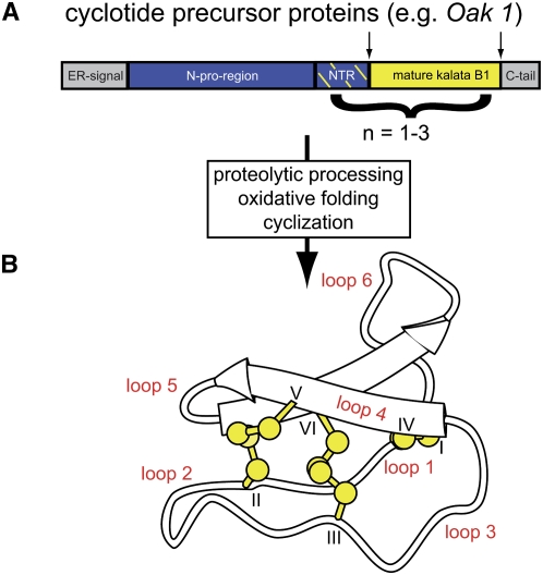
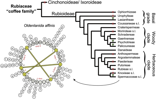
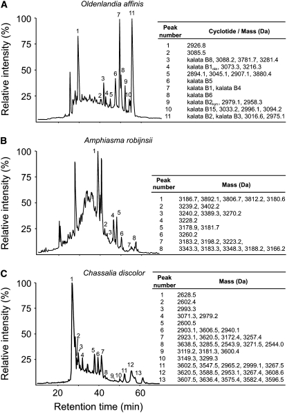
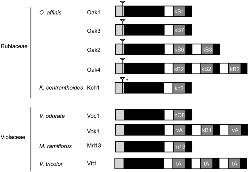
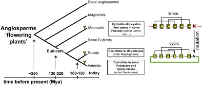
Cyclotides are disulfide-rich miniproteins with the unique structural features of a circular backbone and knotted arrangement of three conserved disulfide bonds. Cyclotides have been found only in two plant families: in every analyzed species of the violet family (Violaceae) and in few species of the coffee family (Rubiaceae). In this study, we analyzed >200 Rubiaceae species and confirmed the presence of cyclotides in 22 species. Additionally, we analyzed >140 species in related plant families to Rubiaceae and Violaceae and report the occurrence of cyclotides in the Apocynaceae. We further report new cyclotide sequences that provide insights into the mechanistic basis of cyclotide evolution. On the basis of the phylogeny of cyclotide-bearing plants and the analysis of cyclotide precursor gene sequences, we hypothesize that cyclotide evolution occurred independently in various plant families after the divergence of Asterids and Rosids ( approximately 125 million years ago). This is strongly supported by recent findings on the in planta biosynthesis of cyclotides, which involves the serendipitous recruitment of ubiquitous proteolytic enzymes for cyclization. We further predict that the number of cyclotides within the Rubiaceae may exceed tens of thousands, potentially making cyclotides one of the largest protein families in the plant kingdom.


Figures

Figure 1.
Figure 1.
Biosynthesis and Structure of Cyclotides. (A) Cyclotides are gene products synthesized on ribosomes and modified to maturation in the secretory pathway (Gruber et al., 2007b). The prototypic cyclotide kalata B1 is synthesized in O. affinis as part of a precursor protein, Oak1 (Oldenlandia affinis kalata B1), comprising an endoplasmic reticulum target signal (gray), N-terminal proregion (blue), an N-terminal repeat (NTR; blue/yellow-dashed), the mature cyclotide domain (yellow), and a C-terminal tail (gray). Other cyclotide precursor proteins contain up to three mature cyclotide domains. Processing of the precursor involves oxidative folding to form three-disulfide bonds, excision of the mature sequence, and head-to-tail cyclization. (B) Mature cyclotides comprise the typical structural CCK motif, characterized by three disulfide bonds (yellow) in a knotted arrangement. Two disulfide bonds and the adjacent backbone segments form a ring (CI-CIV and CII-CV) that is threaded by the third disulfide bond (CIII-CVI). The Cys residues separate the backbone into six loops, labeled loops 1 to 6. The image shows the backbone structure of kalata B1 with the disulfide bonds indicated in ball-stick representation and the small antiparallel β-sheet indicated with arrows.
Figure 2.
Figure 2.
Flowchart for the Screening of Cyclotides. A set of criteria was developed to decide whether a species contains cyclotides or not. In a prescreen, all plant material was extracted by either dichloromethane/methanol/water (1:1:1; v/v/v) or acetonitrile/water (1:1; v/v) and prepurified by C18 chromatography. The main screen was developed to analyze the plant compounds for their chemical and biophysical properties and was divided in three screening parts: (A) hydrophobicity, (B) mass range, and (C) Cys content. Each species was scored for the number of peaks (i.e., peptides) that fulfilled the screening criteria in HPLC and MS spectra. If a species scored ≥1 for each of parts A, B, and C, it was classified as cyclotide-containing species. Selected positive hits were confirmed by amino acid sequencing in a postscreen.
Figure 3.
Figure 3.
Sequence Alignment of Novel Cyclotides CD-1 and PS-1. The novel cyclotides CD-1 and PS-1 from C. discolor and P. suterella, respectively, were aligned to known cyclotide sequences that they are most similar to (kalata B1 and varv F, respectively). The similarity alignment highlights the conserved positions for cyclotides, such as the six highly conserved Cys residues, the Gly and Glu residues in loop 1, the positive residue Arg or Lys in loop 6, and the C-terminal Asn thought to be involved in cyclization of cyclotides. The composition of isobaric residues Leu and Ile of PS-1 were determined by amino acid analysis and their positions differentiated by a chymotrypsin digest. The composition of isobaric residues Leu and Ile of CD-1 were determined by a combination of amino acid analysis and similarity alignment.
Figure 4.
Figure 4.
Distribution of Cyclotides in Rubiaceae. Summary cladogram of the family Rubiaceae based on Bremer and Manen (2000) showing the relationships between the tribes of the subfamily Rubioideae. The tribal delimitations follow Robbrecht and Manen (2006). The woody and herbaceous clades correspond to the supertribes Psychotriidinae and Rubiidinae, respectively. The presence (green triangle) and absence (red cross) of cyclotides are indicated for all analyzed plant tribes/families. The sequence diversity of cyclotides is indicated for peptides found in the prototypic cyclotide-containing plant O. affinis (Spermacoceae). Variations of amino acids for each position to the cyclotide backbone are indicated in a radial formation on the outside of the backbone circle that was generated by alignment of kalata B1–B18.
Figure 5.
Figure 5.
Expression and Numbers of Cyclotides within a Plant Species. LC-MS profiles of the plant extracts from O. affinis (A), A. robijnsii (B), and C. discolor (C) and are shown to highlight the number of unique cyclotides in each plant species. LC elution profiles are shown from 10 to 70 min at 1% min−1 solvent B (solvent B: 90% acetonitrile in Milli-Q H2O with 0.1% formic acid). Elutions of cyclotides, characterized by mass spectrometry, are indicated with Arabic numbers. Cyclotide masses are given to the right of the graph. Some cyclotides coelute from the reverse phase C18 column; hence, multiple masses are indicated. The representative plant species screened from O. affinis contains at least 18, A. robijnsii at least 22, and C. discolor at least 38 unique and novel cyclotides.
Figure 6.
Figure 6.
Structure of Cyclotide Precursor Gene. Cyclotide precursor genes were isolated and characterized from O. affinis (Oak1-4), K. centranthoides (Kch1), Viola odorata (Voc1 and Vok1), Melicytus ramiflorus, (Mrl13), and Viola tricolor (Vtt1). Cyclotide genes have a conserved structure, starting with a signal peptide, followed by a propeptide region and one or more copies of cyclotide coding domains. Introns, indicated by an inverted triangle, are present in the signal region of genes from Rubiaceae plants but are not present in genes from Violaceae plants. The asterisk indicates a premature stop codon.
Figure 7.
Figure 7.
Distribution and Evolution of Cyclotides in Plants. Summary cladogram showing the major evolutionary groups of angiosperms and the occurrence of cyclotides within these groups. The timeline of the evolution of flowering plants is indicated in million years ago (Mya). Cyclization of linear proteins may occur by simple mutations of linear cystine knot ancestor genes, which yield appropriately located Asn/Asp residues.

References

    1. Backlund, M., Oxelman, B., and Bremer, B. (2000). Phylogenetic relationships within the Gentianales based on ndhF and rbcL sequences, with particular reference to the Loganiaceae. Am. J. Bot. 87 1029–1043. - PubMed
    1. Barbeta, B.L., Marshall, A.T., Gillon, A.D., Craik, D.J., and Anderson, M.A. (2008). Plant cyclotides disrupt epithelial cells in the midgut of lepidopteran larvae. Proc. Natl. Acad. Sci. USA 105 1221–1225. - PMC - PubMed
    1. Basse, C.W. (2005). Dissecting defense-related and developmental transcriptional responses of maize during Ustilago maydis infection and subsequent tumor formation. Plant Physiol. 138 1774–1784. - PMC - PubMed
    1. Bokesch, H.R., Pannell, L.K., Cochran, P.K., Sowder, R.C., McKee, T.C., and Boyd, M.R. (2001). A novel anti-HIV macrocyclic peptide from Palicourea condensata. J. Nat. Prod. 64 249–250. - PubMed
    1. Bremer, B. (1996). Phylogenetic studies within Rubiaceae and relationships to other families based on molecular data. Opera Bot. Belg. 7 33–50.

Publication types

LinkOut - more resources