. 2009 Jan;17(1):162-8.
doi: 10.1038/mt.2008.220. Epub 2008 Sep 30.

Chitosan/siRNA nanoparticle-mediated TNF-alpha knockdown in peritoneal macrophages for anti-inflammatory treatment in a murine arthritis model

Affiliations

Chitosan/siRNA nanoparticle-mediated TNF-alpha knockdown in peritoneal macrophages for anti-inflammatory treatment in a murine arthritis model

Kenneth A Howard et al. Mol Ther. 2009 Jan.

Abstract

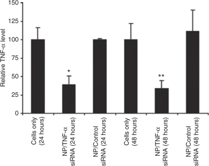
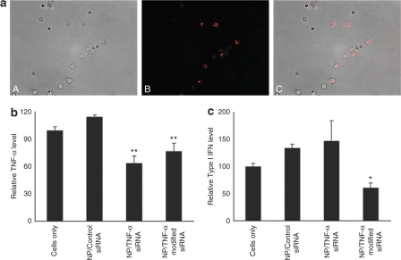
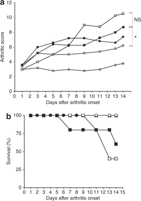
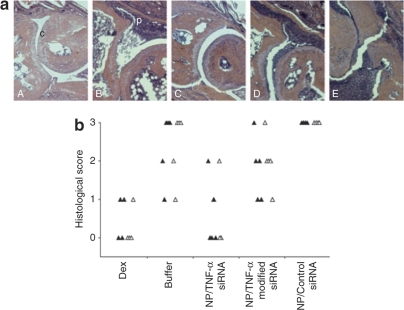
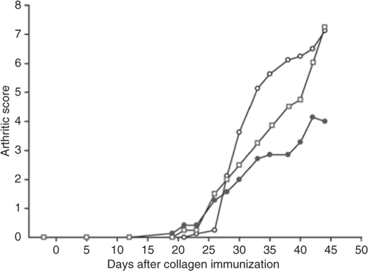
Secretion of tumor necrosis factor-alpha (TNF-alpha) by macrophages plays a predominant role in the development and progression of rheumatoid arthritis. We demonstrate that knockdown of TNF-alpha expression in systemic macrophages by intraperitoneal (i.p.) administration of chitosan/small interfering RNA (siRNA) nanoparticles in mice downregulates systemic and local inflammation. Chitosan nanoparticles containing an unmodified anti-TNF-alpha Dicer-substrate siRNA (DsiRNA) mediated TNF-alpha knockdown (approximately 66%) in primary peritoneal macrophages in vitro. The presence of Cy3-labeled nanoparticles within peritoneal macrophages and specific TNF-alpha knockdown (approximately 44%) with TNF-alpha siRNA after i.p. injection supports our therapeutic approach. Downregulation of TNF-alpha-induced inflammatory responses arrested joint swelling in collagen-induced arthritic (CIA) mice dosed i.p. with anti-TNF-alpha DsiRNA nanoparticles. The use of 2'-O-Me-modified DsiRNA resulted in the lowest arthritic scores and correlated with reduced type I interferon (IFN) activation in macrophages in vivo compared with unmodified DsiRNA. Histological analysis of joints revealed minimal cartilage destruction and inflammatory cell infiltration in anti-TNF-alpha-treated mice. The onset of arthritis could be delayed using a prophylactic dosing regime. This work demonstrates nanoparticle-mediated TNF-alpha knockdown in peritoneal macrophages as a method to reduce both local and systemic inflammation, thereby presenting a novel strategy for arthritis treatment.


Figures

<b>Figure 1</b>
Figure 1
Nanoparticle-mediated TNF-α silencing in murine primary macrophages in vitro. Isolated peritoneal macrophages from C57BL/6J mice were transfected with chitosan/TNF-α-specific siRNA or chitosan/TNF-α control for 4 hours (50 nmol/l siRNA/well). The level of TNF-α in the cellular supernantant, measured 5 hours after LPS stimulation 24 or 48 hours after transfection, was used as a measure of TNF-α knockdown (normalized to untreated macrophages (cells only), arbitrarily set to 100). Error bars represent ±SD. Significant differences between NP/TNF-α and NP/Control siRNA are indicated (*P < 0.01; **P < 0.02; two-sided t-test). Data from single experiment with triplicate samples.
<b>Figure 2</b>
Figure 2
Nanoparticle-mediated transfection and TNF-α knockdown in murine peritoneal macrophages in vivo. The ability of chitosan/siRNA nanoparticles to transfect and silence TNF-α production in peritoneal macrophages after i.p. administration. (a) Fluorescence micrograph showing chitosan nanoparticle uptake in macrophages extracted 4 hours after i.p. administration of 200 μl chitosan/Cy3-siRNA nanoparticles (panel A, light image; panel B, Cy3-labeled siRNA (red); panel C, fluorescence overlay of Cy3-labeled siRNA (red) and cells). (b) Macrophage TNF-α production measured by enzyme-linked immunosorbent assay (ELISA) (normalized to untreated LPS-stimulated macrophages (cells only), set to 100; error bars represent ±SD) in LPS-stimulated peritoneal macrophages extracted 2 hours after i.p. administration of 200 μl of chitosan nanoparticles containing ~5 μg of 2′-O-methyl modified anti-TNF-α DsiRNA (NP/TNF-α modified siRNA) or unmodified anti-TNF-α DsiRNA (NP/TNF-α siRNA) or TNF-α Dicer-substrate control siRNA (NP/Control siRNA) in mice (N = 3). Two-sided t-test statistics relative to chitosan/TNFα-control are given (**P < 0.003). (c) ELISA determination of type I IFN expression in macrophages shown in b (normalized to control untreated LPS-stimulated macrophages (cells only), set to 100; error bars represent ± SD). Two-sided t-test statistics for unmodified versus modified siRNA is shown (*P < 0.03).
<b>Figure 3</b>
Figure 3
Therapeutic effects in collagen-induced arthritic (CIA) mice after i.p. administration of chitosan/siRNA nanoparticles. (a) The therapeutic effect of i.p. administration of chitosan/siRNA-TNF-α nanoparticles on the clinical progression of established collagen-induced arthritis. Animals were scored for clinical symptoms using an arthritic scoring method for level of joint inflammation (scale: 0 = normal, 1 = mild swelling, 2 = moderate swelling, 3 = severe swelling of entire paw, 4 = maximal inflammation in multiple joints; sum total for 4 limbs = 16). Day 1 corresponds to onset of arthritis and assignments to treatment groups (N = 5). Animals were dosed i.p. on days 1, 3, 5, 7, and 9 with 200 μl nanoparticles containing either unmodified (closed circles) or 2′-O-Me modified (open circles) anti-TNF-α DsiRNA or control (closed squares) DsiRNA siRNA, or 0.2 mol/l sodium acetate buffer (open squares). Dexamethasone control group were dosed (400 μg/kg) daily (days 1–14) subcutaneously (open diamonds). Kruskal–Wallis one-way analysis of variance on ranks, followed by pairwise multiple comparison procedures (Dunnett's method) for 2′-O-Me modified versus control siRNA is shown (*P = 0.028), NS, non significant (b) The effect of nanoparticle treatment on CIA mouse survival in groups shown in a. Data from single experiment.
<b>Figure 4</b>
Figure 4
Histopathological evaluation of joints from CIA mice dosed i.p. with chitosan/siRNA nanoparticles. (a) Histological sections from nanoparticle-treated mice. Paws were taken 5 days after treatment with dexamethasone (Panel A), 0.2 mol/l sodium acetate buffer (panel B), chitosan nanoparticles containing unmodified (panel C), 2′-O-Me modified (panel D) anti-TNF-α DsiRNA or control siRNA (panel E). Formalin fixed, decalcified, paraffin-embedded, and hematoxylin and eosin–stained 2-μm sections were assessed for cartilage and bone erosion, and cellular infiltration as evidence of joint inflammation. Representative images of the different groups are shown. Intact cartilage (c) and Panus-forming cellular infiltrates (p) are marked in panels A and B. (b) Histological scoring. Histological scores (scale: 0 = normal, 1 = mild, 2 = moderate, 3 = severe, 4 = maximal) used for cartilage damage (closed triangles) and cellular infiltration (open triangles) evaluation are shown for the different groups (N = 5). Dex, Dexamethasone. Counting was performed using a doublet scoring method.
<b>Figure 5</b>
Figure 5
Prophylactic effects in CIA mice after i.p. administration of chitosan/siRNA nanoparticles. The effect of i.p. administration of chitosan/siRNA-TNF-α nanoparticles on the onset of collagen-induced arthritis. Animals were scored for clinical symptoms using an arthritic scoring method for level of joint inflammation indicated in Figure 3. Animals (N = 8) were dosed i.p. with 200 μl chitosan nanoparticles containing 5 μg of 2′-O-Me modified (open circles) anti-TNF-α DsiRNA or 2′-O-Me modified control siRNA (closed circles) 2 days before collagen immunization and after immunization on days 1, 5, 9, 13, 17, and 21. Untreated group is shown (open squares). Data from single experiment.

References

    1. Arend WP. Physiology of cytokine pathways in rheumatoid arthritis. Arthritis Rheum. 2001;45:101–106. - PubMed
    1. Williams RO, Feldmann M., and , Maini RN. Anti-tumor necrosis factor ameliorates joint disease in murine collagen-induced arthritis. Proc Natl Acad Sci USA. 1992;89:9784–9788. - PMC - PubMed
    1. Williams RO, Mason LJ, Feldmann M., and , Maini RN. Synergy between anti-CD4 and anti-tumor necrosis factor in the amelioration of established collagen-induced arthritis. Proc Natl Acad Sci USA. 1994;91:2762–2766. - PMC - PubMed
    1. Williams RO, Marinova-Mutafchieva L, Feldmann M., and , Maini RN. Evaluation of TNF-α and IL-1 blockade in collagen-induced arthritis and comparison with combined anti-TNF-α/anti-CD4 therapy. J Immunol. 2000;165:7240–7245. - PubMed
    1. Taylor PC, Williams RO., and , Maini RN. Immunotherapy for rheumatoid arthritis. Curr Opin Immunol. 2001;13:611–616. - PubMed

Publication types

LinkOut - more resources