

Fibrosis in diabetes complications: pathogenic mechanisms and circulating and urinary markers
- PMID: 18827908
- PMCID: PMC2515418
- DOI: 10.2147/vhrm.s1991
Fibrosis in diabetes complications: pathogenic mechanisms and circulating and urinary markers
Abstract
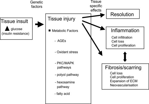
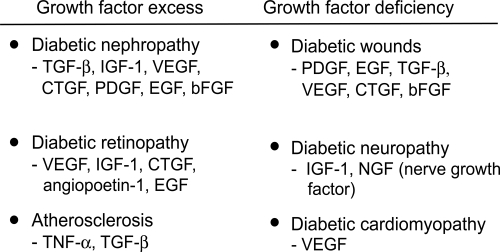
Diabetes mellitus is characterized by a lack of insulin causing elevated blood glucose, often with associated insulin resistance. Over time, especially in genetically susceptible individuals, such chronic hyperglycemia can cause tissue injury. One pathological response to tissue injury is the development of fibrosis, which involves predominant extracellular matrix (ECM) accumulation. The main factors that regulate ECM in diabetes are thought to be pro-sclerotic cytokines and protease/anti-protease systems. This review will examine the key markers and regulators of tissue fibrosis in diabetes and whether their levels in biological fluids may have clinical utility.
Keywords: diabetic complications; extracellular matrix; markers.
Figures


References
-
- Abdel-Wahab N, Weston BS, Roberts T, et al. Connective tissue growth factor and regulation of the mesangial cell cycle: role in cellular hypertrophy. J Am Soc Nephrol. 2002;13:2437–45. - PubMed
-
- Abu El-Asrar AM, Van den Steen PE, Al-Amro SA, et al. Expression of angiogenic and fibrogenic factors in proliferative vitreoretinal disorders. Int Ophthalmol. 2007;27:11–22. - PubMed
-
- Adler SG, Kang SW, Feld S, et al. Glomerular mRNAs in human type 1 diabetes: biochemical evidence for microalbuminuria as a manifestation of diabetic nephropathy. Kidney Int. 2001;60:2330–6. - PubMed
-
- Alessi MC, Juhan-Vague I. Contribution of PAI-1 in cardiovascular pathology. Arch Mal Coeur Vaiss. 2004;97:673–8. - PubMed
Publication types
MeSH terms
Substances
LinkOut - more resources
Full Text Sources
Other Literature Sources
Medical
