. 2008 Oct 1;3(10):e3281.
doi: 10.1371/journal.pone.0003281.

Social modulation during songbird courtship potentiates midbrain dopaminergic neurons

Affiliations

Social modulation during songbird courtship potentiates midbrain dopaminergic neurons

Ya-Chun Huang et al. PLoS One. .

Abstract

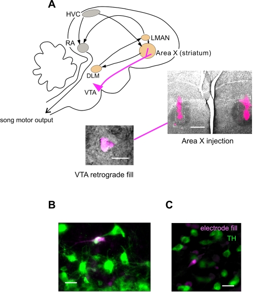
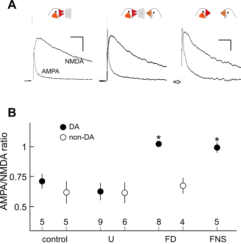
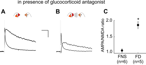
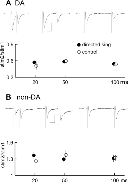
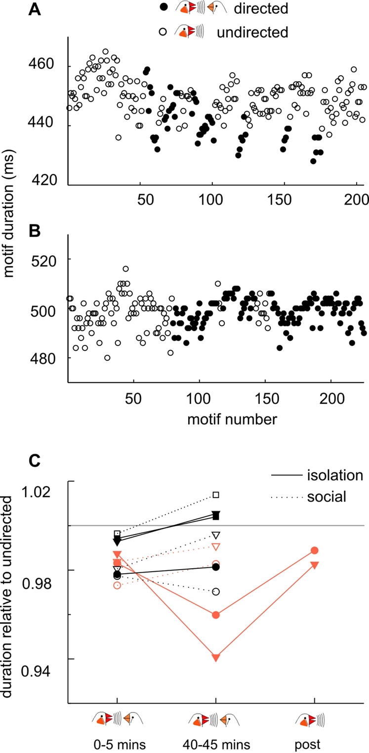
Synaptic transmission onto dopaminergic neurons of the mammalian ventral tegmental area (VTA) can be potentiated by acute or chronic exposure to addictive drugs. Because rewarding behavior, such as social affiliation, can activate the same neural circuitry as addictive drugs, we tested whether the intense social interaction of songbird courtship may also potentiate VTA synaptic function. We recorded glutamatergic synaptic currents from VTA of male zebra finches who had experienced distinct social and behavioral conditions during the previous hour. The level of synaptic transmission to VTA neurons, as assayed by the ratio of alpha-amino-3-hydroxy-5-methyl-4-isoxazolepropionic acid (AMPA) to N-methyl-D-aspartic acid (NMDA) glutamate receptor mediated synaptic currents, was increased after males sang to females, and also after they saw females without singing, but not after they sang while alone. Potentiation after female exposure alone did not appear to result from stress, as it was not blocked by inhibition of glucocorticoid receptors. This potentiation was restricted to synapses of dopaminergic projection neurons, and appeared to be expressed postsynaptically. This study supports a model in which VTA dopaminergic neurons are more strongly activated during singing used for courtship than during non-courtship singing, and thus can provide social context-dependent modulation to forebrain areas. More generally, these results demonstrate that an intense social encounter can trigger the same pathways of neuronal plasticity as addictive drugs.


Conflict of interest statement

Competing Interests: The authors have declared that no competing interests exist.

Figures

Figure 1
Figure 1. Anatomical identification of VTA cell types.
(A) Schematic diagram of sagittal view of singing-related areas in the songbird brain. Motor control nuclei (gray) are critically involved in structuring song output, while nuclei of the anterior forebrain pathway (AFP, orange) are required for song plasticity, and appear to be involved in the communicative function of singing. Area X receives a especially strong dopaminergic input from VTA. Below right is an image of a representative bilateral tracer (Fluoro-ruby) injection in Area X. Image of fluorescent tracer (magenta) is overlaid on image of nissl stain in one brain section including Area X (darkly stained oval nucleus). To the left is an overlay of fluorescently labeled cell body retrogradely labeled by an injection in Area X with IR-DIC image visualized in living brain slice. Scale bars indicate 15 uM (left) and 750 uM (right). (B,C). Post-recording confirmation of cell type by immunohistochemistry and electrode-filling dye. Green label for tyrosine hydroxylase (TH) antibody is overlaid with magenta label of neurons filled with fluorescent dye from the recording electrode (Alexa 568, Molecular Probes) – white signal indicates overlap of two signals. Left panel presents example of post-recording confirmation of TH-positive dopaminergic neuron, and right panel shows example of recording from TH-negative presumptive gamma-aminobutyric acid (GABA) -ergic neuron. Scale bars indicate 30 uM.
Figure 2
Figure 2. AMPA/NMDA ratio of synapses onto VTA dopaminergic neurons is increased after males are exposed to female birds.
(A) Representative plots of average EPSCs mediated by AMPA and NMDA glutamate receptors onto VTA dopaminergic neurons. Left, EPSCs recorded after undirected singing session; Center, after directed singing session; Right, after female exposure without singing. Scale bar in left panel applies to left and middle panels, that in right to the right panel; each indicates 50 ms, 40 pA. (B) Average AMPA/NMDA ratios recorded from dopaminergic (filled) and non-dopaminergic (open) neurons after four behavioral contexts. Mean±s.e.m. are shown for control (colony housed), undirected singing (U), directed singing (FD), and female exposure without singing (FNS) groups (error bar for directed singing FD group is obscured by plot symbol). The number of neurons recorded in each group is indicated above axis.
Figure 3
Figure 3. Inhibition of glucocorticoid receptor activation does not block increase of AMPA/NMDA ratio after exposure to female without singing.
(A, B) Representative plots of average EPSCs mediated by AMPA and NMDA glutamate receptors onto VTA dopaminergic neurons after not singing (A) and singing (B) in the presence of a female, in males treated with mifepristone. Scale bar indicates 50 msec, 40 pA. (C) Average AMPA/NMDA ratios recorded from males treated with mifepristone after exposure to a female and not singing (FNS, n = 6 recorded from 5 birds) or singing (FD, n = 5 recorded from 5 birds; asterisk indicates p<0.001, t-test).
Figure 4
Figure 4. The level of transmitter release is not altered after singing to females.
(A, B) Upper section, average EPSC in response to pairs of stimuli given at 20, 50, and 100 ms intervals, respectively. Lower section, average ratio of EPSC amplitude in response to second stimulus relative to first stimulus for similar experiments (DA; n = 13 directed, n = 7 control; non-DA, n = 8 directed, n = 6 control). Scalebar indicates 30 ms, 50 pA, and error bars indicate s.e.m. for each timepoint. Means for directed and control birds are offset slightly on the x-axis for clarity.
Figure 5
Figure 5. Several days of social isolation can prime short-term song plasticity.
(A,B) Duration of successive song motifs produced while alone (empty circles, undirected singing) and targeted to female birds (filled circles, directed). Experiments plotted in A and B represent examples of high (A) and low (B) levels of short-term alteration of song structure during exposure to females. (C). Summary of results from 5 birds recorded while singing to females after 5 days of social isolation (filled symbols, solid lines) or 5 days of daily exposure to females (open symbols, dotted lines). Each point is the plotted the ratio of motif duration relative to undirected song motifs recorded in 10 minutes prior to female presentation, during the three epochs 0–5 minutes after presentation, 40–45 minutes after presentation, and during undirected singing after females were removed. Data from two birds whose directed song motifs were clearly shortened from early to late during female exposure, only after isolation, are displayed in orange. Isolation and social conditions for each bird are plotted with identical symbols and colors.

References

    1. Ikemoto S, Panksepp J. The role of nucleus accumbens dopamine in motivated behavior: a unifying interpretation with special reference to reward-seeking. Brain Res Brain Res Rev. 1999;31:6–41. - PubMed
    1. Schultz W. Behavioral theories and the neurophysiology of reward. Annu Rev Psychol. 2006;57:87–115. - PubMed
    1. Fields HL, Hjelmstad GO, Margolis EB, Nicola SM. Ventral tegmental area neurons in learned appetitive behavior and positive reinforcement. Annu Rev Neurosci. 2007;30:289–316. - PubMed
    1. Kelley AE, Berridge KC. The neuroscience of natural rewards: relevance to addictive drugs. J Neurosci. 2002;22:3306–3311. - PMC - PubMed
    1. Wise RA. Dopamine, learning and motivation. Nat Rev Neurosc. 2004;5:483–94. - PubMed

Publication types