Review
. 2009;43(1-3):15-24.
doi: 10.1007/s12026-008-8046-9.

Amplification mechanisms for the enhancement of antigen-mediated mast cell activation

Affiliations
Review

Amplification mechanisms for the enhancement of antigen-mediated mast cell activation

Alasdair M Gilfillan et al. Immunol Res. 2009.

Abstract

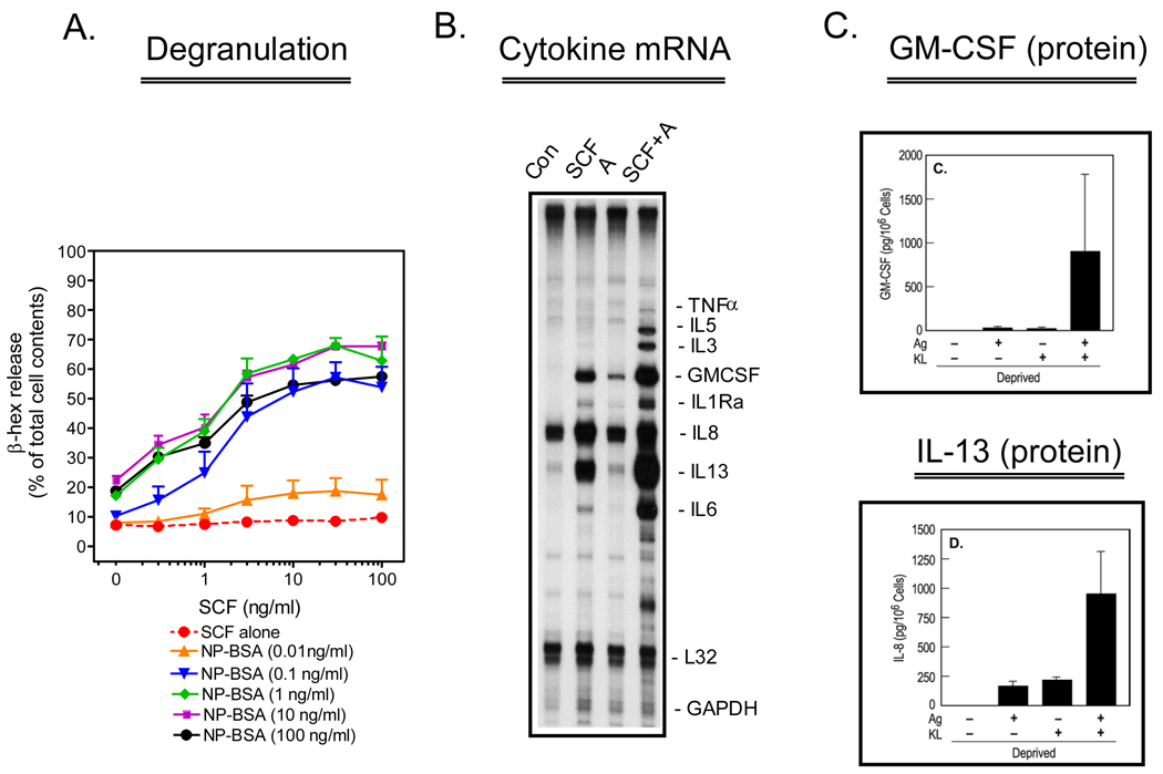
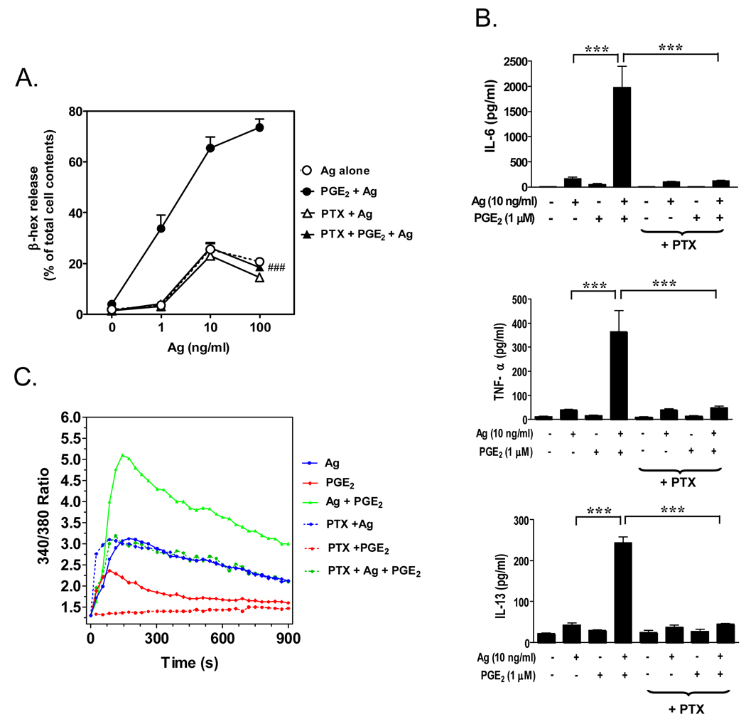
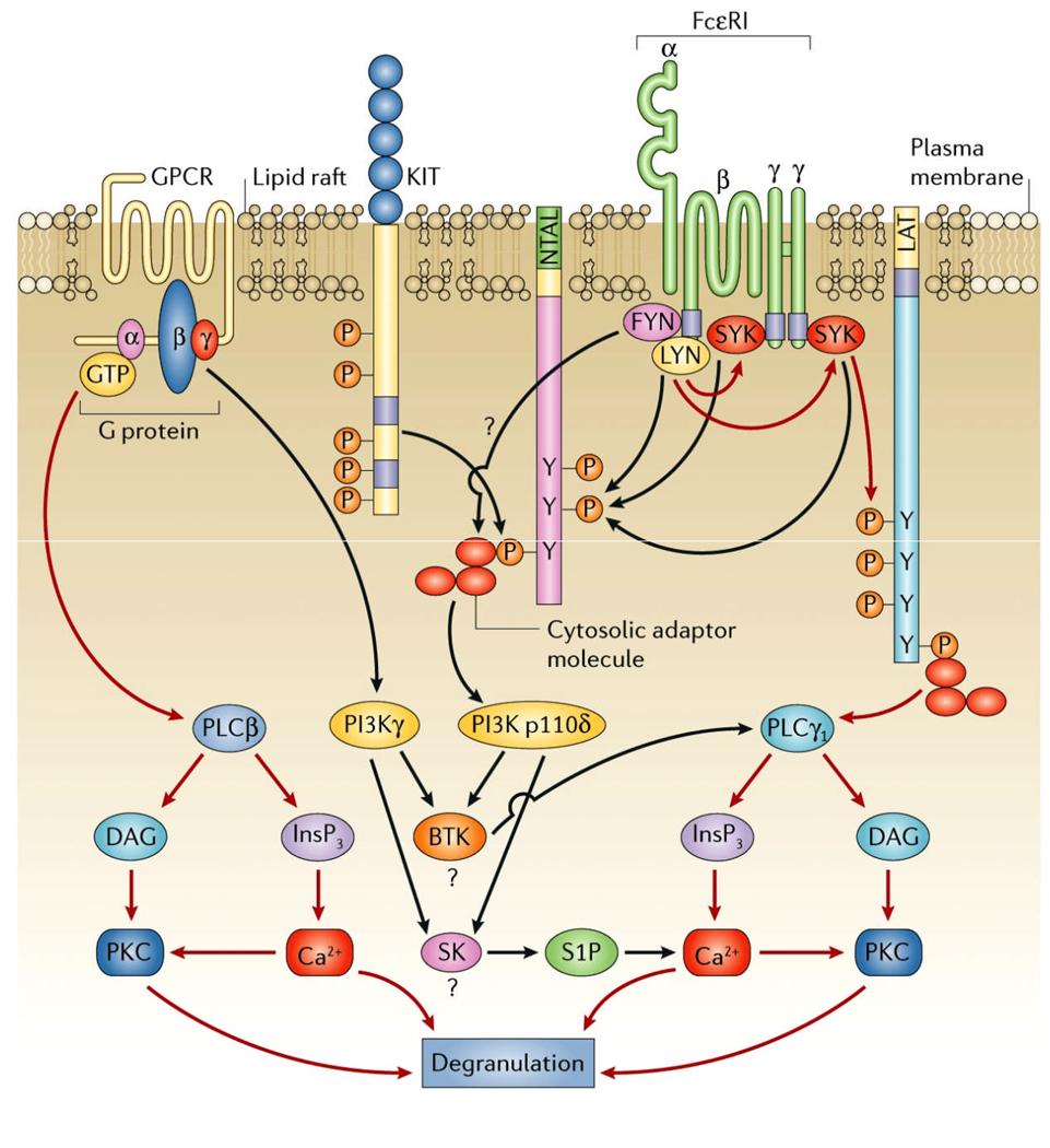
Activation of mast cells in the allergic inflammatory response occurs via the high affinity receptor for IgE (FcepsilonRI) following receptor aggregation induced by antigen-mediated cross-linking of IgE-occupied FcepsilonRI. Recent observations suggest this response is profoundly influenced by other factors that reduce the threshold for, and increase the extent of, mast cell activation. For example, under experimental conditions, cell surface receptors such as KIT and specific G protein-coupled receptors synergistically enhance FcepsilonRI-mediated mast cell degranulation and cytokine production. Activating mutations in critical signaling molecules may also contribute to such responses. In this review, we describe our research exploring the mechanisms regulating these synergistic interactions and, furthermore, discuss the relevance of our observations in the context of clinical considerations.


Figures

Figure 1
Figure 1
Enhancement of antigen-induced mast cell activation by SCF. Human mast cells, derived from CD34+ progenitors, were sensitized overnight with anti-NP-human IgE (1 µg/mL) in the absence of cytokines. In A., sensitized cells were triggered for 30 minutes with the indicated concentrations of NP-BSA and SCF and degranulation monitored by the release of β-hexosaminidase (β-hex). The data are means+/− S.E.M. of n=2–5 experiments conducted in duplicate. In B., the cells were triggered for 2 hours with NP-BSA (A) (100 ng/mL), SCF (100 ng/mL) or both, then cytokine mRNA determined by RNase protection assay. In C., Cells were triggered as above for 16 hours then the release of the cytokines, GM-CSF and IL-13, was determined by ELISA. The date are means+/− S.E.M. of n=3 following correction for the spontaneous release. This research was originally published in Blood (A.: Tkaczyk et al., Blood. 2004;104:207–14; and B., C.: Hundley et al., Blood. 2004;104:2410–7).
Figure 2
Figure 2
Enhancement of antigen-induced mast cell activation by the GPCR agonist PGE2. Mouse bone marrow-derived mast cells (BMMCs) were sensitized overnight with mouse monoclonal anti-DNP IgE (100 ng/mL) then pre-incubated with or without pertussis toxin (PTX) (1 µg/mL) for 4 hours before antigen (Ag) (DNP-HSA) or PGE2 or both were added. Degranulation (A.) was monitored by β-hexosaminidase (β-hex) release after 30 minutes incubation. Cytokine release (B.) was measured after 6 hours by ELISA. Changes in [Ca2+]i over the indicated intervals were monitored following loading the cells with FURA2-AM then challenging with Ag (10 ng/mL) or PGE2 (1 µM) or Ag together with PGE2. The Ca2+ data are representative of n=3 experiments conducted in duplicate. The data in A and B are presented as means +/− S.E. of (n=3–4) separate experiments conducted in duplicate. ###, p < 0.001 for comparison with Ag/PGE2 (A.). ***, p < 0.001 by Student’s t-test (B.). The ability of pertussis toxin to block the PGE2 responses indicates that they were mediated via the Gαi-linked EP3 receptor. This research was originally published in Cellular Signaling (Kuehn et al., Cell Signal. 2008;20:625–36).
Figure 3
Figure 3
Integration of the signaling pathways induced by G protein coupled receptors, Kit, and the FcεRI. The red lines represent the principal signaling pathways initiated by these receptors and the black lines represent the complementary/amplification pathways utilized by these receptors for degranulation. This figure was originally published in Nature Reviews Immunology (Gilfillan and Tkaczyk, Nat Rev Immunol. 2006;6:218–30).

References

    1. Metcalfe DD. Mast cells and mastocytosis. Blood. 2008;112:946–956. - PMC - PubMed
    1. Mekori YA, Metcalfe DD. Mast cells in innate immunity. Immunol Rev. 2000;173:131–140. - PubMed
    1. Marshall JS. Mast-cell responses to pathogens. Nat Rev Immunol. 2004;4:787–799. - PubMed
    1. Galli SJ, Nakae S, Tsai M. Mast cells in the development of adaptive immune responses. Nat Immunol. 2005;6:135–142. - PubMed
    1. Tkaczyk C, Jensen BM, Iwaki S, Gilfillan AM. Adaptive and innate immune reactions regulating mast cell activation: from receptor-mediated signaling to responses. Immunol Allergy Clin North Am. 2006;26:427–450. - PubMed

Publication types

MeSH terms

Substances

LinkOut - more resources