. 2008 Oct 1:8:165.
doi: 10.1186/1471-2180-8-165.

Identification and characterization of domains responsible for self-assembly and cell wall binding of the surface layer protein of Lactobacillus brevis ATCC 8287

Affiliations

Identification and characterization of domains responsible for self-assembly and cell wall binding of the surface layer protein of Lactobacillus brevis ATCC 8287

Silja Avall-Jääskeläinen et al. BMC Microbiol. .

Abstract

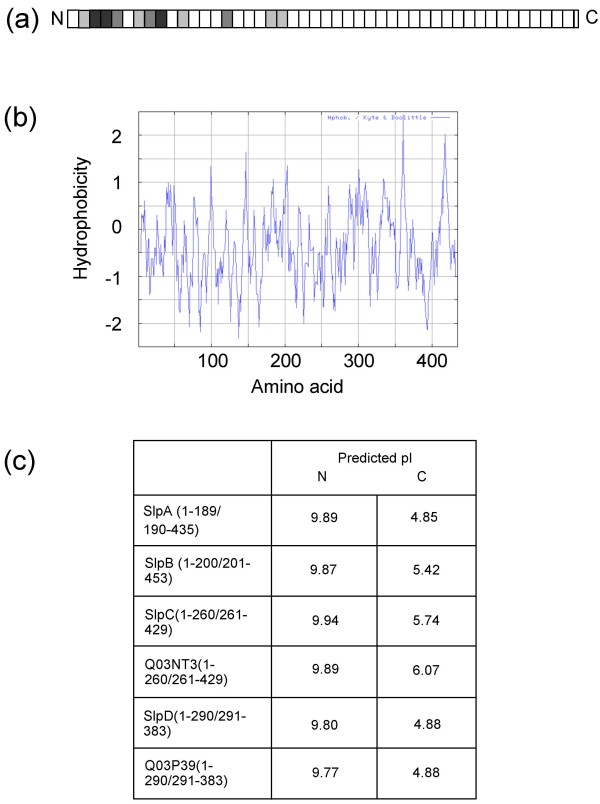
Background: Lactobacillus brevis ATCC 8287 is covered by a regular surface (S-) layer consisting of a 435 amino acid protein SlpA. This protein is completely unrelated in sequence to the previously characterized S-layer proteins of Lactobacillus acidophilus group.

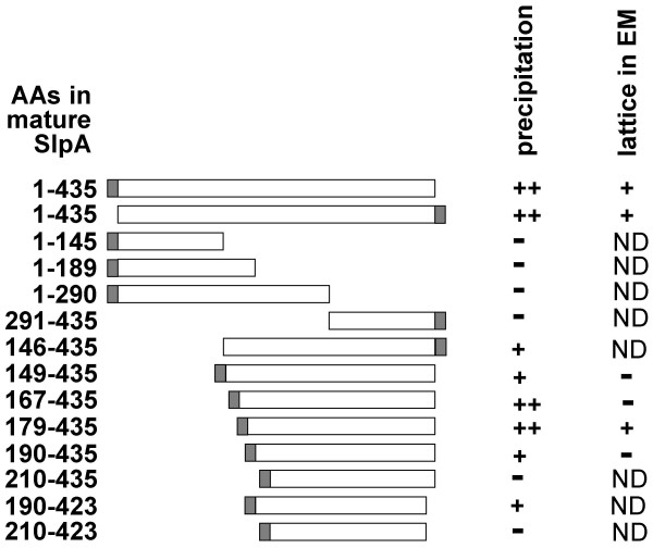
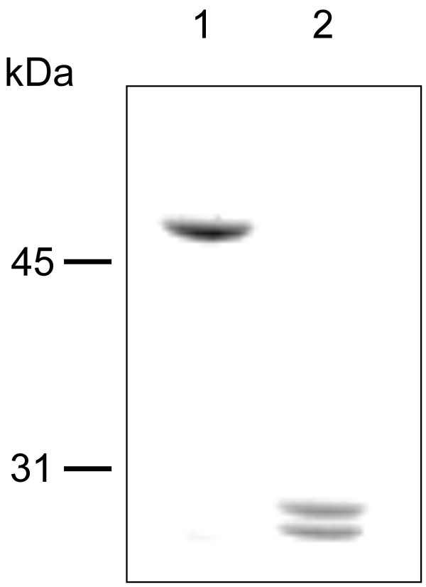
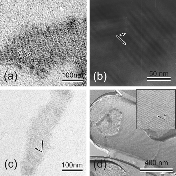
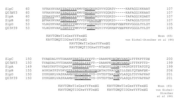
Results: In this work, the self-assembly and cell wall binding domains of SlpA were characterized. The C-terminal self-assembly domain encompassed residues 179-435 of mature SlpA, as demonstrated by the ability of N-terminally truncated recombinant SlpA to form a periodic structure indistinguishable from that formed by full length SlpA. Furthermore, a trypsin degradation analysis indicated the existence of a protease resistant C-terminal domain of 214 amino acids. By producing a set of C-terminally truncated recombinant SlpA (rSlpA) proteins the cell wall binding region was mapped to the N-terminal part of SlpA, where the first 145 amino acids of mature SlpA alone were sufficient for binding to isolated cell wall fragments of L. brevis ATCC 8287. The binding of full length rSlpA to the cell walls was not affected by the treatment of the walls with 5% trichloroacetic acid (TCA), indicating that cell wall structures other than teichoic acids are involved, a feature not shared by the Lactobacillus acidophilus group S-layer proteins characterized so far. Conserved carbohydrate binding motifs were identified in the positively charged N-terminal regions of six Lactobacillus brevis S-layer proteins.

Conclusion: This study identifies SlpA as a two-domain protein in which the order of the functional domains is reversed compared to other characterized Lactobacillus S-layer proteins, and emphasizes the diversity of potential cell wall receptors despite similar carbohydrate binding sequence motifs in Lactobacillus S-layer proteins.


Figures

Figure 1
Figure 1
1(a) – Alignment of L. brevis S-layer protein sequences. The mature forms of S-layer proteins were aligned by ClustalW and the alignments were divided into stretches of 10 amino acids, from which the percentage of identical amino acids and amino acids with conserved substitutions were calculated. The following colours are used to indicate the percentages of identical and similar amino acids in each calculated stretch: white, 0–20%, light gray, 21–40%, medium gray, 41–60%, dark gray, 61–80%. 1(b) &#8211 Hydrophobicity of mature SlpA. The hydrophobicity was calculated according to Kyte and Doolittle [34] with a window of seven amino acids. 1(c) &#8211 Predicted pI values of the N- and C-terminal regions of L. brevis S-layer proteins. The lengths of the N- and C-terminal regions as well as the full lengths of the mature forms of the proteins are indicated in brackets.
Figure 2
Figure 2
Schematic presentation of the recombinant SlpA proteins expressed and their self-assembly properties. Shaded bars, tags consisting of a 13 amino acid linker and six histidine residues.
Figure 3
Figure 3
SDS-PAGE analysis of SlpA fragments obtained after trypsin digestion. Lane 1, undigested SlpA. Lane 2, SlpA after digestion with trypsin. Numbers on the left indicate molecular weights in kilodaltons.
Figure 4
Figure 4
Self-assembly of SlpA, rSlpA and rSlpA C-terminal domain. (a-c) Transmission electron micrographs showing self-assembly products of (a) nontruncated rSlpA, (b) rSlpA179–435, and (c) wild type SlpA isolated from L. brevis ATCC 8287 cells. (d) Transmission electron micrograph showing a freeze-etched preparation of L. brevis ATCC 8287 cells completely covered with the oblique S-layer lattice formed by SlpA. Arrows indicate the base vectors of the oblique lattice.
Figure 5
Figure 5
Transmission electron micrograph of isolated native cell walls of L. brevis ATCC 8287.
Figure 6
Figure 6
6(a) – Binding of truncated rSlpA proteins to isolated native cell walls of L. brevis ATCC 8287. Full-length rSlpA (lanes 1–4), rSlpA1–145 (lanes 5–8), rSlpA1–189 (lanes 9–12), rSlpA190–435 (lanes 13–16) and rSlpA167–435 (lanes 17–20) were incubated with (lanes 1–2, 5–6, 9–10, 13–14 and 17–18) or without (lanes 3–4, 7–8, 11–12, 15–16 and 19–20) cell walls (CWF), and the pellets and supernatants (*) recovered by centrifugation were analyzed by SDS-PAGE. Lanes 21–22, CWF incubated alone; *, supernatant. Numbers on the left indicate molecular weights in kilodaltons. 6(b) &#8211 Effect of treatment of cell walls with TCA at +4&#176C (b1) or at +37&#176C (b2) on the binding of rSlpA. Full length rSlpA was incubated with native CWF (lanes 1–2) or with CWF treated with 5% TCA (lanes 3–4) and the pellets and supernatants (*) were analyzed by SDS-PAGE. Lanes 5–6, full-length rSlpA incubated alone, lanes 7–8, TCA-treated CWF incubated alone; *, supernatant. Numbers on the left indicate molecular weights in kilodaltons.
Figure 7
Figure 7
Similarity of the N-terminal regions of L. brevis S-layer proteins with carbohydrate binding motifs. The motifs were determined by Wren [45] and von Eichel-Streiber et al [44]. In the consensus sequences upper case letters indicate highly conserved residues [44] or residues with an identity of 50% or higher [45]. X, variable residue. A broken underline indicates potential carbohydrate-binding motifs at different locations: YFRAYG of SlpA corresponds to YFDxNG of the consensus sequence of Ref [44]; KAYRGW of SlpB corresponds to KAVTGW of the consensus sequences of References [44] and [45]; LSNKSYY of SlpD and Q03P39 corresponds to IDGkwYY of the consensus sequence of Ref [44].

References

    1. Sará M, Sleytr UB. S-layer proteins. J Bacteriol. 2000;182:859–868. doi: 10.1128/JB.182.4.859-868.2000. - DOI - PMC - PubMed
    1. Sleytr U, Messner P. Crystalline surface layers in procaryotes. J Bacteriol. 1988;170:2891–2897. - PMC - PubMed
    1. Lupas A, Engelhardt H, Peters J, Santarius U, Volker S, Baumeister W. Domain structure of the Acetogenium kivui surface layer revealed by electron crystallography and sequence analysis. J Bacteriol. 1994;176:1224–1233. - PMC - PubMed
    1. Mesnage S, Fontaine T, Mignot T, Delepierre M, Mock M, Fouet A. Bacterial SLH domain proteins are non-covalently anchored to the cell surface via a conserved mechanism involving wall polysaccharide pyruvylation. EMBO J. 2000;19:4473–4484. doi: 10.1093/emboj/19.17.4473. - DOI - PMC - PubMed
    1. Schäffer C, Messner P. The structure of secondary cell wall polymers: how Gram-positive bacteria stick their cell walls together. Microbiology. 2005;151:643–651. doi: 10.1099/mic.0.27749-0. - DOI - PubMed

Publication types

MeSH terms