. 2008 Dec;195(2):219-25.
doi: 10.1016/j.jmr.2008.09.012. Epub 2008 Sep 17.

Clinical NOE 13C MRS for neuropsychiatric disorders of the frontal lobe

Affiliations

Clinical NOE 13C MRS for neuropsychiatric disorders of the frontal lobe

Napapon Sailasuta et al. J Magn Reson. 2008 Dec.

Abstract

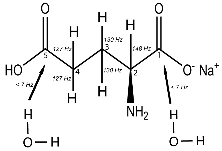
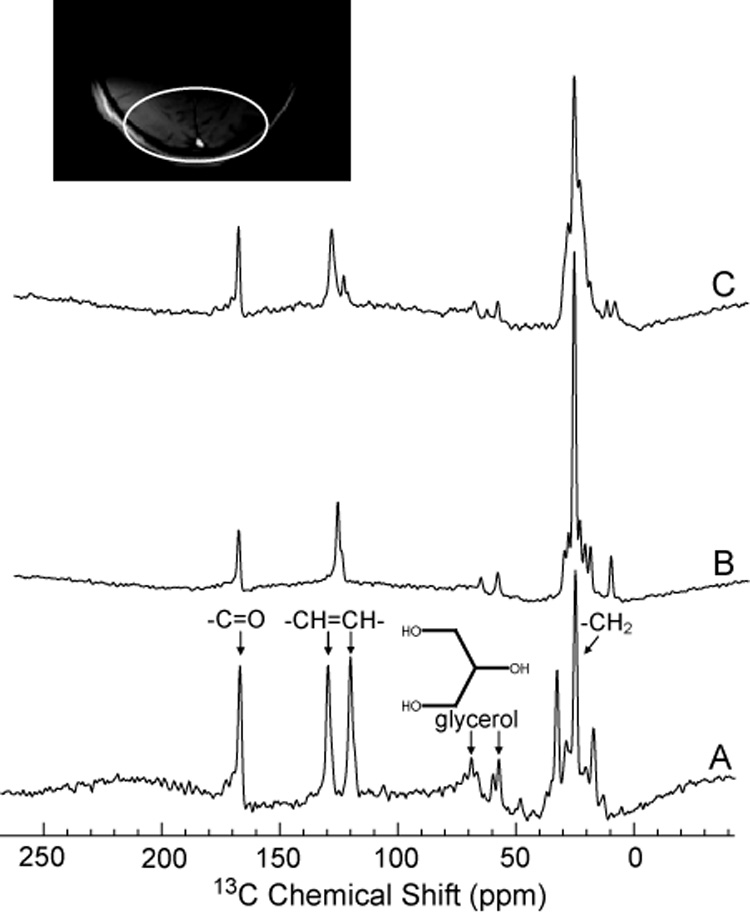
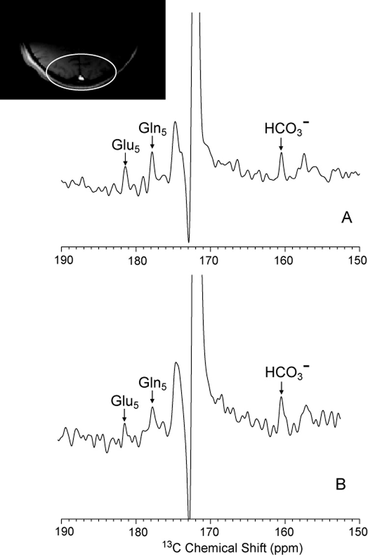
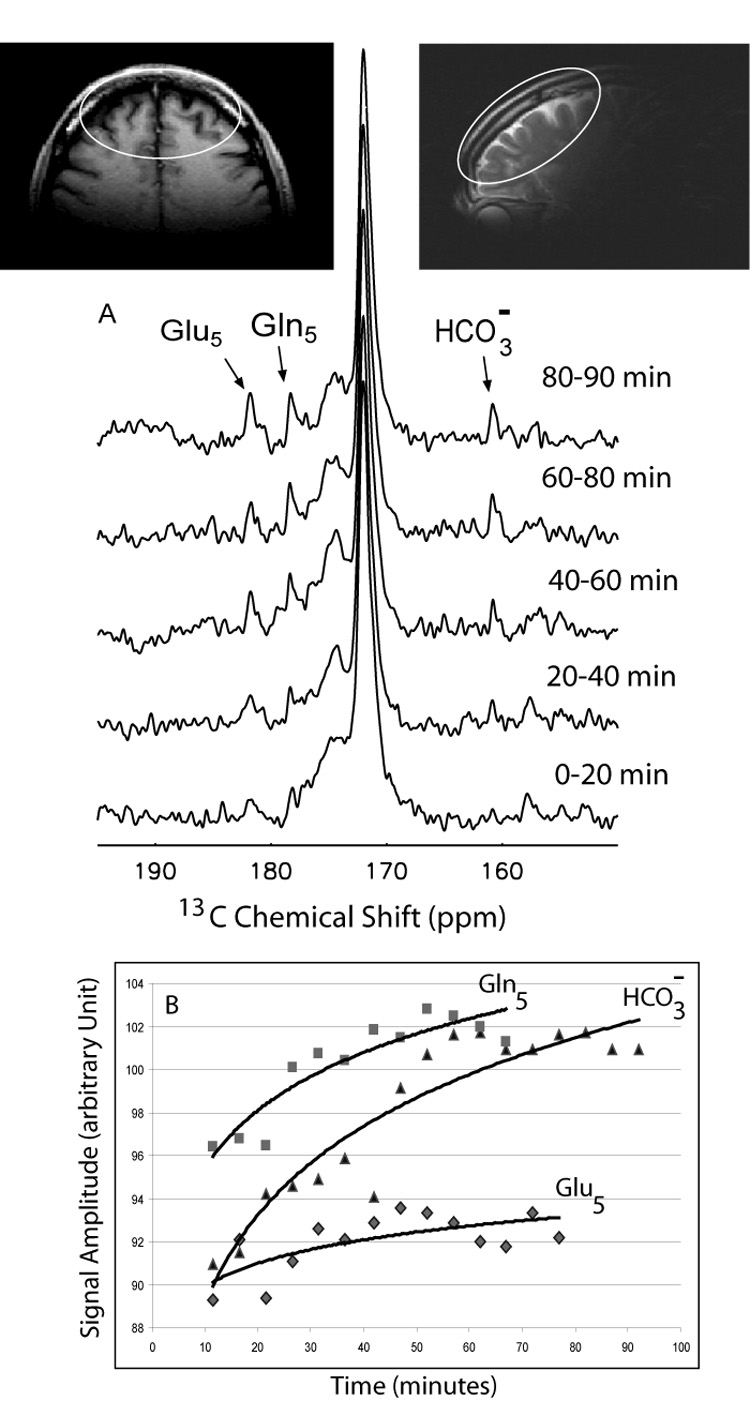
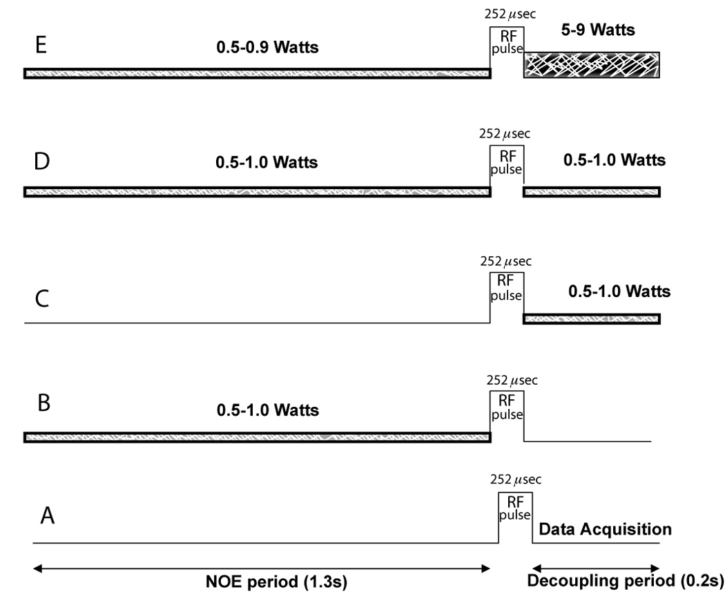
In this communication, a scheme is described whereby in vivo (13)C MRS can safely be performed in the frontal lobe, a human brain region hitherto precluded on grounds of SAR, but important in being the seat of impaired cognitive function in many neuropsychiatric and developmental disorders. By combining two well known features of (13)C NMR-the use of low power NOE and the focus on (13)C carbon atoms which are only minimally coupled to protons, we are able to overcome the obstacle of SAR and develop means of monitoring the (13)C fluxes of critically important metabolic pathways in frontal brain structures of normal volunteers and patients. Using a combination of low-power WALTZ decoupling, variants of random noise for nuclear overhauser effect enhancement it was possible to reduce power deposition to 20% of the advised maximum specific absorption rate (SAR). In model solutions (13)C signal enhancement achieved with this scheme were comparable to that obtained with WALTZ-4. In human brain, the low power procedure effectively determined glutamine, glutamate and bicarbonate in the posterior parietal brain after [1-(13)C] glucose infusion. The same (13)C enriched metabolites were defined in frontal brain of human volunteers after administration of [1-(13)C] acetate, a recognized probe of glial metabolism. Time courses of incorporation of (13)C into cerebral glutamate, glutamine and bicarbonate were constructed. The results suggest efficacy for measurement of in vivo cerebral metabolic rates of the glutamate-glutamine and tricarboxylic acid cycles in 20 min MR scans in previously inaccessible brain regions in humans at 1.5 T. We predict these will be clinically useful biomarkers in many human neuropsychiatric and genetic conditions.


Figures

Figure 1
Figure 1
Chemical structure of glutamate showing C2, C3 and C4 carbons with their scalar couplings (J-coupling constant indicated) to protons whereas C5 and C1 carbons are not directly coupled to protons but may interact via dipolar coupling with water molecule.
Figure 2
Figure 2
Proton Decoupled 13C spectra of dioxane. Experiments were performed in a 1.5 Tesla clinical MR scanner using dual tuned half-head coil previously described [2]. The various decoupling and NOE schemes described in Methods, (Figure 8), resulted in collapse of the coupled spins with incremental signal enhancement of the resulting 13C singlet resonance. The greatest signal enhancement was obtained with low power noise decoupling together with Gauss filter (Spectrum B). All spectra were scaled relative to the highest peak (Spectrum A). A). High power WALTZ-4 NOE (0.6Watts) and decoupling (6 Watts), B). Low power noise NOE (0.5 Watts) and decoupling (0.5 Watts) with Gaussian filter C). Low power noise (0.5 Watts) and decoupling (0.5 Watts) with Sinc filter, D). Low power noise NOE (0.5 Watts) and decoupling (0.5 Watts) without filter.
Figure 3
Figure 3
Decoupling efficiency for 1,4 dioxane acquired using NOE and decoupling scheme in figure 8D at different decoupling powers.
Figure 4
Figure 4
Impact of NOE and decoupling on carbon atoms of glutamate and bicarbonate. Comparison of A). no decoupling or NOE (Figure 8A), B). low power noise decoupling alone (Figure 8C), C). low power noise NOE alone (Figure 8B), D). low power decoupling and NOE (LPND, Figure 8D), and E). high power WALTZ-4 decoupling and low power NOE (Figure 8E) of 1M glutamate and 1M Bicarbonate (HCO3 ) indicates that in natural abundance glutamate, signal is almost equally enhanced by either high power proton decoupling or low power decoupling and NOE.
Figure 5
Figure 5
Natural abundance 13C MRS lipid spectra from the human head in vivo. A): no decoupling or NOE, B): high power proton decoupling and NOE (WALTZ-4 at 8W, figure 8E), C): low power NOE (figure 8D) (0.9W). Proton frequency for both B) and C) was set to 100Hz from water (approximately 3 ppm). Note that Spectrums B and C are scaled to each other; peak intensities can be directly compared. Spectrum A has not been scaled and is provided as a chemical shift reference.
Figure 6
Figure 6
Comparison of NOE + DC (Figure 8E scheme) (A) and low power NOE (LPND, figure 8D scheme) (B) of the posterior parietal human brain in an [1-13C] glucose infusion protocol. The enrichment of 13C in glutamate, glutamine and bicarbonate region is shown.
Figure 7
Figure 7
Enrichment of 13C in glutamate and glutamine in the anterior (frontal) brain during 1-13C acetate infusion. A). Sequentially acquired 13C spectra are shown for 20 minutes increments from the start of the infusion, with the appearance of 13C5 glutamate and 13C5 glutamine and bicarbonate resonances (Figure 7A). B). The complete time course of accumulation of enriched metabolites over time are shown at 5 minute intervals. (Note: Signal amplitudes for Gln5, Glu5 and HCO3 are not to scale) Inserts are axial and sagittal images showing that Broadmann areas 9–11, 46 and possibly 47 of frontal cortex are included in the MRS field of view.
Figure 8
Figure 8
Different proton decoupling and NOE schemes employed in this study. A). No decoupling and NOE, B). Low power noise (1.0-0.5 Watts) NOE only, C). Low power noise (1.0-0.4 Watts) decoupling only, D). Low power noise (1.0-0.5 Watts) for NOE and for decoupling (LPND), E). Low power WALTZ-4 NOE (0.9-0.5 Watts) and decoupling (5–9 Watts). Bandwidth of NOE applied was 250Hz and of Waltz-4, 1000Hz.

References

    1. Rothman DL, Novotny EJ, Shulman GI, Howseman AM, Petroff OA, Mason G, Nixon T, Hanstock CC, Prichard JW, Shulman RG. 1H-[13C] NMR measurements of [4-13C]glutamate turnover in human brain. Proc Natl Acad Sci U S A. 1992;89:9603–9606. - PMC - PubMed
    1. Bluml S, Moreno-Torres A, Ross BD. [1-13C] glucose MRS in chronic hepatic encephalopathy in man. Magn Reson Med. 2001;45:981–993. - PubMed
    1. Shen J, Petersen KF, Behar KL, Brown P, Nixon TW, Mason GF, Petroff OA, Shulman GI, Shulman RG, Rothman DL. Determination of the rate of the glutamate/glutamine cycle in the human brain by in vivo 13C NMR. Proc Natl Acad Sci U S A. 1999;96:8235–8240. - PMC - PubMed
    1. Mason GF, Pan JW, Chu WJ, Newcomer BR, Zhang Y, Orr R, Hetherington HP. Measurement of the tricarboxylic acid cycle rate in human grey and white matter in vivo by 1H-[13C] magnetic resonance spectroscopy at 4.1T. J Cereb Blood Flow Metab. 1999;19:1179–1188. - PubMed
    1. Bluml S, Moreno A, Hwang JH, Ross BD. 1-(13)C glucose magnetic resonance spectroscopy of pediatric and adult brain disorders. NMR Biomed. 2001;14:19–32. - PubMed

Publication types