

Initial characterization of Chlamydophila (Chlamydia) pneumoniae cultured from the late-onset Alzheimer brain
- PMID: 18829386
- PMCID: PMC2730674
- DOI: 10.1016/j.ijmm.2008.07.002
Initial characterization of Chlamydophila (Chlamydia) pneumoniae cultured from the late-onset Alzheimer brain
Abstract
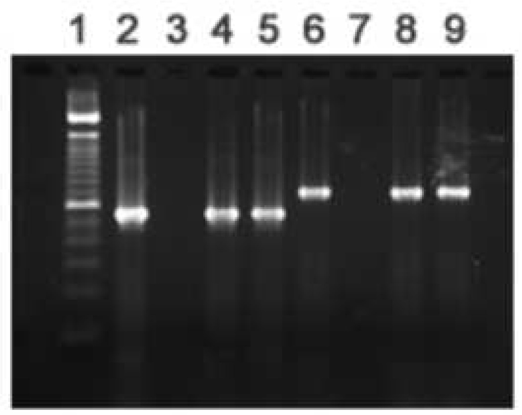
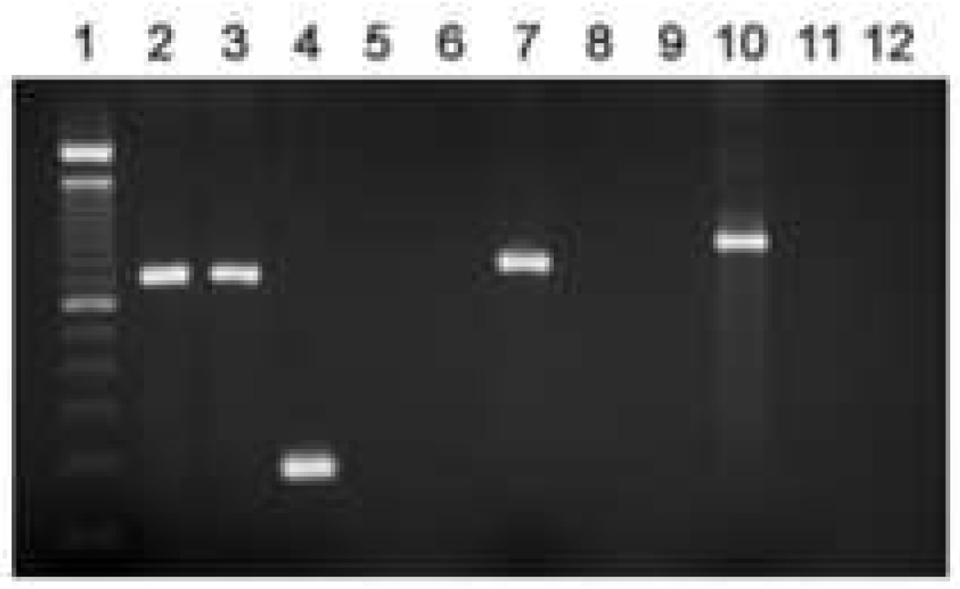
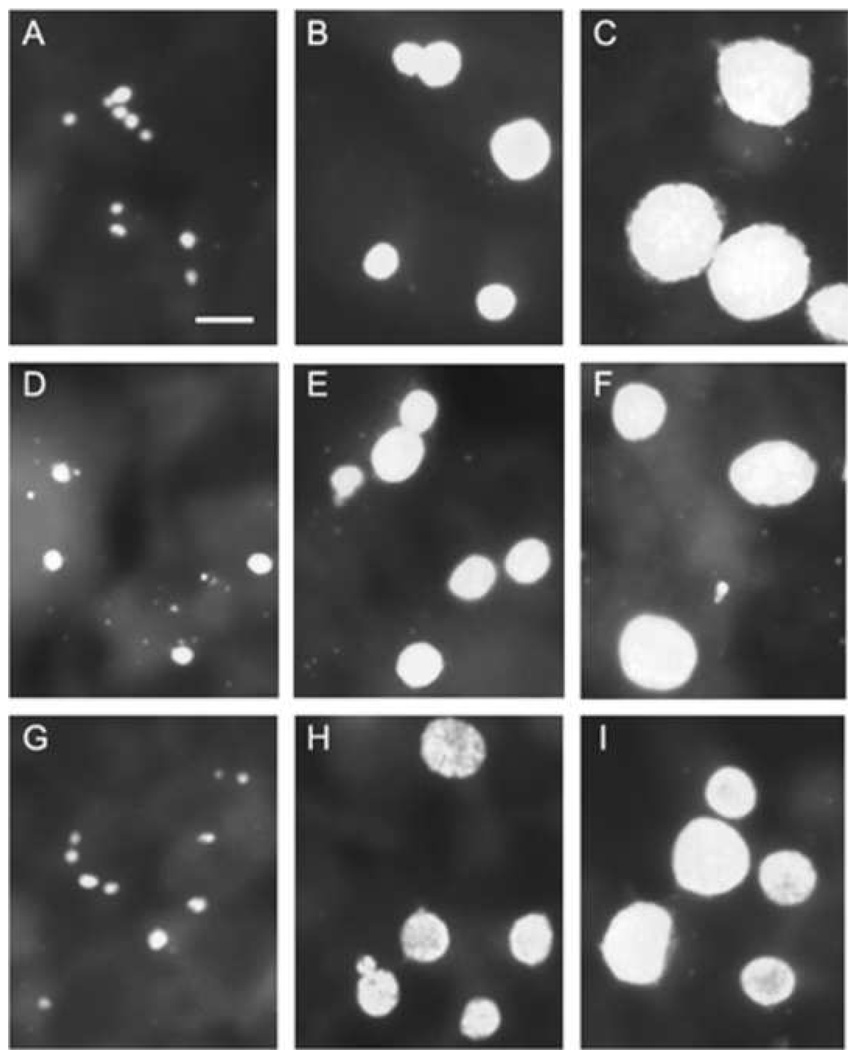
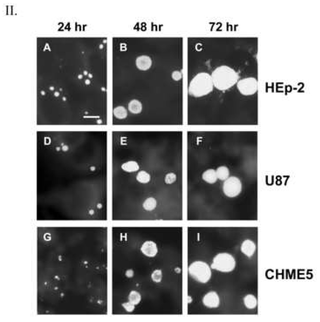
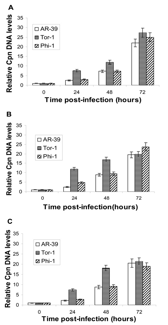
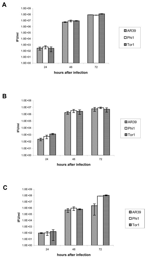
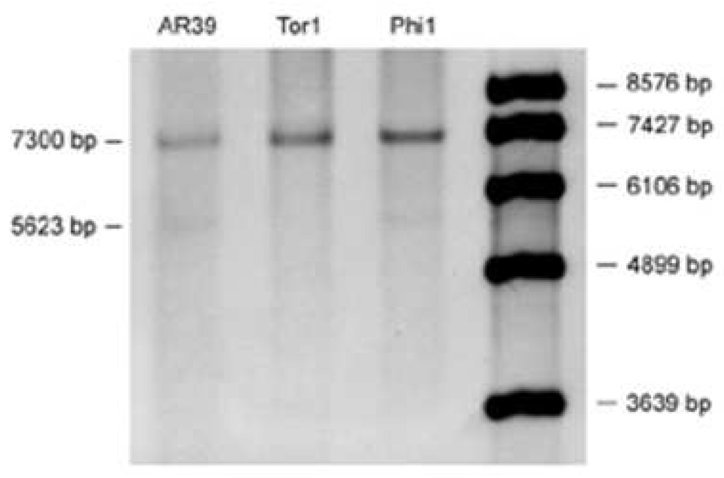
Previous studies from this laboratory provided evidence that the intracellular bacterial pathogen Chlamydophila (Chlamydia) pneumoniae is present in the late-onset Alzheimer's disease (AD) brain. Here we report culture of the organism from two AD brain samples, each of which originated from a different geographic region of North America. Culturable organisms were detectable after one and two passages in HEp-2 cells for the two samples. Both isolates, designated Tor-1 and Phi-1, were demonstrated to be authentic C. pneumoniae using PCR assays targeting the C. pneumoniae-specific genes Cpn0695, Cpn1046, and tyrP. Assessment of inclusion morphology and quantitation of infectious yields in epithelial (HEp-2), astrocytic (U-87 MG), and microglial (CHME-5) cell lines demonstrated an active, rather than a persistent, growth phenotype for both isolates in all host cell types. Sequencing of the omp1 gene from each isolate, and directly from DNA prepared from several additional AD brain tissue samples PCR-positive for C. pneumoniae, revealed genetically diverse chlamydial populations. Both brain isolates carry several copies of the tyrP gene, a triple copy in Tor-1, and predominantly a triple copy in Phi-1 with a minor population component having a double copy. This observation indicated that the brain isolates are more closely related to respiratory than to vascular/atheroma strains of C. pneumoniae.
Figures
Similar articles
-
Chlamydophila (Chlamydia) pneumoniae infection of human astrocytes and microglia in culture displays an active, rather than a persistent, phenotype.Am J Med Sci. 2006 Oct;332(4):168-74. doi: 10.1097/00000441-200610000-00003. Am J Med Sci. 2006. PMID: 17031241
-
Chlamydophila (Chlamydia) pneumoniae in the Alzheimer's brain.FEMS Immunol Med Microbiol. 2006 Dec;48(3):355-66. doi: 10.1111/j.1574-695X.2006.00154.x. Epub 2006 Oct 18. FEMS Immunol Med Microbiol. 2006. PMID: 17052268
-
Identification and localization of Chlamydia pneumoniae in the Alzheimer's brain.Med Microbiol Immunol. 1998 Jun;187(1):23-42. doi: 10.1007/s004300050071. Med Microbiol Immunol. 1998. PMID: 9749980
-
[Diagnostic tests: Chlamydia trachomatis, Chlamydophila pneumoniae].Nihon Rinsho. 2005 Jul;63 Suppl 7:220-6. Nihon Rinsho. 2005. PMID: 16111232 Review. Japanese. No abstract available.
-
Assessment of evidence for or against contributions of Chlamydia pneumoniae infections to Alzheimer's disease etiology.Brain Behav Immun. 2020 Jan;83:22-32. doi: 10.1016/j.bbi.2019.10.014. Epub 2019 Oct 15. Brain Behav Immun. 2020. PMID: 31626972 Review.
Cited by
-
Chlamydia pneumoniae-specific intrathecal oligoclonal antibody response is predominantly detected in a subset of multiple sclerosis patients with progressive forms.J Neurovirol. 2009 Sep;15(5-6):425-33. doi: 10.3109/13550280903475580. J Neurovirol. 2009. PMID: 20053141
-
Can infections cause Alzheimer's disease?Epidemiol Rev. 2013;35(1):161-80. doi: 10.1093/epirev/mxs007. Epub 2013 Jan 24. Epidemiol Rev. 2013. PMID: 23349428 Free PMC article. Review.
-
The Possibility of an Infectious Etiology of Alzheimer Disease.Mol Neurobiol. 2019 Jun;56(6):4479-4491. doi: 10.1007/s12035-018-1388-y. Epub 2018 Oct 18. Mol Neurobiol. 2019. PMID: 30338482 Review.
-
Chlamydia pneumoniae is genetically diverse in animals and appears to have crossed the host barrier to humans on (at least) two occasions.PLoS Pathog. 2010 May 20;6(5):e1000903. doi: 10.1371/journal.ppat.1000903. PLoS Pathog. 2010. PMID: 20502684 Free PMC article.
-
Phylogenetic analysis of human Chlamydia pneumoniae strains reveals a distinct Australian indigenous clade that predates European exploration of the continent.BMC Genomics. 2015 Dec 22;16:1094. doi: 10.1186/s12864-015-2281-y. BMC Genomics. 2015. PMID: 26694618 Free PMC article.
References
-
- Apfalter P, Loidl M, Nadrchal R, Makristathis A, Rotter M, Bergmann M, Polterauer P, Hirschl AM. Isolation and continuous growth of Chlamydia pneumoniae from arterectomy specimens. Eur. J. Clin. Microbiol. Infect. Dis. 2000;19:305–308. - PubMed
-
- Alvesalo J, Greco D, Leinonen M, Raitila T, Vuorela P, Auvinen P. Microarray analysis of a Chlamydia pneumoniae-infected human epithelial cell line by use of gene ontology hierarchy. J. Infect. Dis. 2008;197:156–162. - PubMed
-
- Balin BJ, Gerard HC, Arking EJ, Appelt DM, Branigan PJ, Abrams JT, Whittum-Hudson JA, Hudson AP. Identification and localization of Chlamydia pneumoniae in the Alzheimer’s brain. Med. Microbiol. Immunol. 1998;187:23–42. - PubMed
Publication types
MeSH terms
Substances
Grants and funding
LinkOut - more resources
Full Text Sources
Medical
