

Structure, chemical composition and mechanical properties of human and rat cementum and its interface with root dentin
- PMID: 18829402
- PMCID: PMC2685077
- DOI: 10.1016/j.actbio.2008.08.013
Structure, chemical composition and mechanical properties of human and rat cementum and its interface with root dentin
Abstract
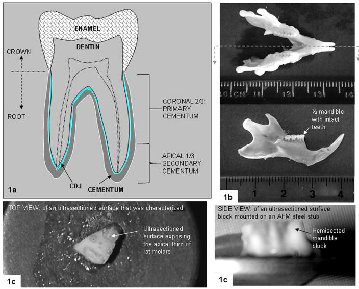
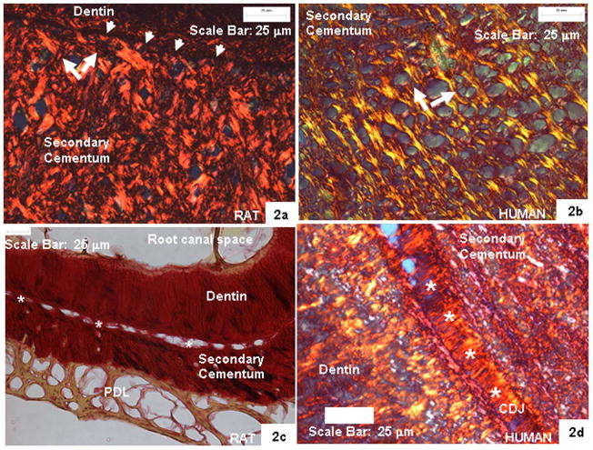
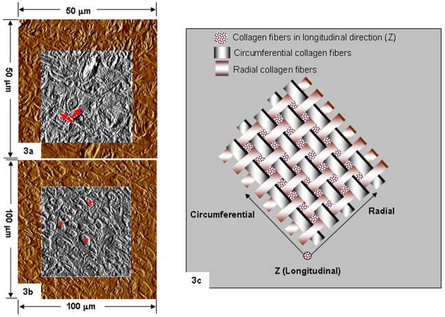
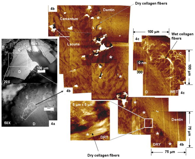
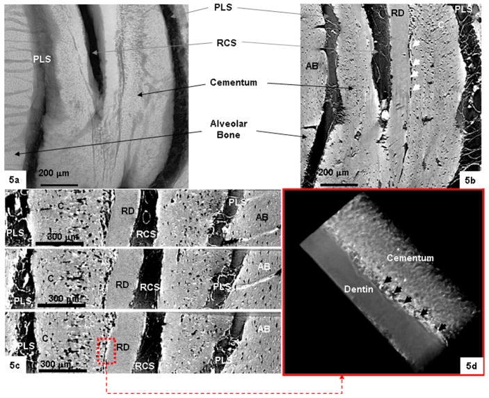
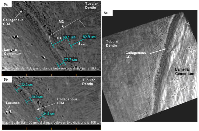
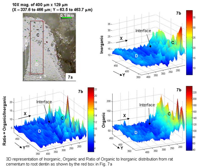
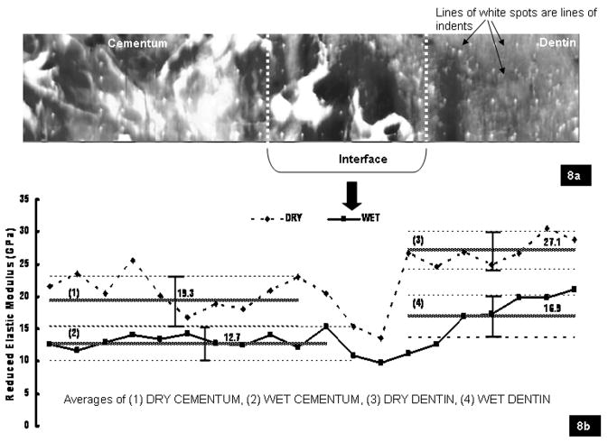
This work seeks to establish comparisons of the physical properties of rat and human cementum, root dentin and their interface, including the cementum-dentin junction (CDJ), as a basis for future studies of the entire periodontal complex using rats as animal models. In this study the structure, site-specific chemical composition and mechanical properties of cementum and its interface with root dentin taken from 9- to 12-month-old rats were compared to the physiologically equivalent 40- to 55-year-old human age group using qualitative and quantitative characterization techniques, including histology, atomic force microscopy (AFM), micro-X-ray computed tomography, Raman microspectroscopy and AFM-based nanoindentation. Based on results from this study, cementum taken from the apical third of the respective species can be represented as a woven fabric with radially and circumferentially oriented collagen fibers. In both species the attachment of cementum to root dentin is defined by a stiffness-graded interface (CDJ/cementum-dentin interface). However, it was concluded that cementum and the cementum-dentin interface from a 9- to 12-month-old rat could be more mineralized, resulting in noticeably decreased collagen fiber hydration and significantly higher modulus values under wet conditions for cementum and CDJ (E(rat-cementum)=12.7+/-2.6 GPa; E(rat-CDJ)=11.6+/-3.2 GPa) compared to a 40- to 55-year-old human (E(human-cementum)=3.73+/-1.8 GPa; E(human-CDJ)=1.5+/-0.7 GPa). The resulting data illustrated that the extensions of observations made from animal models to humans should be justified with substantial and equivalent comparison of data across age ranges (life spans) of mammalian species.
Figures
Similar articles
-
The tooth attachment mechanism defined by structure, chemical composition and mechanical properties of collagen fibers in the periodontium.Biomaterials. 2007 Dec;28(35):5238-45. doi: 10.1016/j.biomaterials.2007.08.031. Epub 2007 Sep 17. Biomaterials. 2007. PMID: 17870156 Free PMC article.
-
Ultrastructure and nanomechanical properties of cementum dentin junction.J Biomed Mater Res A. 2004 Feb 1;68(2):343-51. doi: 10.1002/jbm.a.20061. J Biomed Mater Res A. 2004. PMID: 14704976
-
Adaptive properties of human cementum and cementum dentin junction with age.J Mech Behav Biomed Mater. 2014 Nov;39:184-96. doi: 10.1016/j.jmbbm.2014.07.015. Epub 2014 Jul 24. J Mech Behav Biomed Mater. 2014. PMID: 25133753 Free PMC article.
-
Methods for studying tooth root cementum by light microscopy.Int J Oral Sci. 2012 Sep;4(3):119-28. doi: 10.1038/ijos.2012.57. Epub 2012 Sep 21. Int J Oral Sci. 2012. PMID: 22996273 Free PMC article. Review.
-
Cementogenesis reviewed: a comparison between human premolars and rodent molars.Anat Rec. 1996 Jun;245(2):267-92. doi: 10.1002/(SICI)1097-0185(199606)245:2<267::AID-AR12>3.0.CO;2-N. Anat Rec. 1996. PMID: 8769668 Review.
Cited by
-
Review of research on the mechanical properties of the human tooth.Int J Oral Sci. 2014 Jun;6(2):61-9. doi: 10.1038/ijos.2014.21. Int J Oral Sci. 2014. PMID: 24743065 Free PMC article. Review.
-
Healing sequelae following tooth extraction and dental implant placement in an aged, ovariectomy model.JBMR Plus. 2024 Aug 31;8(10):ziae113. doi: 10.1093/jbmrpl/ziae113. eCollection 2024 Oct. JBMR Plus. 2024. PMID: 39347482 Free PMC article.
-
Periodontal ligament entheses and their adaptive role in the context of dentoalveolar joint function.Dent Mater. 2017 Jun;33(6):650-666. doi: 10.1016/j.dental.2017.03.007. Epub 2017 May 2. Dent Mater. 2017. PMID: 28476202 Free PMC article. Review.
-
The biomechanical characteristics of the bone-periodontal ligament-cementum complex.Biomaterials. 2010 Sep;31(25):6635-46. doi: 10.1016/j.biomaterials.2010.05.024. Epub 2010 Jun 11. Biomaterials. 2010. PMID: 20541802 Free PMC article.
-
Age-related adaptation of bone-PDL-tooth complex: Rattus-Norvegicus as a model system.PLoS One. 2012;7(4):e35980. doi: 10.1371/journal.pone.0035980. Epub 2012 Apr 30. PLoS One. 2012. PMID: 22558292 Free PMC article.
References
-
- Bosshardt DD. Are cementoblasts a subpopulation of osteoblasts or a unique phenotype? Critical Reviews in Oral Biology & Medicine, Journal of Dental Research. 2005;84(5):390–406. - PubMed
-
- Klausen B. Microbiological and immunological aspects of experimental periodontal disease in rats: a review article. Journal of Periodontology. 1991;62:59–73. - PubMed
-
- Page RC, Schroeder E. Periodontitis in Man and Other Animals. Basel: Karger; 1982.
-
- Yamamoto T, Domon T, Takahashi S, Islam MD, Suzuki R, Wakita M. The regulation of fiber arrangement in advanced cellular cementogenesis of human tooth. Journal of Periodontal Research. 1998;33:83–90. - PubMed
-
- Yamamoto T, Domon T, Takahashi S, Islam NM, Suzuki R. Wakita M Twisted plywood structure of an alternating lamellar pattern in cellular cementum of human teeth. Anatomical Embryology. 2000;202(1):25–30. - PubMed
Publication types
MeSH terms
Grants and funding
LinkOut - more resources
Full Text Sources
Other Literature Sources
Miscellaneous
