. 2008 Oct 7;105(40):15382-7.
doi: 10.1073/pnas.0807771105. Epub 2008 Sep 30.

Single-particle kinetics of influenza virus membrane fusion

Affiliations

Single-particle kinetics of influenza virus membrane fusion

Daniel L Floyd et al. Proc Natl Acad Sci U S A. .

Abstract

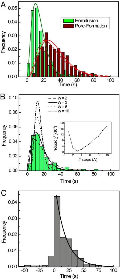
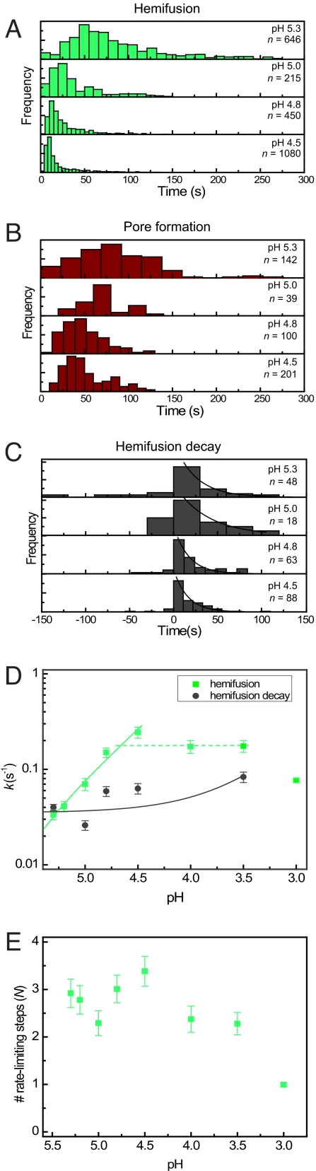
Membrane fusion is an essential step during entry of enveloped viruses into cells. Conventional fusion assays are generally limited to observation of ensembles of multiple fusion events, confounding more detailed analysis of the sequence of the molecular steps involved. We have developed an in vitro, two-color fluorescence assay to monitor kinetics of single virus particles fusing with a target bilayer on an essentially fluid support. Analysis of lipid- and content-mixing trajectories on a particle-by-particle basis provides evidence for multiple, long-lived kinetic intermediates leading to hemifusion, followed by a single, rate-limiting step to pore formation. We interpret the series of intermediates preceding hemifusion as a result of the requirement that multiple copies of the trimeric hemagglutinin fusion protein be activated to initiate the fusion process.


Conflict of interest statement

The authors declare no conflict of interest.

Figures

Fig. 1.
Fig. 1.
Proposed mechanism for viral fusion proteins. (A) In the prefusion state, the protein, anchored in the viral membrane by a C-terminal transmembrane segment, folds so that the fusion peptide (green) is sequestered. (B) A ligand-dependent trigger (e.g., proton binding, for HA and many other viral fusion proteins) induces a conformational change in which the fusion peptide projects toward the target membrane, forming an extended intermediate that bridges the two membranes. (C) The intermediate collapses, by zipping up of the C-terminal part of the ectodomain (blue) alongside the trimer-clustered N-terminal part (red). (D) The collapse pulls the two membranes together, leading to formation of a hemifusion stalk. (E) A fusion pore opens up, and snapping into place of the membrane-proximal and transmembrane segments of the protein completes the conformational transition and stabilizes the fusion pore. (Figure adapted from ref. .)
Fig. 2.
Fig. 2.
Experimental design. (A) Virus particles are labeled with two fluorescent dyes to monitor the kinetics of hemifusion and fusion pore formation. Fluorescence is collected by a high-NA microscope objective and imaged onto a CCD. (B) Fluorescence images before (Left) and during (Right) the fusion of individual viral particles. B Upper and Lower correspond to the red and green fluorescence, respectively, of the same ≈70 × 140-μm2 area of the supported bilayer. Dequenching of the Rh110C18 membrane dye upon hemifusion gives rise to the transient brightening of individual particles. (C) The fluorescence intensity of the red SRB viral content tracer (upper trace), the green Rh110C18 membrane dye (middle trace), and the fluorescein pH sensor (lower trace) provide exact times elapsed between pH drop, hemifusion, and fusion.
Fig. 3.
Fig. 3.
Fusion kinetics of fluorescently labeled influenza virus. (A) Time elapsed between pH decrease and hemifusion (green) and pore formation (red) of individual particles. The presence of intermediate states before hemifusion is clearly visible as a rise and decay in the histograms. Solid lines are best fits to a gamma function with N transitions [N = 3 for hemifusion (green); N = 4 for pore formation (red)]. The dashed line represents a convolution of the N = 3 gamma distribution of hemifusion times with the experimentally observed single-exponential transition between hemifusion and pore formation. (B) Hemifusion histogram from A compared with gamma distribution fits with varying numbers of steps. (Inset) Fitting error for fits with 1–10 transitions. (C) Distribution of lag times between hemifusion and pore formation of individual particles. The solid line represents a single-exponential fit with a rate constant of 0.55 ± 0.04 s−1. See also Figs. S2 and S3.
Fig. 4.
Fig. 4.
Fusion kinetics under varying pH conditions. (A–C) Hemifusion (A), pore-formation (B), and hemifusion decay (C) histograms for events recorded at varying acidic pH conditions. (D) Kinetic rate constants for transitions between prehemifusion intermediates (green squares) and decay of hemifusion to formation of fusion pores (gray circles) plotted as a function of proton concentration. The solid lines are plotted from least-squares fit (y = a + bx) of the rate constants as a function of proton concentration (green line: a = −0.01135, b = 8114, R = 0.987; gray line: a = 0.03522, b = 160.6, R = 0.635). A horizontal dotted green line has been included to emphasize the plateau in the hemifusion rate constants below pH 4.8. The solid lines appear curved in the log–log plot as a result of the nonzero y-intercepts. (E) Number of transitions N preceding hemifusion as a function of proton concentration. Values for N are obtained by fitting the hemifusion histograms with gamma distributions.

References

    1. Chernomordik LV, Kozlov MM. Protein-lipid interplay in fusion and fission of biological membranes. Annu Rev Biochem. 2003;72:175–207. - PubMed
    1. Harrison SC. Mechanism of membrane fusion by viral envelope proteins. Adv Virus Res. 2005;64:231–261. - PMC - PubMed
    1. Skehel JJ, Wiley DC. Receptor binding and membrane fusion in virus entry: The influenza hemagglutinin. Annu Rev Biochem. 2000;69:531–569. - PubMed
    1. Matlin KS, Reggio H, Helenius A, Simons K. Infectious entry pathway of influenza virus in a canine kidney cell line. J Cell Biol. 1981;91:601–613. - PMC - PubMed
    1. Wilson IA, Skehel JJ, Wiley DC. Structure of the haemagglutinin membrane glycoprotein of influenza virus at 3 Å resolution. Nature. 1981;289:366–373. - PubMed

Publication types