. 2008 Nov 28;283(48):33321-8.
doi: 10.1074/jbc.M805717200. Epub 2008 Sep 30.

The solution structure of DNA-free Pax-8 paired box domain accounts for redox regulation of transcriptional activity in the pax protein family

Affiliations

The solution structure of DNA-free Pax-8 paired box domain accounts for redox regulation of transcriptional activity in the pax protein family

Luca Codutti et al. J Biol Chem. .

Abstract

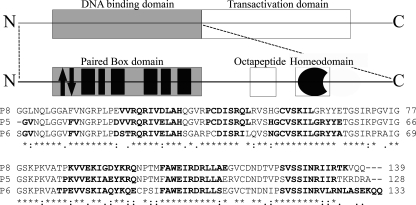
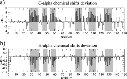
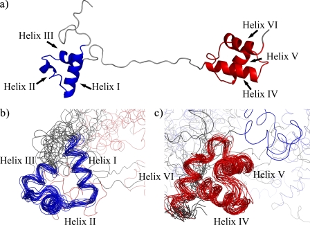
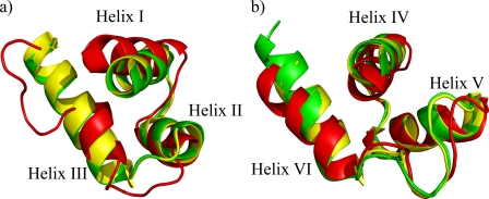
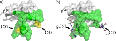
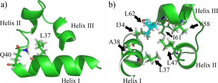
Pax-8 is a transcription factor belonging to the PAX genes superfamily and its crucial role has been proven both in embryo and in the adult organism. Pax-8 activity is regulated via a redox-based mechanism centered on the glutathionylation of specific cysteines in the N-terminal region (Cys45 and Cys57). These residues belong to a highly evolutionary conserved DNA binding site: the Paired Box (Prd) domain. Crystallographic protein-DNA complexes of the homologues Pax-6 and Pax-5 showed a bipartite Prd domain consisting of two helix-turn-helix (HTH) motifs separated by an extended linker region. Here, by means of nuclear magnetic resonance, we show for the first time that the HTH motifs are largely defined in the unbound Pax-8 Prd domain. Our findings contrast with previous induced fit models, in which Pax-8 is supposed to largely fold upon DNA binding. Importantly, our data provide the structural basis for the enhanced chemical reactivity of residues Cys45 and Cys57 and explain clinical missense mutations that are not obviously related to the DNA binding interface of the paired box domain. Finally, sequence conservation suggests that our findings could be a general feature of the Pax family transcription factors.


Figures

FIGURE 1.
FIGURE 1.
Schematic representation of Pax-8 protein organization. The upper frame enlightens the dualistic organization of this protein, with a N-terminal DNA binding region and a C-terminal transactivation region. The central frame is a magnification of the N-terminal region, showing the paired box domain, the evolutionary conserved octapeptide, and the non-functional homeodomain. The Paired domain black filled boxes represent α-helices, whereas the arrows indicate the β-hairpin. The lower part shows the primary structure of the Pax-8 Prd domain (P8) aligned with homologues Pax-5 (P5) and Pax-6 (P6) by means of ClustalW (44). Bold fonts highlight the residues found to be organized in a secondary structure. The investigated Pax-8 construct C-terminal linker plus the His6 tag are omitted for sake of clarity.
FIGURE 2.
FIGURE 2.
Residue by residue chemical shift deviation from random coil for the secondary structure diagnostic Cα and Hα resonances. Gray columns highlight the validated α-helices (60).
FIGURE 3.
FIGURE 3.
Structure of the free Pax-8 Prd domain. a, schematic representation of the lowest energy structure; b and c, backbone traces of the ensemble of structures superimposed on the PAI subdomain (in blue) (b) and the RED subdomain (in red) (c).
FIGURE 4.
FIGURE 4.
PAI (a) and RED subdomain (b) superimposition among the closest to mean structures of Pax-8 (red) and the crystal structures of Pax-6 (green) and Pax-5 (yellow).
FIGURE 5.
FIGURE 5.
Model of Pax8 glutathionylated at Cys45 and Cys57. Panel a shows the surface of the unbound Pax-8 Prd domain where the green surface represents the DNA binding interface. Panel b shows how glutathionylation of Cys45 and Cys57 hinders part of the DNA interaction surface. The model was built using PyMol (52).
FIGURE 6.
FIGURE 6.
Schematic representations of Pax-8 PAI subdomain. Panel a shows the hydrogen bonds found between the Leu37 carbonyl oxygen and Gln40 side chain and backbone amide nitrogens (Nε-2 and HN). Panel b depicts the hydrophobic residue side chains around Leu62 (in cyan), i.e. 5 Å apart. The cluster is an important part of the hydrophobic core of the PAI subdomain.

Similar articles

Cited by

References

    1. Kozmik, Z., Kurzbauer, R., Dörfler, P., and Busslinger, M. (1993) Mol. Cell. Biol. 13 6024-6035 - PMC - PubMed
    1. Poleev, A., Fickenscher, H., Mundlos, S., Winterpacht, A., Zabel, B., Fidler, A., Gruss, P., and Plachov, D. (1992) Development 116 611-623 - PubMed
    1. Zannini, M., Francis-Lang, H., Plachov, D., and Di Lauro, R. (1992) Mol. Cell. Biol. 12 4230-4241 - PMC - PubMed
    1. Espinoza, C., R., Schmitt, T., L., and Loos, U. (2001) J. Mol. Endocrinol. 27 59-67 - PubMed
    1. Ohno, M., Zannini, S., Levy, O., Carrasco, N., and Di Lauro, R. (1999) Mol. Cell. Biol. 19 2051-2060 - PMC - PubMed

Publication types

MeSH terms

Associated data

LinkOut - more resources