. 2008 Nov 28;283(48):33544-53.
doi: 10.1074/jbc.M805534200. Epub 2008 Sep 30.

IRBIT, inositol 1,4,5-triphosphate (IP3) receptor-binding protein released with IP3, binds Na+/H+ exchanger NHE3 and activates NHE3 activity in response to calcium

Affiliations

IRBIT, inositol 1,4,5-triphosphate (IP3) receptor-binding protein released with IP3, binds Na+/H+ exchanger NHE3 and activates NHE3 activity in response to calcium

Peijian He et al. J Biol Chem. .

Abstract

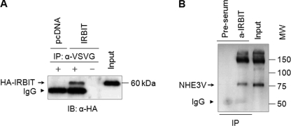
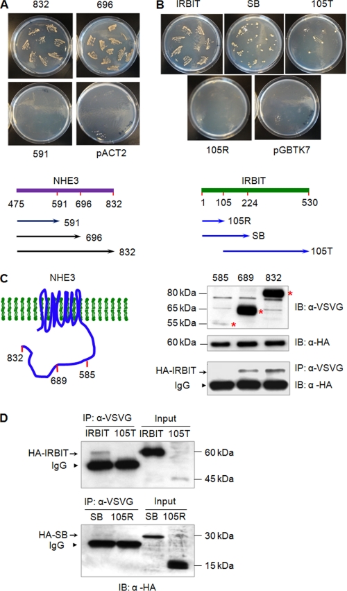
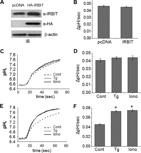
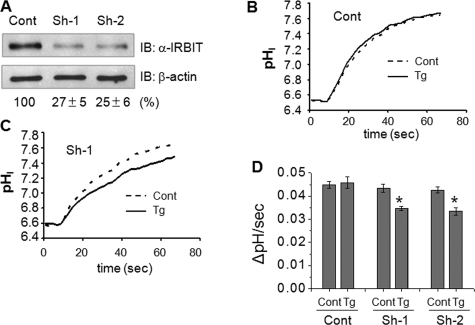
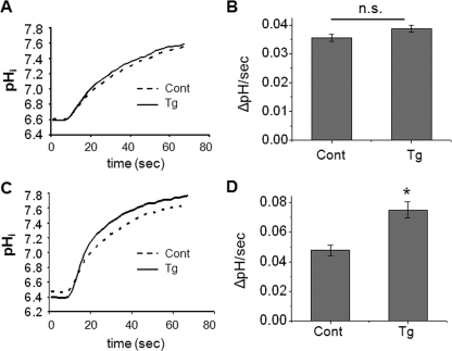
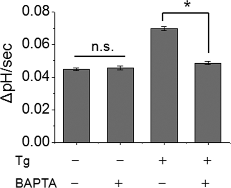
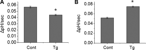
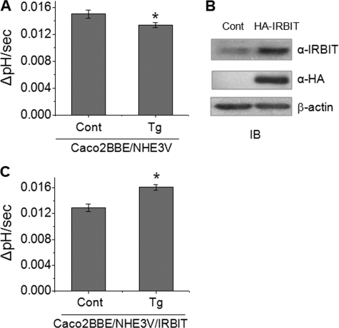
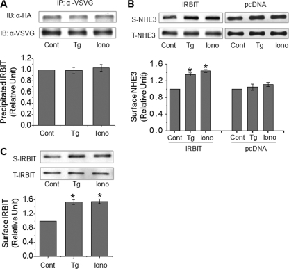
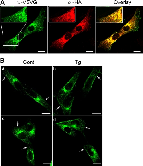
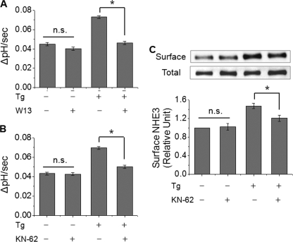
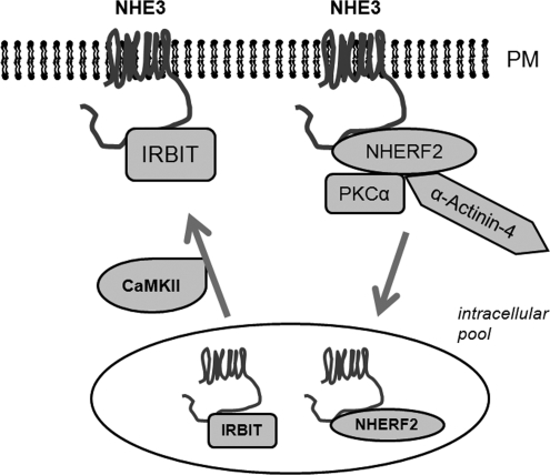
Calcium (Ca2+) is a highly versatile second messenger that regulates various cellular processes. Previous studies showed that elevation of intracellular Ca2+ regulates the activity of Na+/H+ exchanger 3 (NHE3). However, the effect of Ca2+-dependent signaling on NHE3 activity varies depending on cell types. In this study, we report the identification of IP3 receptor-binding protein released with IP3 (IRBIT) as a NHE3 interacting protein and its role in regulation of NHE3 activity. IRBIT bound to the carboxyl-terminal domain of NHE3, which is necessary for acute regulation of NHE3. Ectopic expression of IRBIT resulted in Ca2+-dependent activation of NHE3 activity, whereas silencing of endogenous IRBIT resulted in inhibition of NHE3 activity. Ca2+-dependent stimulation of NHE3 activity was dependent on the binding of IRBIT to NHE3. Previously Ca2+-dependent inhibition of NHE3 was demonstrated in the presence of NHERF2. Co-expression of IRBIT was able to reverse the NHERF2-dependent inhibition of NHE3. We also showed that IRBIT-dependent activation of NHE3 involves exocytic trafficking of NHE3 to the plasma membrane and this activation was blocked by inhibition of calmodulin (CaM) or CaM-dependent kinase II. These results suggest that the overall effect of Ca2+ on NHE3 activity is balanced by IRBIT-dependent activation and NHERF2-dependent inhibition.


Figures

FIGURE 1.
FIGURE 1.
NHE3 interacts with IRBIT. A, PS120/NHE3V cells were transfected with HA-IRBIT or pcDNA. Cell lysates were incubated with or without monoclonal anti-VSVG antibodies and bound proteins were subjected to Western blotting using an anti-HA antibody. B, cell lysates from PS120/NHE3V/IRBIT were incubated with anti-IRBIT antibody or pre-serum and co-immunoprecipitated NHE3 was detected by Western blotting with an anti-VSVG antibody. Representative immunoblots from three independent experiments are shown.
FIGURE 2.
FIGURE 2.
The region between aa 585 and 696 is necessary for the interaction with IRBIT. A, yeast two-hybrid assay was used to determine domains within the NHE3 COOH terminus that are necessary for the interaction with IRBIT. pACT2-IRBIT was co-transformed into host strain AH109 with C832, C696, or C591 in pGBKT7 and selected on SD/-Leu/-Trp for double transformants. 8-9 colonies were re-streaked on SD/-Ade/-His/-Leu/-Trp to verify the interaction between IRBIT and NHE3. A schematic diagram of IRBIT constructs is shown at the bottom. Similar results were obtained from three independent transformation and subsequent screens by auxotrophic growth. B, pGBKT7-C832 was co-transformed with pACT2 harboring truncated constructs of IRBIT into AH109 cells and protein interaction was determined by growth of double transformants on SD/-Ade/-His/-Leu/-Trp. A schematic diagram of IRBIT constructs is shown at the bottom. Similar results were obtained from three independent transformations and subsequent screen by auxotrophic growth. C, HA-IRBIT was expressed in PS120/NHE3V, PS120/NHE3V-689, or PS120/NHE3V-585 cells. Left panel illustrates NHE3V and its truncated constructs. NHE3V and truncated constructs were immunoprecipitated with an anti-VSVG antibody and co-immunoprecipitated IRBIT was detected by Western blotting using an anti-HA antibody. Top and middle panels show input NHE3V and the variants (*) and HA-IRBIT proteins, respectively, in the cell lysate. Bottom panel shows co-immunoprecipitated HA-IRBIT. Arrow, co-immunoprecipitated IRBIT. Arrowheads, IgG. D, HA-IRBIT and its variants were expressed in PS120/NHE3V cells. NHE3V was immunoprecipitated with an anti-VSVG antibody and co-immunoprecipitated HA-IRBIT and its truncated proteins were detected by an anti-HA antibody. Right two lanes show input and left two lanes are pulled down immunocomplex. Arrow, co-immunoprecipitated IRBIT. Arrowheads, IgG. Representative immunoblots from three separate experiments are shown.
FIGURE 3.
FIGURE 3.
Expression of IRBIT results in Ca2+-dependent activation of NHE3 activity. A, Western blot (IB, immunoblot) shows ectopic expression of HA-IRBIT in PS120/NHE3V cells. Total IRBIT was detected by anti-IRBIT and ectopic expression of IRBIT was determined with an anti-HA antibody. B, serum-starved PS120/NHE3V/pcDNA or PS120/NHE3V/IRBIT cells loaded with BCECF and NHE3 activity was determined fluorometically as described under “Experimental Procedures.” NHE3 activity was represented as the rate of Na+-dependent pH recovery, ΔpH/s. n = 8. C, representative traces of Na+-dependent pH recovery in PS120/NHE3V/pcDNA cells treated with 100 nm thapsigargin, 500 nm ionomycin, or carrier are shown. D, NHE3 activities in PS120/NHE3V/pcDNA cells treated with thapsigargin, ionomycin, or carrier are shown. The rates of pHi recovery at pHi 6.7 are shown. n = 8. E, representative traces of Na+-dependent pH recovery in PS120/NHE3V/HA-IRBIT cells are shown. F, NHE3 activities in PS120/NHE3V/IRBIT cells are shown. The rates of pHi recovery at pHi 6.7 are shown. n = 12 or more. *, p < 0.01 compare with the control.
FIGURE 4.
FIGURE 4.
Knockdown of IRBIT results in Ca2+-dependent inhibition of NHE3 activity. A, PS120/NHE3V cells were transfected with shRNA targeting IRBIT or control shRNA. Each effective shRNA (sh-1 or sh-2) knocked down IRBIT expression in PS120 cells by ∼75%. Representative traces of Na+-dependent pH recovery in PS120/NHE3V cells treated with control (B) or sh-1 (C) in the presence of absence of thapsigargin (Tg) are shown. D, NHE3 activities in PS120/NHE3V cells transfected with control, sh-1, or sh-2 determined at pHi 6.6 are shown. *, p < 0.01 compare with the control. IB, immunoblot.
FIGURE 5.
FIGURE 5.
The presence of IRBIT activates NHE3V-689 but not NHE3V-585. A and B, PS120/NHE3V-585; or C and D, PS120/NHE3V-689 cells were transfected with HA-IRBIT and the effect of thapsigargin (Tg) on NHE3 activity was determined. A and C show representative traces. B and D show the rate of pH recovery at pHi at 6.7. n = 8 or more. n.s., not significant. *, p < 0.01. IB, immunoblot.
FIGURE 6.
FIGURE 6.
Ca2+-dependent activation of NHE3 is blocked by BAPTA. PS120/NHE3V/IRBIT cells were pretreated with the Ca2+ chelator, BAPTA (40 μm), for 20 min and NHE3 activity was determined with or without thapsigargin (Tg) treatment. The rates of pH recovery at pHi 6.6 are shown. n.s., not significant. n = 8 or more. *, p < 0.01.
FIGURE 7.
FIGURE 7.
Expression of IRBIT counteracts the effect of NHERF2 on NHE3 activity. A, PS120/NHE3V/NHERF2 cells were treated with thapsigargin (Tg) or carrier and NHE3 activity was determined. B, NHE3 activity in PS120/NHE3V/NHERF2/IRBIT cells with or without thapsigargin treatment was determined. The rates of pH recovery at pHi 6.7 are shown. n = 8. *, p < 0.01.
FIGURE 8.
FIGURE 8.
Expression of IRBIT induces Ca2+-dependent activation of NHE3 in Caco2BBE cells. A, NHE3 activity in Caco2BBE/NHE3V cells with or without thapsigargin (Tg) treatment was determined in the presence of 50 μm HOE694. The rate of pH recovery at pHi 6.9 are shown. B, HA-IRBIT was ectopically expressed in Caco2BBE/NHE3V cells. Total and exogenous IRBIT proteins were detected by anti-IRBIT and anti-HA antibodies, respectively. C, NHE3 activity, represented as the rate of pH recovery at pHi 6.9, was determined in Caco2BBE/NHE3V/IRBIT cells. n = 8. *, p < 0.05 compare with the control.
FIGURE 9.
FIGURE 9.
Elevated Ca2+ increases surface NHE3 protein abundance in the presence of IRBIT. A, NHE3V and IRBIT were co-immunoprecipitated with anti-VSVG in PS120/NHE3V/IRBIT cells treated with thapsigargin (Tg), ionomycin, or carrier. Relative amounts of co-immunoprecipitated HA-IRBIT proteins was quantified. B, PS120/NHE3V/pcDNA and PS120/NHE3V/IRBIT cells were treated with thapsigargin, ionomycin, or carrier for 10 min, and the amount of NHE3V protein on the plasma membrane was determined by surface biotinylation as detailed under “Experimental Procedures.” Aliquots of surface and total protein were resolved by SDS-PAGE and the amount of surface (S) NHE3 and total (T) NHE3 proteins were determined by Western blot using an anti-VSVG antibody. The amount of surface NHE3 was normalized to total NHE3 and relative changes are shown in the graph. C, the amount of IRBIT in surface and total fractions was determined. n = 4. *, p < 0.01 compare with the control. IB, immunoblot.
FIGURE 10.
FIGURE 10.
NHE3 and IRBIT co-localize in PS120/NHE3V cells. A, cellular distribution of NHE3 and IRBIT in PS120/NHE3V/IRBIT cells was determined by indirect immunofluorescence using anti-VSVG and anti-HA antibodies, respectively. NHE3 (green) and IRBIT (red) were analyzed by a confocal microscope using a ×63 lens. The overlaid image of the double staining is shown on the right. Insets show magnified images. B, cellular localization of NHE3 in the presence or absence of IRBIT under basal conditions or after 10 min of thapsigargin (Tg) treatment was determined. Arrows point to NHE3 proteins on the plasma membrane. Bars, 10 μm.
FIGURE 11.
FIGURE 11.
Ca2+-mediated activation of NHE3 is dependent on CaM and CaMKII. The effect of thapsigargin (Tg) on NHE3 activity was determined with or without pretreatment of cells with (A) CaM inhibitor W13 (50 μm) or (B) CaMKII inhibitor KN-62 (20 μm). C, the effect of KN-62 on the amount of NHE3 protein at the surface membrane was determined by surface biotinylation. n.s., not significant. *, p < 0.05 compare with the absence of KN-62.
FIGURE 12.
FIGURE 12.
A tentative model of regulation of NHE3 by Ca2+. Elevation of intracellular Ca2+ regulates NHE3 in two opposing pathways. NHE3 is retrieved into a cytoplasmic pool by a mechanism dependent on NHERF2-PKCα-Actinin-4. IRBIT stimulates NHE3 activity by enhancing exocytic trafficking of NHE3 to the surface membrane and this process is dependent of CaM/CaMKII. PM, plasma membrane.

Similar articles

Cited by

References

    1. Berridge, M. J., Bootman, M. D., and Roderick, H. L. (2003) Nat. Rev. Mol. Cell. Biol. 4 517-529 - PubMed
    1. Berridge, M. J., Lipp, P., and Bootman, M. D. (2000) Nat. Rev. Mol. Cell. Biol. 1 11-21 - PubMed
    1. Mikoshiba, K. (2007) J. Neurochem. 102 1426-1446 - PubMed
    1. Ando, H., Mizutani, A., Matsu-ura, T., and Mikoshiba, K. (2003) J. Biol. Chem. 278 10602-10612 - PubMed
    1. Ando, H., Mizutani, A., Kiefer, H., Tsuzurugi, D., Michikawa, T., and Mikoshiba, K. (2006) Mol. Cell 22 795-806 - PubMed

Publication types

MeSH terms