. 2008 Nov 28;283(48):33516-26.
doi: 10.1074/jbc.M805358200. Epub 2008 Sep 30.

Potent and selective disruption of protein kinase D functionality by a benzoxoloazepinolone

Affiliations

Potent and selective disruption of protein kinase D functionality by a benzoxoloazepinolone

Elizabeth R Sharlow et al. J Biol Chem. .

Abstract

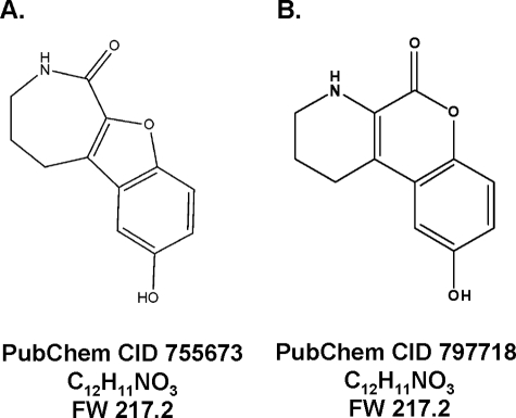
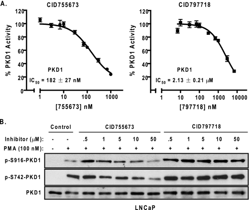
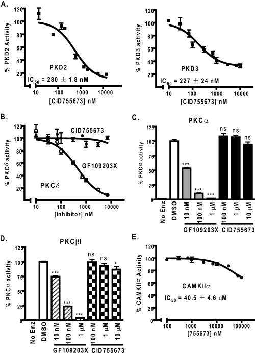
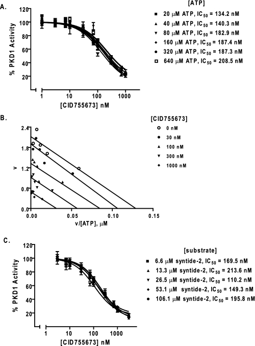
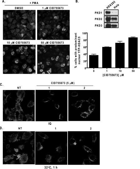
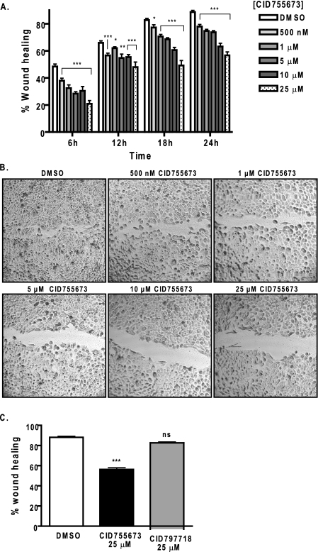
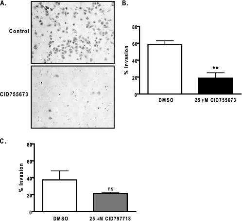
Protein kinase D (PKD) is a novel family of serine/threonine kinases targeted by the second messenger diacylglycerol. It has been implicated in many important cellular processes and pathological conditions. However, further analysis of PKD in these processes is severely hampered by the lack of a PKD-specific inhibitor that can be readily applied to cells and in animal models. We now report the discovery of the first potent and selective cell-active small molecule inhibitor for PKD, benzoxoloazepinolone (CID755673). This inhibitor was identified from the National Institutes of Health small molecule repository library of 196,173 compounds using a human PKD1 (PKCmu)-based fluorescence polarization high throughput screening assay. CID755673 suppressed half of the PKD1 enzyme activity at 182 nm and exhibited selective PKD1 inhibition when compared with AKT, polo-like kinase 1 (PLK1), CDK activating kinase (CAK), CAMKIIalpha, and three different PKC isoforms. Moreover, it was not competitive with ATP for enzyme inhibition. In cell-based assays, CID755673 blocked phorbol ester-induced endogenous PKD1 activation in LNCaP cells in a concentration-dependent manner. Functionally, CID755673 inhibited the known biological actions of PKD1 including phorbol ester-induced class IIa histone deacetylase 5 nuclear exclusion, vesicular stomatitis virus glycoprotein transport from the Golgi to the plasma membrane, and the ilimaquinone-induced Golgi fragmentation. Moreover, CID755673 inhibited prostate cancer cell proliferation, cell migration, and invasion. In summary, our findings indicate that CID755673 is a potent and selective PKD1 inhibitor with valuable pharmacological and cell biological potential.


Figures

FIGURE 1.
FIGURE 1.
Chemical structures of CID755673 and CID797718. A, chemical structure of CID755673, a compound identified and confirmed as a PKD1 inhibitor after interrogation of the PMLSC library. B, chemical structure of CID797718, an analogue of CID755673 obtained during the synthesis of the latter structure.
FIGURE 2.
FIGURE 2.
The inhibitory activities of CID755673 and CID797718. A, inhibition of recombinant human PKD1 in vitro. The kinase activity of PKD1 was assayed in the presence of 10 different concentrations of CID755673 by a radiometric PKD kinase assay. The IC50 values were the mean ± S.E. of at least three independent experiments with triplicate determinations at each drug concentration in each experiment. The data were plotted as a function of drug concentration and a representative graph is shown. B, inhibition of endogenous PKD1 activity by CID755673 in cells. LNCaP cells were pretreated with different doses of CID755673 or CID797718 for 45 min, followed by PMA stimulation at 100 nm for 20 min. Cell lysates were subjected to immunoblotting for p-S742-PKD1 and p-S916-PKD1. PKD1 was blotted as loading control. Note that the low p-S916-PKD1 signal in lane 2 (PMA alone) was likely caused by uneven loading. The experiment was repeated five times and a representative blot is shown.
FIGURE 3.
FIGURE 3.
Selectivity of CID755673. A, PKD isoform selectivity of CID755673. Inhibition of human recombinant PKD2 and PKD3 by CID755673 was assayed by the radiometric PKD kinase assay. The experiment was repeated three times and a representative graph is shown. B, CID755673 did not inhibit PKCδ. Inhibitory activity of CID755673 for PKCδ was measured using a radiometric PKC kinase assay. Inhibition of PKCδ by GF109203X was assayed as control. Data are from one of three representative experiments. C and D, inhibition of PKCα (C) or PKCβI (D) was determined at 100 nm, 1 μm, and 10 μm CID755673. As controls, the PKC inhibitor GF109203X potently inhibited PKCα or PKCβI activity. Data are the mean ± S.E. of two independent experiments. ns, not statistically significant; *, p < 0.05; ***, p < 0.001. E, inhibition of CAMKIIα was measured by the radiometric CAMK kinase assay. The experiment was repeated twice and a representative curve is shown.
FIGURE 4.
FIGURE 4.
CID755673 was not competitive with ATP or substrate for PKD1 inhibition. A, ATP competition analysis. PKD1 kinase activity was measured as a function of increasing doses of CID755673 in the presence of varying concentrations of ATP. B, Eadie-Hofstee plot analysis. The reaction velocity (v) was plotted as a function of the velocity versus ATP concentration ratio (v/[ATP]) for each concentration of CID755673. The points were fitted by linear regression analysis. C, substrate competition analysis. Inhibitory activity of CID755673 was determined in the presence of varying concentrations of substrate peptide syntide-2. Each point of the curves represent the average of triplicate determinations.
FIGURE 5.
FIGURE 5.
Effects of CID755673 on HDAC5 localization and VSVG-GFP trafficking. A, effect of CID755673 on PMA-induced nuclear exclusion of HDAC5. HeLa cells were transiently transfected with YFP-HDAC5. Two days after transfection, cells were pretreated with or without different concentrations of CID755673 for 45 min, following by the addition of PMA at 1 μm for 4 h. Cells were fixed and imaged under fluorescence confocal microscope. B, quantitative analysis of YFP-HDAC5 nuclear localization. Percent cells with predominant nuclear YFP-HDAC5 were determined. Data were obtained from a total of 400 randomly captured cells in a given experimental condition. Upper panel, the expression of endogenous PKD isoforms in HeLa cells was evaluated by Western blot analysis. HEK293 lysates were probed as positive control. C and D, effect of CID755673 on ilimaquinone-induced Golgi fragmentation and on trafficking of VSVG-GFP from the Golgi to the plasma membrane. HeLa cells were transiently transfected with the VSVG-GFP plasmid. Two days after transfection, cells were incubated for 2 h at 40 °C, followed by incubation at 20 °C for 30 min to trap the VSVG-GFP protein to the Golgi. Cells were then pretreated with or without CID755673 (5 μm) at 20 °C, followed by a 2-h incubation at 20 °C with 30 μm ilimaquinone to induce Golgi fragmentation (C) or by a 1-h incubation at 32 °C to allow protein transport to the cell surface (D). Images of VSVG-GFP distribution were captured under a fluorescence confocal microscope. The experiments were repeated at least three times and representative images are shown. NT, no inhibitor treatment or DMSO only.
FIGURE 6.
FIGURE 6.
Effects of CID755673 on tumor cell migration. A, wound healing assay. DU145 cells were grown to confluence in 6-well plates. Monolayer was wounded and imaged immediately (0 h). Growth media containing a vehicle (DMSO) or varying concentrations of CID755673 was added. Wound closure was recorded every 6 h up to 24 h. The width of the wound is the average of 9 determinations per time point. Percent wound healing was calculated at each time point as described under “Experimental Procedures.” B, after 24 h, cells were stained with 0.25% crystal violet. Phase-contrast images of the final wounds were taken at ×100 magnification. The experiment was repeated three times and a representative experiment is shown. C, the weaker analogue CID797718 did not inhibit wound closure. DU145 monolayer was wounded and incubated in the presence of the vehicle (DMSO), 25 μm CID755673 or CID797718. Wound closure was measured after 24 h. Data represent one of two independent experiments. ns, not statistically significant; *, p < 0.05; **, p < 0.01; ***, p < 0.001.
FIGURE 7.
FIGURE 7.
Effects of CID755673 on tumor cell invasion. A, tumor cell invasion was evaluated by a Matrigel invasion assay. A fixed number of DU145 cells (1.0 × 10⁵/ml) were seeded into the upper control or invasion chamber. After 22 h, non-invading cells were removed and cells that invaded through the Matrigel were fixed, stained, and photographed under a microscope. Magnification, ×200. B, percent invasion is expressed as the number of cells that invaded through the Matrigel matrix relative to the number of cells that migrated through the control insert. Cell number is determined by counting total cell number in 10 random fields. The experiment was repeated twice and a representative experiment is shown. C, the weaker analogue CID797718 did not significantly inhibit tumor cell invasion. DU145 cell invasion was measured using Matrigel invasion chambers after incubating with DMSO or 25 μm CID797718 for 22 h. ns, not statistically significant; **, p < 0.01.
FIGURE 8.
FIGURE 8.
CID755673 inhibited cell proliferation in LNCaP and PC3 cells. A and B, LNCaP (A) or PC3 (B) cells were plated in triplicates in 24-well plates. Cells were allowed to attach overnight. A cell count at day 1 was made, and then either a vehicle (DMSO) or CID755673 at the indicated concentrations was added. Cells were counted daily for a total of 6 days. Fresh media and inhibitor were added every 2 days. The means of triplicate determinations were plotted over time. The experiment was repeated twice and results from one representative experiment are shown. Right panel (B), the effect of the weaker analogue CID797718 on PC3 cell proliferation. Results from one of two independent experiments are shown. *, p < 0.05; **, p < 0.01; ***, p < 0.001. C, CID755673 induced apparent morphological changes. Phase-contrast images of PC3 cells after 6 days of CID755673 treatment are shown. D, CID755673-induced G2/M phase cell cycle arrest. PC3 cells were incubated with a vehicle (DMSO) or varying concentrations of CID755673 for 6 days. Media with fresh inhibitors were replenished daily. Cell cycle distribution was evaluated by flow cytometry after propidium iodide labeling of fixed cells. One of two independent experiments is shown.

Similar articles

Cited by

References

    1. Manning, G., Whyte, D. B., Martinez, R., Hunter, T., and Sudarsanam, S. (2002) Science 298 1912-1934 - PubMed
    1. Wang, Q. J. (2006) Trends Pharmacol. Sci. 27 317-323 - PubMed
    1. Hayashi, A., Seki, N., Hattori, A., Kozuma, S., and Saito, T. (1999) Biochim. Biophys. Acta 1450 99-106 - PubMed
    1. Johannes, F. J., Prestle, J., Eis, S., Oberhagemann, P., and Pfizenmaier, K. (1994) J. Biol. Chem. 269 6140-6148 - PubMed
    1. Sturany, S., Van Lint, J., Muller, F., Wilda, M., Hameister, H., Hocker, M., Brey, A., Gern, U., Vandenheede, J., Gress, T., Adler, G., and Seufferlein, T. (2001) J. Biol. Chem. 276 3310-3318 - PubMed

Publication types

MeSH terms

LinkOut - more resources