. 2008 Dec 12;283(50):35154-63.
doi: 10.1074/jbc.M802592200. Epub 2008 Sep 30.

Structural basis of the migfilin-filamin interaction and competition with integrin beta tails

Affiliations

Structural basis of the migfilin-filamin interaction and competition with integrin beta tails

Yatish Lad et al. J Biol Chem. .

Abstract

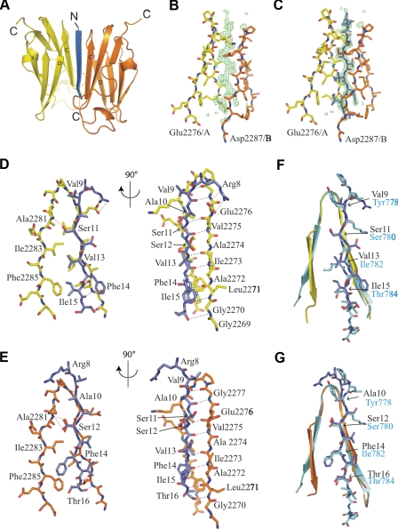
A link between sites of cell adhesion and the cytoskeleton is essential for regulation of cell shape, motility, and signaling. Migfilin is a recently identified adaptor protein that localizes at cell-cell and cell-extracellular matrix adhesion sites, where it is thought to provide a link to the cytoskeleton by interacting with the actin cross-linking protein filamin. Here we have used x-ray crystallography, NMR spectroscopy, and protein-protein interaction studies to investigate the molecular basis of migfilin binding to filamin. We report that the N-terminal portion of migfilin can bind all three human filamins (FLNa, -b, or -c) and that there are multiple migfilin-binding sites in FLNa. Human filamins are composed of an N-terminal actin-binding domain followed by 24 immunoglobulin-like (IgFLN) domains and we find that migfilin binds preferentially to IgFLNa21 and more weakly to IgFLNa19 and -22. The filamin-binding site in migfilin is localized between Pro(5) and Pro(19) and binds to the CD face of the IgFLNa21 beta-sandwich. This interaction is similar to the previously characterized beta 7 integrin-IgFLNa21 interaction and migfilin and integrin beta tails can compete with one another for binding to IgFLNa21. This suggests that competition between filamin ligands for common binding sites on IgFLN domains may provide a general means of modulating filamin interactions and signaling. In this specific case, displacement of integrin tails from filamin by migfilin may provide a mechanism for switching between different integrin-cytoskeleton linkages.


Figures

FIGURE 1.
FIGURE 1.
The N-terminal 85 amino acids of migfilin bind to filamins A, B, and C. A-D, pull-down assays were performed using lysates of Chinese hamster ovary cells transfected with (A) FLNa-GFP, (B) FLNb-GFP, (C) FLNc-GFP, or (D) FLNa-GFP fragments, FLNa-(1-1761) (ABD + rod 1) or FLNa-(1762-2647) (rod 2) to beads coated with migfilin-(1-85) or -(86-373). Bound proteins were detected by immunoblotting with anti-GFP antibodies and loading with anti-His antibodies. 20% lanes represent corresponding percentage of the starting material in the binding assay.
FIGURE 2.
FIGURE 2.
Migfilin binds IgFLNa21 directly. A and B, direct pull-down assays were performed using (A) purified GST-IgFLNa19, -20, -21, -22, or -23 to migfilin-(1-85)-coated beads or (B) purified GST-IgFLNa21 or GST to migfilin-(1-85), migfilin K7T/R8G 1-85 or integrin β7-coated beads. Bound proteins were detected by immunoblotting with anti-GST antibodies. C, protein binding was quantified by densitometry and expressed as filamin binding (arbitrary units) was calculated as the ratio of filamin bound to filamin in the loading control, normalized to maximal filamin binding in each experiment (mean ± S.E.; n ≥ 3). D, pull-down assays were performed using purified (D) GST-IgFLNa21 containing mutations in strands C (A2272D,A2274K or A2272S,A2274T) or D (I2283C or I2283M) to migfilin-(1-85)-coated beads. 10% lanes represent the corresponding percentage of the starting material in the binding assay.
FIGURE 3.
FIGURE 3.
Migfilin residues 5-19 mediate binding to FLNa. A, alignment of the N terminus of migfilin with the IgFLNa21 binding region of β7, β2, and IgFLNa20. B, comparison of IgFLNa21 HSQC overlaid spectra showing the effect of addition of the migfilin 5Pro-19Pro-derived peptide; the observed behavior generally corresponds to the slow exchange regime except for a few resonances with small shifts, outlined in black boxes. The color scheme of the overlaid spectral layers is indicated. C, normalized induced chemical shifts of those resonances with typical fast exchange behavior were plotted against the ligand-to-protein ratio for IgFLNa21. Due to the relatively large error for those small shifts and the relatively high affinity, the data fitting was not very successful. Comparison with the theoretical simulated binding curves for Kd at 0.1, 1, and 10 μm, the affinity for migfilin binding to IgFLNa21 is estimated to be ≤1 μm. D, overlaid HSQC spectra of IgFLNa21 upon titration by migfilin-(1-85); the corresponding induced shifts are nearly identical to the above short migfilin peptide (C), although some peaks are broadened, probably due to aggregation. E, migfilin added to IgFLNa19 gives many more fast exchange peaks, and, in that case, the induced chemical shifts could be readily fitted to a Kd of about 107 μm.
FIGURE 4.
FIGURE 4.
Structure of IgFLNa21/Pro5-Pro19 migfilin peptide complex. A, overall structure of the IgFLNa21 Pro5-Pro19 migfilin peptide complex shown as a schematic. N and C termini are indicated. Migfilin peptide (blue) is surrounded by two IgFLNa21 domains, chain A (yellow) and chain B (orange). B, electron density map (Fo - Fc) of the migfilin peptide without peptide calculated from the final model without the peptide and shown at σ level 3.0. C, the same panel as B but with migfilin peptide. D, details of residues participating in the interaction of IgFLNa21 chain A and migfilin peptide. Most side chains of peptide residues interact with residues of the IgFLNa21 D-strand. Residues likely to be important for the interaction are named. The backbone hydrogen bonds are shown with blue dashed lines and side chain hydrogen bonds with red. Ser11 and Val13 were modeled in 2 alternative conformations. E, details of the side chain interactions between IgFLNa21 chain B and migfilin peptide. F and G, superposition of the IgFLNa21-β7 integrin complex (PDB 2BRQ) with the IgFLNa21 chain A-migfilin (F), or IgFLNa21 chain B-migfilin (G) structures. IgFLNa21 strands C and D are shown as schematics with the integrin and migfilin peptides as stick models. Colors of the IgFLNa21-migfilin complex are as in panel A, the IgFLNa21-β7 complex is shown in pale blue.
FIGURE 5.
FIGURE 5.
Migfilin binds to the CD face of IgFLNa21 in a manner similar to the integrin β7. A, combined chemical shift perturbations of resonances in the U-15N-IgFLNa21 amide H-Ns induced by a 4-fold excess of migfilin Pro5—Pro19-derived peptide are mapped onto the crystal structure of IgFLNa21 from the IgFLNa21-β7 integrin complex (PDB 2BRQ). Residues are colored according to the size of the shift with the largest shifts shown in red and no shifts in blue. B, chemical shift perturbations are also represented in a two-dimensional bar chart, strongly perturbed butnon-assigned residues are denoted by solidblue bars. C, extracted HSQC spectra of IgFLNa21 residue Ser2279 upon addition of migfilin-(1-85)/5-19 WT or S11E, S12E, F14E, and I15E peptides. The color scheme of the overlaid spectra is shown in the figure. (When the binding is tight the peaks are in slow exchange with one peak decreasing and the other increasing on addition of ligand. With weaker binding (S11E and I15E) the peaks are in fast exchange and migrate from one position to another.) D, normalized induced chemical shifts of resonances with typical fast exchange behavior and relatively large shifts were plotted against the ligand-to-protein ratio for IgFLNa21 binding to migfilin S11E with a theoretically simulated binding curve shown as a dashed line. E, direct pull-down assays were performed using GST-IgFLNa21 to migfilin-(1-85) WT, S11D, or I15E. F, pull-down assays were performed using lysates of Chinese hamster ovary cells transfected with FLNa-GFP to beads coated with migfilin-(1-85) wild type, S11D, or I15E. G, binding of FLNa, FLNa-(1-1761) (ABD + rod 1), or FLNa-(1762-2647) (rod 2) to beads coated with migfilin-(1-85) wild type or I15E. 10% lanes represent corresponding percentage of the starting material in the binding assay.
FIGURE 6.
FIGURE 6.
Migfilin competes with integrinβ tails for binding to IgFLNa21. Direct pull-down assays were performed using purified GST-IgFLNa21 to β7 tails in the presence of migfilin-(5-19) or control peptide. Protein binding quantified by densitometry and expressed as filamin binding (arbitrary units) was calculated as the ratio of filamin bound to filamin bound in the absence of peptide in each experiment (mean ± S.E.; n ≥ 3). Bound proteins were detected by immunoblotting with anti-GST antibodies. 10% lanes represent the corresponding percentage of the starting material in the binding assay.
FIGURE 7.
FIGURE 7.
Migfilin-filamin interactions target migfilin to stress fibers. Immunofluorescence of GFP-migfilin or 1-85 mutant-transfected NIH3T3 cells attached to fibronectin-coated coverslips for 4 h prior to fixation. A, cells were stained for actin with phalloidin-Alexa 568; or B, endogenous FLNa.

Similar articles

Cited by

References

    1. Evans, E. A., and Calderwood, D. A. (2007) Science 316 1148-1153 - PubMed
    1. Zaidel-Bar, R., Itzkovitz, S., Ma'ayan, A., Iyengar, R., and Geiger, B. (2007) Nat. Cell Biol. 9 858-867 - PMC - PubMed
    1. Tu, Y., Wu, S., Shi, X., Chen, K., and Wu, C. (2003) Cell 113 37-47 - PubMed
    1. Takafuta, T., Saeki, M., Fujimoto, T. T., Fujimura, K., and Shapiro, S. S. (2003) J. Biol. Chem. 278 12175-12181 - PubMed
    1. Wu, C. (2005) J. Cell Sci. 118 659-664 - PubMed

Publication types