. 2008 Oct 1;14(19):6062-72.
doi: 10.1158/1078-0432.CCR-08-0979.

Androgen-regulated and highly tumorigenic human prostate cancer cell line established from a transplantable primary CWR22 tumor

Affiliations

Androgen-regulated and highly tumorigenic human prostate cancer cell line established from a transplantable primary CWR22 tumor

Ayush Dagvadorj et al. Clin Cancer Res. .

Abstract

Purpose: One of the major obstacles in understanding the molecular mechanisms underlying the transition of prostate cancer growth from androgen dependency to a hormone-refractory state is the lack of androgen-regulated and tumorigenic human prostate cancer cell lines.

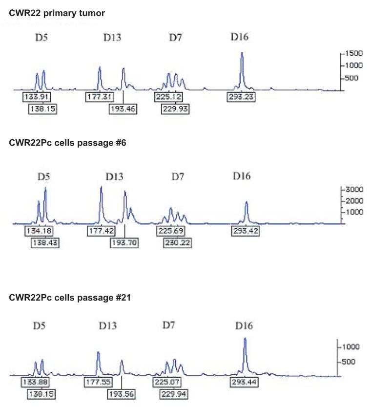
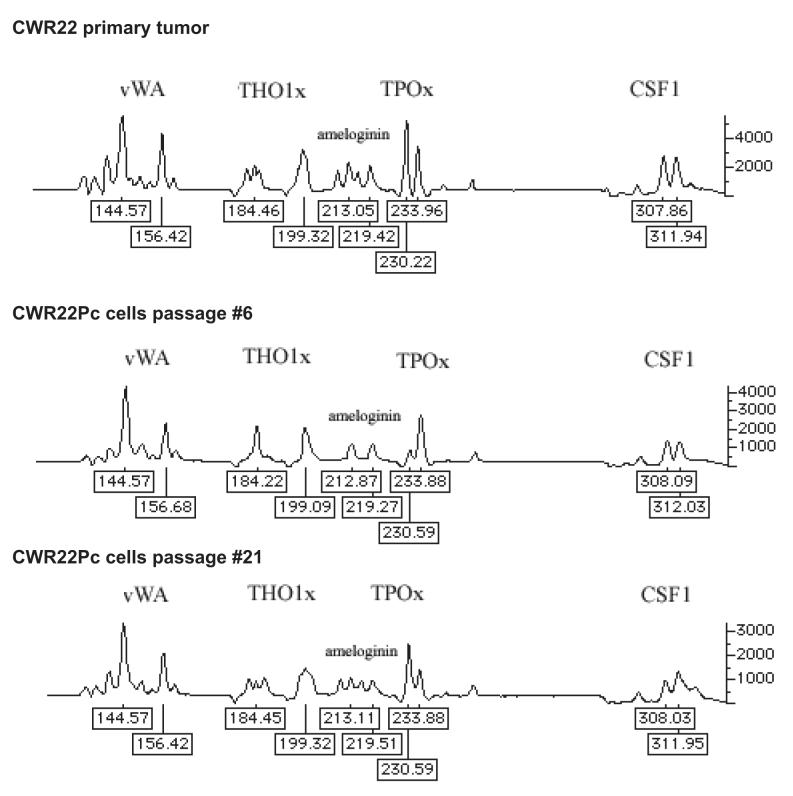
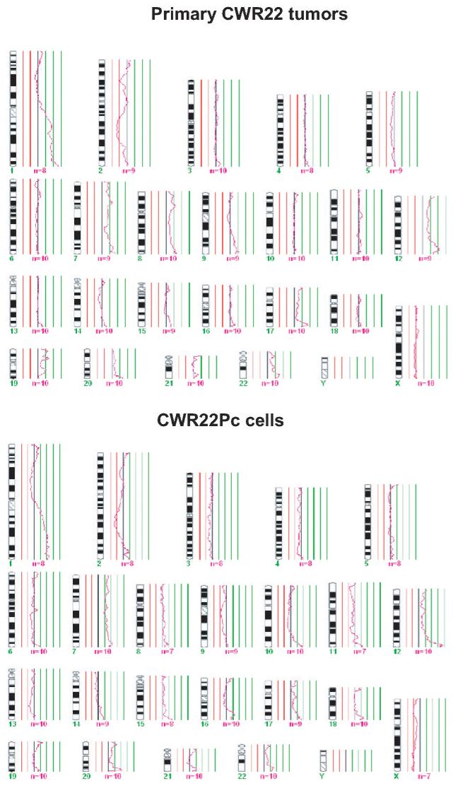
Experimental design: We have established and characterized a new human prostate cancer cell line, CWR22Pc, derived from the primary CWR22 human prostate xenograft tumors.

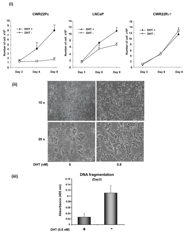
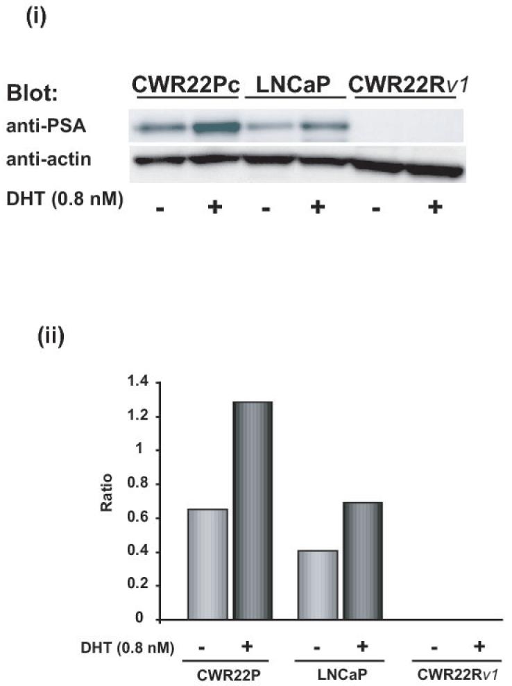
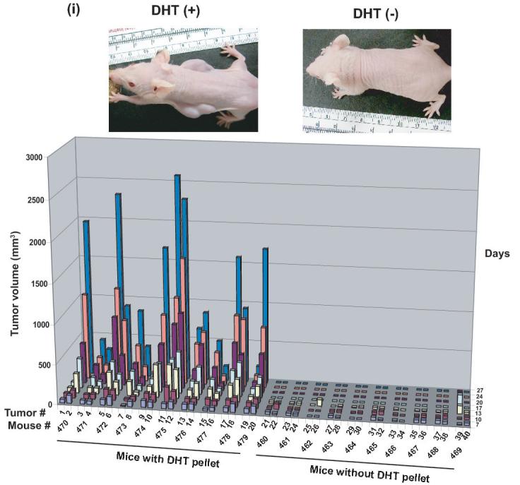
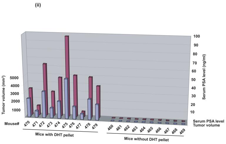
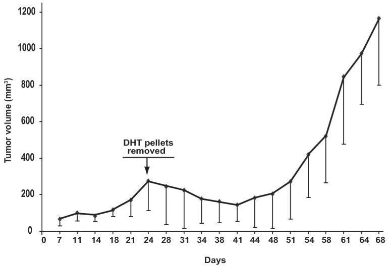
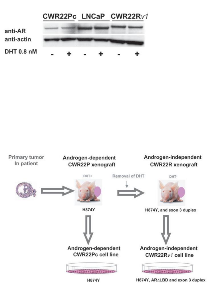
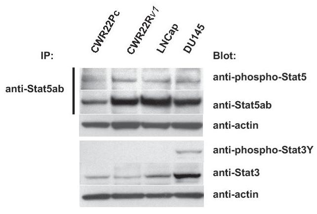
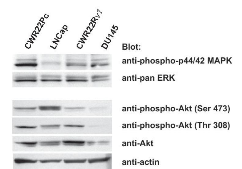
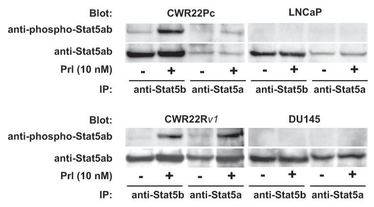
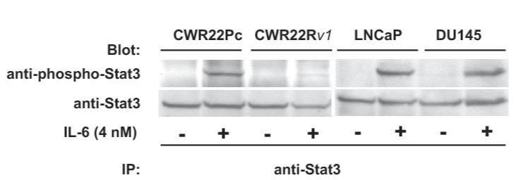
Results: The growth of CWR22Pc cells is induced markedly by dihydrotestosterone, and CWR22Pc cells express high levels of androgen receptor (AR) and prostate-specific antigen (PSA). Importantly, PSA expression in CWR22Pc cells is regulated by androgens. Stat5a/b, Stat3, Akt, and mitogen-activated protein kinase were constitutively active or cytokine inducible in CWR22Pc cells. The AR in CWR22Pc cells contains the H874Y mutation, but not the exon 3 duplication or other mutations. When inoculated subcutaneously into dihydrotestosterone-supplemented castrated nude mice, large tumors formed rapidly in 20 of 20 mice, whereas no tumors developed in mice without circulating dihydrotestosterone. Moreover, the serum PSA levels correlated with the tumor volumes. When androgens were withdrawn from the CWR22Pc tumors grown in nude mice, the tumors initially shrank but regrew back as androgen-independent tumors.

Conclusions: This androgen-regulated and tumorigenic human prostate cancer cell line provides a valuable tool for studies on androgen regulation of prostate cancer cells and on the molecular mechanisms taking place in growth promotion of prostate cancer when androgens are withdrawn from the growth environment. CWR22Pc cells also provide a model system for studies on the regulation of transcriptional activity of mutated H874YAR in a prostate cancer cell context.


Figures

Fig. 1
Fig. 1. The genetic lineage identity of CWR22Pc cells is similar to that of CWR22 primary prostate tumors
DNA fingerprinting analysis of the CWR22 primary tumor, and the cell line CWR22Pc (passages 6 and 21) show a similar pattern for all nine markers analyzed. A, Patterns of alleles at markers D5S818 (D5), D13S317 (D13), D7S820 (D7), and D16S539 (D16). B, The results at markers vWA (vWA), TH01 (THO1x), Amelogenin, TPOX (TPOx), and CSF1PO (CSF1). The allele sizes are indicated under each allele. C. The comparative genomic hybridization analysis shows gains at 1q, 7, 8p and 12, and losses of chromosomes 2 and X in both primary CWR22 tumors and in CWR22Pc cells. D. Flow cytometric analysis of CWR22Pc cells shows that the cell line consists of a mixed population of cells with DNA indices of 1.11 and 2.23. #1 represents the G1 peak of the human peripheral blood lymphocytes (added as a control). #2 represents the G1 peak of the cells with a lower ploidy (DNA index = 1.1). #3 represents the G1 peak of the cells with a higher ploidy (DNA index = 2.23).
Fig. 2
Fig. 2. The growth of CWR22Pc cells is increased by androgens
A, CWR22Pc, LNCaP and CWR22Rv1 cells were grown in the medium containing 3% CS-FBS in the presence or absence of 0.8 nM dihydrotestosterne (DHT) for 9 days. Cells were counted every third day. The means of three independent experiments are presented with SDs (panel i). The absence of DHT had marked effects on the cell morphology of CWR22Pc cells. In the absence of DHT, stereomicroscope photographs show that the majority of the cells were dead and floating (panel ii) with increased DNA fragmentation (panel iii). B, Prostate specific antigen (PSA) expression is regulated by androgens in CWR22Pc and LNCaP cells. CWR22Pc, LNCaP and CWR22Rv1 cell lines were cultured in RPMI with or without 0.8 nM DHT for 12 days. Cells were harvested, lysed and immunoblotted with anti-PSA pAb. Stripped filters were re-blotted with anti-actin pAb to demonstrate equal loading (panel i). Densitometric normalization and comparison of the PSA levels (panel ii). C, The growth of CWR22Pc cells as subcutaneous xenograft tumors in athymic nude mice is regulated by androgens. CWR22Pc cells were inoculated subcutaneously into flanks of castrated nude mice supplied with sustained-release 5α-dihydrotestosterone (DHT)-pellets (n=10/group, 2 tumors/mouse, 20 × 106 CWR22Pc cells per site). The tumor incidence and growth were measured twice a week for 36 days. Tumor volumes were calculated using the formula (3.14 x length x width x depth/6) (panel i). Serum PSA levels in mice carrying CWR22Pc tumors correlated with the volumes of the tumors (panel ii). PSA levels in the sera of mice carrying CWR22Pc xenograft tumors were determined using a PSA ELISA assay. The total tumor burden (the sum of both right and left tumor volumes) were compared to the serum PSA levels. D, CWR22Pc tumors recur after androgen-deprivation induced regression of the tumors. CWR22Pc cells were inoculated subcutaneously into flanks of castrated athymic nude mice supplied with sustained-release 5α-DHT-pellets (n=4 mice, 1 tumor/mouse, 20 ×106 CWR22Pc cells per site). Once the tumors reached 10 mm in diameter in size, the DHT-pellets were removed and the tumor growth was measured twice a week. Tumor volumes were calculated as described above.
Fig. 3
Fig. 3. Androgen receptor expression in CWR22Pc cells
A, Androgen receptor (AR) expression in CWR22Pc, LNCaP and CWR22Rv1 cells. Cells were cultured in the presence or absence of 0.8 nM dihydrotestosterone (DHT), lysed and immunoblotted with an anti-AR mAb. Filters were re-blotted with anti-actin pAb to demonstrate the protein loading. AR protein expressed in CWR22Rv1 is of higher molecular weight than in CWR22Pc and LNCaP cells. B, Schematic presentation of AR mutations in primary CWR22 prostate tumors (CWR22P), CWR22Pc cells, recurrent CWR22 tumors (CWR22R) and CWR22Rv1 cells. C, AR gene sequencing primers. PCR primers were designed to amplify and sequence four overlapping segments (I, II, III, IV) encompassing the entire AR coding region plus 5′ and 3′ untranslated sequences. The Segment I covers a part of the 5′- UTR and the 5-prime-end part of the exon 1, the segment II covers the 3-prime-end of the exons 1 and 2, the segment III covers the exons 2, 3, 5 and a part of the exon 6. The segment IV covers a part of the exons 5, 6, 7, 8 and a part of 3′- UTR. D, RT-PCR products of the AR segment III in different human prostate cancer xenograft tumors and prostate cancer cell lines. The RT-PCR product for the segment III yielded the expected amplicon size of 673 bp in CWR22PC cells, primary CWR22 xenograft tumors and in LNCaP cells, while the CWR22Rv1 RT-PCR reaction yielded an approximately 100-bp larger RT-PCR product.
Fig. 4
Fig. 4. Protein kinase signaling pathways are activated in CWR22Pc cells
Transcription factor Stat5a/b, MAPK(p44/42) and AKT are constitutively active in CWR22Pc cells. A, Stat5ab or Stat3 were immunoprecipitated (IP) from exponentially growing CWR22Pc, CWR22Rv1, LNCaP and DU145 cells using anti-Stat5ab or anti-Stat3 pAb, respectively, and blotted with anti-phopho-Stat5a/b or anti-phospho-Stat3 antibody as indicated. Filters were stripped and re-blotted with anti-Stat5ab mAb or anti-Stat3 mAb and whole cell lysates before IP were immunoblotted with anti-actin pAb. B, Whole cell lysates of exponentially growing CWR22Pc, LNCaP, CWR22Rv1 and DU145 cells were immunoblotted with antibodies to phosphorylated MAPK (anti-phospho-p44/42 MAPK), ERK (antipanERK), serine-phosphorylated AKT (anti-phospho-AKTSer473), threonine-phosphorylated AKT (antiphospho-AKTThr308), AKT (anti-AKT) or actin (anti-actin). To investigate cytokine activation of Stat5a/b and Stat3 in CWR22Pc cells, all four cell lines (CWR22Pc, LNCaP, CWR22Rv1 and DU145) were serum-starved for 16 h and stimulated for 15 min with 10 nM human prolactin (hPrl) (C) or 4 nM interleukin-6 (IL-6) (D) and harvested. Stat5a, Stat5b and Stat3 were immunoprecipated and blotted with anti-phospho-Stat5ab or anti-phospho-Stat3 antibodies as indicated. The filters were stripped and re-blotted with anti-Stat5ab mAb or anti-Stat3 mAb to demonstrate equal loading.

Similar articles

Cited by

References

    1. Trachtenberg J, Walsh PC. Correlation of prostatic nuclear androgen receptor content with duration of response and survival following hormonal therapy in advanced prostatic cancer. J Urol. 1982;127:466–71. - PubMed
    1. DeMarzo AM, Nelson WG, Isaacs WB, Epstein JI. Pathological and molecular aspects of prostate cancer. Lancet. 2003;361:955–64. - PubMed
    1. Horoszewicz JS, Leong SS, Kawinski E, et al. LNCaP model of human prostatic carcinoma. Cancer Res. 1983;43:1809–18. - PubMed
    1. Igawa T, Lin FF, Lee MS, et al. Establishment and characterization of androgen-independent human prostate cancer LNCaP cell model. Prostate. 2002;50:222–35. - PubMed
    1. Pretlow TG, Wolman SR, Micale MA, et al. Xenografts of primary human prostatic carcinoma. J Natl Cancer Inst. 1993;85:394–8. - PubMed

Publication types