. 2008 Oct 1;14(19):6125-36.
doi: 10.1158/1078-0432.CCR-08-1146.

Dissociation of its opposing immunologic effects is critical for the optimization of antitumor CD8+ T-cell responses induced by interleukin 21

Affiliations

Dissociation of its opposing immunologic effects is critical for the optimization of antitumor CD8+ T-cell responses induced by interleukin 21

Sascha Ansén et al. Clin Cancer Res. .

Abstract

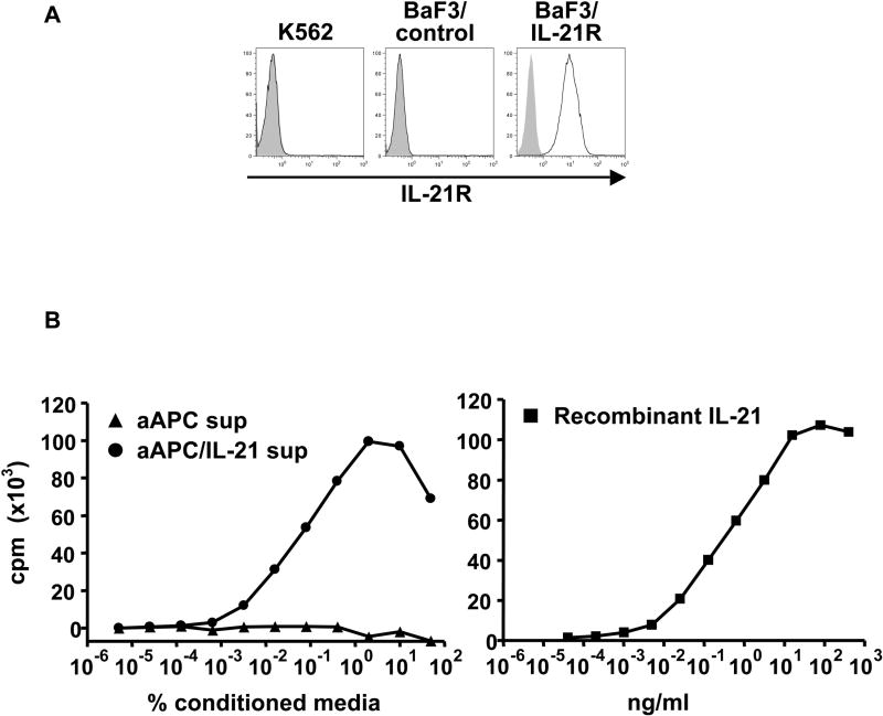
Purpose: Interleukin 21 (IL-21) is a promising new cytokine, which is undergoing clinical testing as an anticancer agent. Although IL-21 provides potent stimulation of CD8(+) T cells, it has also been suggested that IL-21 is immunosuppressive by counteracting the maturation of dendritic cells. The dissociation of these two opposing effects may enhance the utility of IL-21 as an immunotherapeutic. In this study, we used a cell-based artificial antigen-presenting cell (aAPC) lacking a functional IL-21 receptor (IL-21R) to investigate the immunostimulatory properties of IL-21.

Experimental design: The immunosuppressive activity of IL-21 was studied using human IL-21R(+) dendritic cells. Antigen-specific CD8(+) T cells stimulated with human cell-based IL-21R(-)aAPC were used to isolate the T-cell immunostimulatory effects of IL-21. The functional outcomes, including phenotype, cytokine production, proliferation, and cytotoxicity were evaluated.

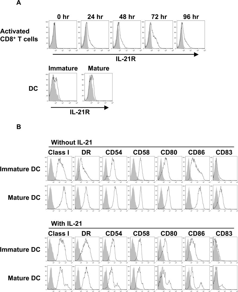
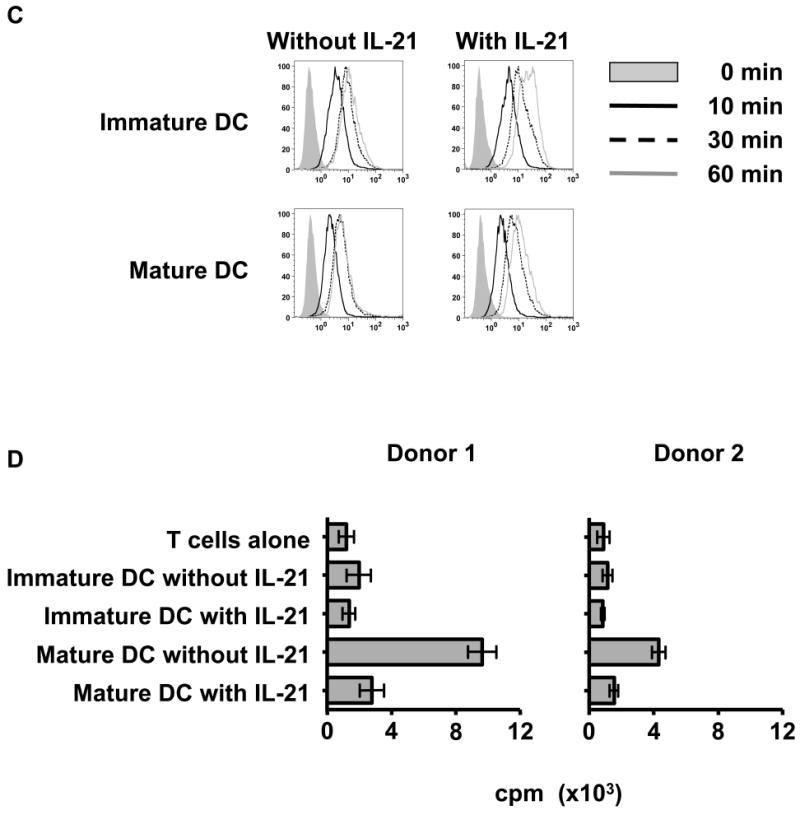
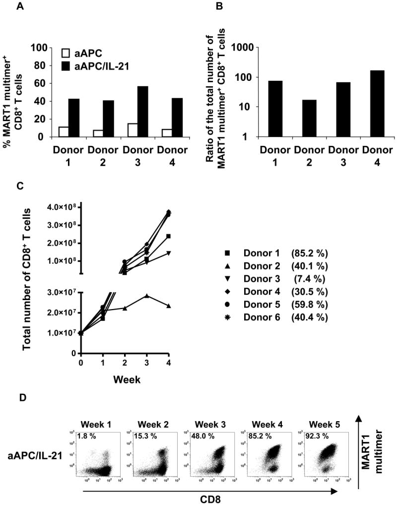
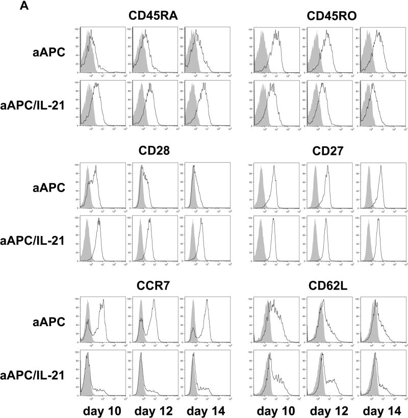
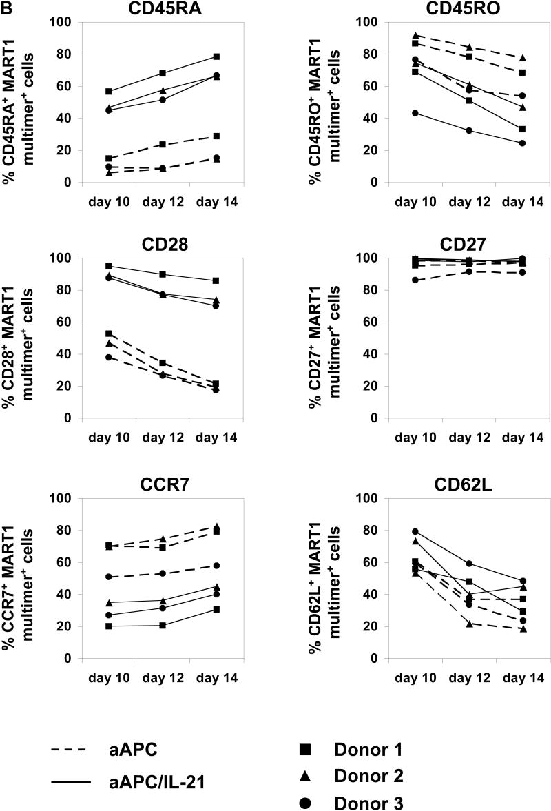
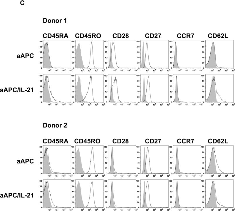
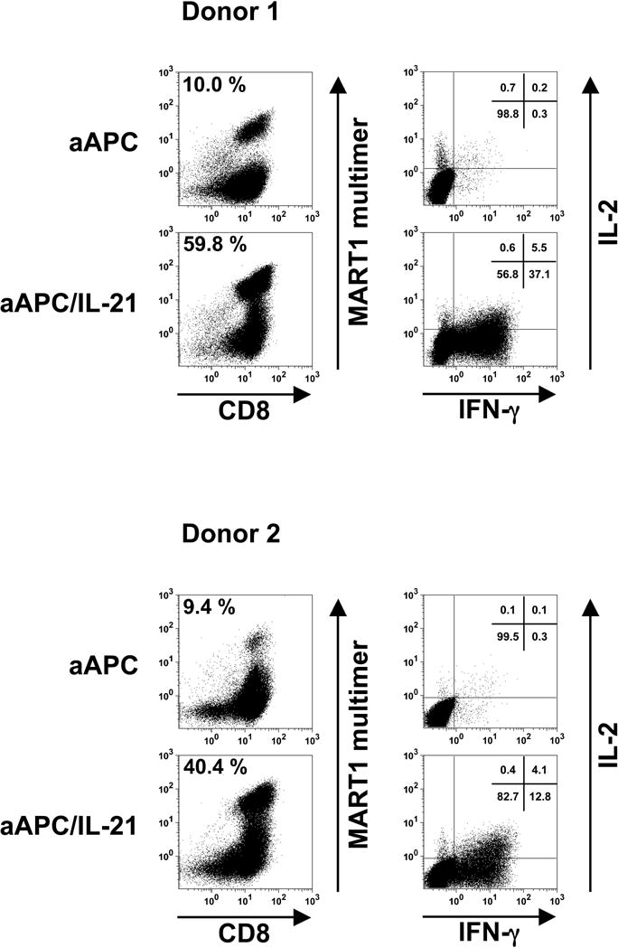
Results: IL-21 limits the immune response by maintaining immunologically immature dendritic cells. However, stimulation of CD8(+) T cells with IL-21R(-) aAPC, which secrete IL-21, results in significant expansion. Although priming in the presence of IL-21 temporarily modulated the T-cell phenotype, chronic stimulation abrogated these differences. Importantly, exposure to IL-21 during restimulation promoted the enrichment and expansion of antigen-specific CD8(+) T cells that maintained IL-2 secretion and gained enhanced IFN-gamma secretion. Tumor antigen-specific CTL generated in the presence of IL-21 recognized tumor cells efficiently, demonstrating potent effector functions.

Conclusions: IL-21 induces opposing effects on antigen-presenting cells and CD8(+) T cells. Strategic application of IL-21 is required to induce optimal clinical effects and may enable the generation of large numbers of highly avid tumor-specific CTL for adoptive immunotherapy.


Figures

Figure 1
Figure 1. IL-21 counteracts the full maturation of DC
(A) IL-21R is not expressed on resting CD8+ T cells but induced upon activation by phorbol myristate acetate and calcium ionophore A23187. In contrast, DC constitutively express IL-21R. Surface expression of IL-21R on purified CD8+ T cells, immature, and mature DC from healthy donors was determined by flow cytometry with specific antibody (open curves) and isotype control (shaded curves). One result of at least 6 (CD8+ T cells) and 4 (DC) independent experiments is shown. (B) IL-21 inhibits the upregulation of adhesion and costimulatory molecules by DC upon exposure to maturation signals. Immature DC were generated from purified monocytes using IL-4 and GM-CSF. Where indicated, DC cultures were also supplemented with rIL-21 at 50 ng/ml every two days. Surface phenotype of immature and mature DC generated in the presence or absence of IL-21 is shown. Open curves, staining for indicated molecules; shaded curves, isotype controls. A representative result of 4 independent experiments is given. (C) IL-21 enhances the endocytic activity of DC. Endocytic activity of immature and mature DC generated in the presence or absence of IL-21 was measured by FITC-dextran uptake and quantified by flow cytometry. Note that DC lose endocytic activity in response to maturation signals despite the presence of IL-21. Shaded curve, 0 min; solid black line, 10 min; dashed line, 30 min; solid grey line, 60 min. Three independent experiments were performed with similar results. (D) T cell stimulatory capacity of IL-21 treated mature DC is markedly suppressed. Purified CD8+ T cells from four healthy donors were cocultured with irradiated allogeneic immature or mature DC that had been generated in the absence or presence of IL-21. Allogeneic CD8+ T cell response was measured using a standard [3H]thymidine proliferation assay. Results from 2 representative donors are shown, and each value displayed is the average of six replicates (±SD). Note that since purified CD8+ T cells are used as responders, the measured values are lower in comparison to values typically obtained when CD4+ or CD3+ T cells are used as responder cells. However, the results obtained for CD8+ T cell proliferation in this assay are reproducible.
Figure 2
Figure 2. Generation of IL-21R negative aAPC constitutively secreting IL-21
(A) K562, the parental cell line of aAPC and aAPC/IL-21, lacks the expression of IL-21R. To create an IL-21 dependent cell line, mouse BaF3 cells were engineered to express human IL-21R. Surface expression of IL-21R on wild-type K562, BaF3 transduced with vector (BaF3 control), and BaF3 cells transduced with human IL-21R (BaF3/IL-21R) was determined by flow cytometry with specific antibody (open curves) and isotype control (shaded curves). (B) aAPC/IL-21 but not aAPC constitutively secretes large amounts of biologically active IL-21. Induction of BaF3/IL-21R proliferation by supernatants derived from aAPC and aAPC/IL-21, and rIL-21 was determined using a standard thymidine proliferation assay. Baseline values for proliferation observed in the absence of conditioned medium or rIL-21 were subtracted. Neither supernatant from aAPC nor aAPC/IL-21 was able to support the growth of BaF3/control cells (data not shown).
Figure 3
Figure 3. Stimulation in the presence of IL-21 induces robust enrichment and expansion of antigen-specific CD8+ T cells
(A) IL-21 augments the expansion and enrichment of antigen-specific CD8+ T cells. Purified CD8+ T cells from HLA-A2+ healthy donors were primed with aAPC pulsed with MART1 peptide in the absence of IL-21. After one week, T cell cultures were split by half and stimulated three times on a weekly basis with either peptide-pulsed aAPC or aAPC/IL-21. Between stimulations T cells were supplemented with IL-2 (10 IU/ml) and IL-15 (10 ng/ml). Resulting T cell cultures were stained with HLA/peptide multimer to determine the percentage of MART1-specific CD8+ T cells. (B) The number of MART1-specific T cells was determined by calculating the product of the total number of T cells and the percentage of multimer positive cells. Shown here is the ratio of the total number of MART1-specific CD8+ T cells generated in the presence versus the absence of IL-21 for each donor. (C) Generation of CTL by both priming and restimulation with MART1 peptide-pulsed aAPC/IL-21 was performed. Purified CD8+ T cells from six A2+ healthy donors were stimulated once a week. Between stimulations T cells were supplemented with IL-2 (10 IU/ml) and IL-15 (10 ng/ml). The calculated total number of CD8+ T cells generated is shown. The percentage of MART1 multimer positive CD8+ T cells after 4 stimulations is also presented. (D) Representative MART1 multimer staining performed weekly after each stimulation is presented (donor 1 from Figure 3C).
Figure 4
Figure 4. Priming in the presence of IL-21 generates antigen-specific CD8+ T cells with distinct phenotype
Purified CD8+ T cells from three A2+ healthy donors were primed with either MART1 peptide-pulsed aAPC or aAPC/IL-21. Without any restimulation, each culture was supplemented with IL-2 (10 IU/ml) and IL-15 (10 ng/ml) every three days. After 10, 12, and 14 days, immunophenotype of MART1–specific T cells was analyzed by flow cytometry. (A) A representative result of one donor out of three is presented. Open curves, staining for indicated molecules; shaded curves, isotype controls. Similar results were obtained using aAPC versus aAPC plus rIL-21. (B) A summary of the phenotypic analysis of MART1 multimer positive T cells from all three donors on all three days studied is shown. (C) The effect of chronic stimulation in the presence of IL-21 on the phenotype of antigen-specific T cells was also evaluated. Purified CD8+ T cells from two different healthy donors were stimulated once a week with either MART1 peptide-pulsed aAPC or aAPC/IL-21. Between stimulations T cells were supplemented with IL-2 (10 IU/ml) and IL-15 (10 ng/ml). After three and five stimulations, MART1 multimer positive T cells were found to have a similar phenotype. Results after 5 stimulations are shown. Open curves, staining for indicated molecules; shaded curves, isotype controls. Two independent experiments were performed with similar results.
Figure 5
Figure 5. IL-21 promotes the acquisition of IFN-γ secretion while preserving IL-2 secretion by antigen-specific CD8+ T cells
Purified CD8+ T cells from A2+ healthy donors were primed and restimulated with either aAPC or aAPC/IL-21 pulsed with MART1 peptide on a weekly basis. Between stimulations T cells were supplemented with IL-2 (10 IU/ml) and IL-15 (10 ng/ml). After a total of four stimulations, T cells were analyzed for antigen specificity by HLA/peptide multimer staining and antigen-specific effector function. The panels in the left column depict MART1 multimer staining and the percentages in the upper left corner indicate MART1 multimer+ cells within CD8+ T cells. The panels in the right column show the antigen-specific intracellular release of IL-2 and IFN-γ on gated CD8+ T cells of the corresponding T cell cultures. The percentages of cells present in each quadrant are shown. Results of two different donors out of three are shown.
Figure 6
Figure 6. IL-21 can enhance the generation of anti-tumor CD8+ T cells with low pCTL frequency and/or growth potential
Survivin-specific CTL were generated using aAPC/IL-21. Purified CD8+ T cells from A2+ healthy donors were stimulated on a weekly basis with aAPC/IL-21 pulsed with Sur9 peptide. Between stimulations T cells were supplemented with IL-2 (10 IU/ml) and IL-15 (10 ng/ml). After 4 stimulations, survivin-specific effector functions were evaluated by IFN-γ ELISPOT and standard 51Cr release assay. (A) Survivin-specific IFN-γ secretion was studied by IFN-γ ELISPOT. CD8+ T cells were incubated with T2 cells pulsed with either Sur9 or HTLV-I-derived TAX control peptide. (B) Cytotoxicity assay was performed using radiolabeled T2 cells pulsed with Sur9 peptide (■) or HIV-derived Pol peptide (●). (C) Sur9 peptide-specific CD8+ T cells generated by aAPC/IL-21 possessed high TCR avidity and recognized hematologic tumor cells in an HLA-A2-restricted manner. Cytotoxicity assay was performed using radiolabeled hematologic cancer cells from different lineages. HLA-A2 negative cell lines were engineered to express HLA-A2 as previously described(26). The cell lines used as targets were HSS (HS-Sultan, myeloma cell line); Sup-T1 (T lymphoblastic lymphoma cell line); Jurkat (T cell leukemia cell line), MEG-01 (megakaryoblastic cell line); and KG-1 (acute myelogenous leukemia cell line).

Similar articles

Cited by

References

    1. Gattinoni L, Powell DJ, Jr, Rosenberg SA, Restifo NP. Adoptive immunotherapy for cancer: building on success. Nature reviews. 2006;6:383–93. - PMC - PubMed
    1. Leen AM, Rooney CM, Foster AE. Improving T cell therapy for cancer. Annual review of immunology. 2007;25:243–65. - PubMed
    1. June CH. Principles of adoptive T cell cancer therapy. J Clin Invest. 2007;117:1204–12. - PMC - PubMed
    1. Banchereau J, Palucka AK. Dendritic cells as therapeutic vaccines against cancer. Nature reviews. 2005;5:296–306. - PubMed
    1. Figdor CG, de Vries IJ, Lesterhuis WJ, Melief CJ. Dendritic cell immunotherapy: mapping the way. Nature medicine. 2004;10:475–80. - PubMed

Publication types

MeSH terms