. 2008 Oct 1;68(19):7947-55.
doi: 10.1158/0008-5472.CAN-08-0971.

ATM and the Mre11-Rad50-Nbs1 complex respond to nucleoside analogue-induced stalled replication forks and contribute to drug resistance

Affiliations

ATM and the Mre11-Rad50-Nbs1 complex respond to nucleoside analogue-induced stalled replication forks and contribute to drug resistance

Brett Ewald et al. Cancer Res. .

Abstract

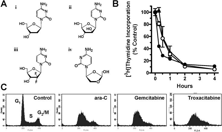
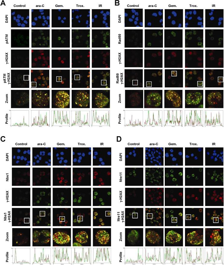
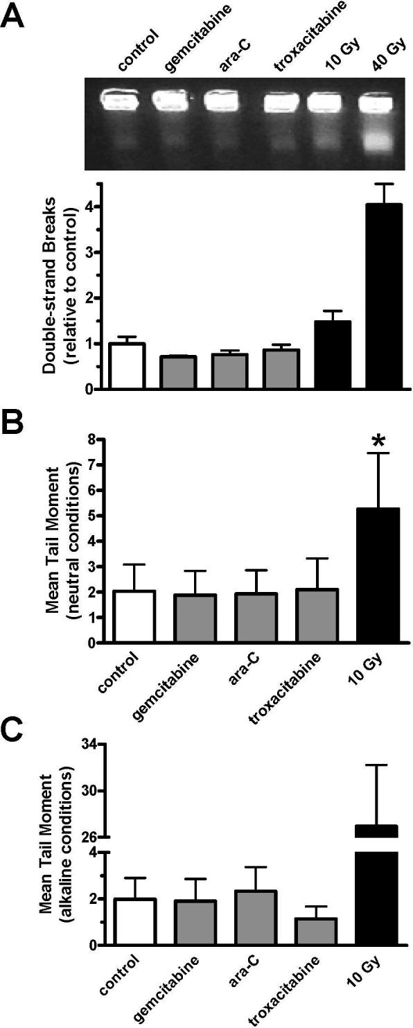
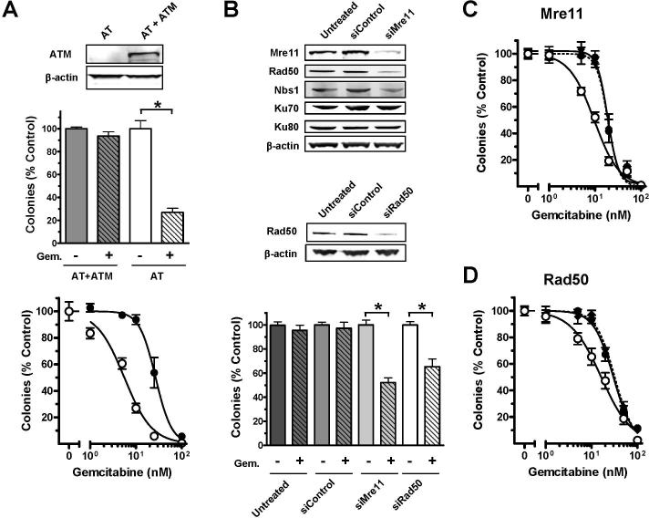
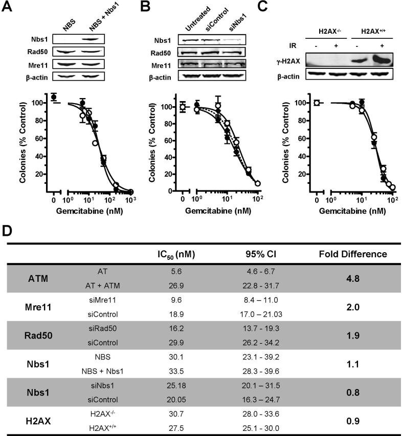
The Mre11-Rad50-Nbs1 complex and autophosphorylated Ser(1981)-ATM are involved in recognizing and repairing DNA damage, such as double-strand breaks (DSB). However, the role of these factors in response to stalled replication forks is not clear. Nucleoside analogues are agents that are incorporated into DNA during replication, which cause stalling of replication forks. The molecular mechanisms that sense these events may signal for DNA repair and contribute to survival but are poorly understood. Cellular responses to both DSBs and stalled replication forks are marked by H2AX phosphorylation on Ser(139) (gamma-H2AX), which forms nuclear foci at sites of DNA damage. Here, concentrations of the nucleoside analogues 1-beta-d-arabinofuranosylcytosine (cytarabine; ara-C), gemcitabine, and troxacitabine, which inhibited DNA synthesis by 90% within 2 hours, were determined for each agent. Using gamma-H2AX as a marker for changes in chromatin structure, we show that Mre11, Rad50, Nbs1, and phosphorylated ATM respond to nucleoside analogue-induced stalled replication forks by forming nuclear foci that colocalize with gamma-H2AX within 2 hours. Because neither DSBs nor single-strand breaks were detectable after nucleoside analogue exposure, we conclude that this molecular response is not due to the presence of DNA breaks. Deficiencies in ATM, Mre11, or Rad50 led to a 2- to 5-fold increase in clonogenic sensitization to gemcitabine, whereas Nbs1 and H2AX deficiency did not affect reproductive growth. Taken together, these results suggest that ATM, Mre11, and Rad50 are required for survival after replication fork stalling, whereas Nbs1 and H2AX are inconsequential.


Figures

Figure 1
Figure 1. The effect of nucleoside analogues on DNA synthesis
(A) Chemical structures of (i) 2’-deoxycytidine, (ii) ara-C, (iii) gemcitabine, and (iv) troxacitabine. (B) Effect of 0.5 μM ara-C (•), 0.1 μM gemcitabine (○), or 2 μM troxacitabine (▲) on DNA synthesis in OCI-AML3 cultures, as measured by [3H]thymidine incorporation after a 30-minute pulse. Error bars represent the standard deviation of the mean. (C) Accumulation of OCI-AML3 cells in early S-phase after exposure to 1 μM ara-C, 10 nM gemcitabine, or 250 nM troxacitabine for 24 h, as measured by DNA content (propidium iodide). Cell populations representing G1, S, and G2/M phases are appropriately marked.
Figure 2
Figure 2. Co-localization of phosphorylated Ser1981-ATM, Rad50, Nbs1, and Mre11 at sites of stalled replication forks
Exponentially growing OCI-AML3 cultures were incubated with 0.5 μM ara-C, 0.1 μM gemcitabine, or 2 μM troxacitabine for 2 h or exposed to 10 Gy ionizing radiation, harvested, and subjected to fluorescent staining. Nuclei were stained with 4',6-diamidino-2-phenylindole (DAPI) and are shown in the upper panels in blue. A single line was randomly drawn through the center of nuclei to generate a red and green fluorescence profile, as shown in the bottom panels. Overlapping peaks illustrate overlapping red/green nuclear foci. Representative images of two independent experiments taken by confocal microscopy of (A) pSer1981-ATM (green)/γ-H2AX (red), (B) Rad50 (green)/γ-H2AX (red), (C) Nbs1 (red)/γ-H2AX (green), and (D) Mre11 (green)/γ-H2AX (red), are shown.
Figure 3
Figure 3. Undetectable levels of DNA breaks after nucleoside analogue exposure
OCIAML3 cultures were incubated with 0.1 μM gemcitabine, 0.5 μM ara-C, or 2 μM troxacitabine for 2 h or exposed to 10−40 Gy ionizing radiation, harvested, and examined for DNA break induction. (A) Representative gel image of DNA stained with ethidium bromide in a typical experiment after PFGE (upper). The lower band represents the fraction of DNA with DSBs. The DNA in the lower band divided by the total DNA in each lane determined by [14C]-labeling, as compared to untreated controls is graphed. Error bars represent the standard error of the mean for two independent experiments performed in duplicate. (B-C) Calculated mean tail moment after exposure to nucleoside analogues or ionizing radiation, as determined by comet assay under neutral (B) or alkaline (C) conditions. Error bars represent the standard deviation of the mean for two independent experiments in which 100 cells per experiment were analyzed. *, P<0.001
Figure 4
Figure 4. Effect of ATM, Mre11, and Rad50 on colony growth after exposure to gemcitabine
(A) Exponentially growing AT and AT+ATM fibroblasts were analyzed for ATM protein level by immunoblotting of whole cell lysates (upper), exposed to 10 nM gemcitabine for 24 h (middle) or a range of gemcitabine concentrations (lower) prior to analysis of clonogenic survival after 10 days. (B-D) Exponentially growing AT+ATM cultures were untreated, transfected with control siRNA, Mre11 siRNA, or Rad50 siRNA. Seventy-two hours after transfection, cells were harvested and split into 2 samples. One sample was lysed and saved for immunoblotting (B). Cells from the other sample were replated and allowed to grow undisturbed for 8 h before being exposed to 10 nM gemcitabine (B, lower) or 1−100 nM gemcitabine (C, D) for 24 h. Then, gemcitabine was washed out of the medium and colonies of untreated (◆), control siRNA (•), and Mre11/Rad50 siRNA (○) were counted after 10 days of undisturbed growth. The mean of two independent experiments performed in triplicate (n=6) ± S.E. is graphed. *, P<0.001. Gem.: 10 nM gemcitabine
Figure 5
Figure 5. Effect of DNA damage response molecules on colony growth after exposure to gemcitabine
Exponentially growing (A) NBS (○) and NBS+Nbs1 (•) human fibroblasts, (B) AT+ATM human fibroblasts that were untreated (◆), transfected with control siRNA (•), or exposed to Nbs1 siRNA (○) prior to drug exposure, and (C) H2AX−/− (○) and H2AX+/+ (•) mouse embryonic fibroblasts were exposed to a range of gemcitabine concentrations for one cell cycle (15−24 h) prior to fresh medium replacement. Cells were allowed to grow undisturbed for 5−10 days in normal growing conditions before colonies of ≥50 cells were counted. Mean of two independent experiments performed in triplicate are shown. Error bars represent the standard error of the mean. IR: 10 Gy ionizing radiation. (D) IC50 values, 95% confidence intervals (CI), and fold differences between compared conditions is summarized.

Similar articles

Cited by

References

    1. Paulsen RD, Cimprich KA. The ATR pathway: fine-tuning the fork. DNA Repair (Amst) 2007;6:953–66. - PubMed
    1. Sampath D, Rao VA, Plunkett W. Mechanisms of apoptosis induction by nucleoside analogs. Oncogene. 2003;22:9063–74. - PubMed
    1. Sampath D, Shi Z, Plunkett W. Inhibition of cyclin-dependent kinase 2 by the Chk1-Cdc25A pathway during the S-phase checkpoint activated by fludarabine: dysregulation by 7-hydroxystaurosporine. Mol Pharmacol. 2002;62:680–8. - PubMed
    1. Ewald B, Sampath D, Plunkett W. H2AX phosphorylation marks gemcitabine-induced stalled replication forks and their collapse upon S-phase checkpoint abrogation. Mol Cancer Ther. 2007;6:1239–48. - PubMed
    1. Shiloh Y. The ATM-mediated DNA-damage response: taking shape. Trends Biochem Sci. 2006;31:402–10. - PubMed

Publication types

MeSH terms