. 2008 Oct 1;68(19):7966-74.
doi: 10.1158/0008-5472.CAN-08-1333.

Autophagy: a novel mechanism of synergistic cytotoxicity between doxorubicin and roscovitine in a sarcoma model

Affiliations

Autophagy: a novel mechanism of synergistic cytotoxicity between doxorubicin and roscovitine in a sarcoma model

Laura A Lambert et al. Cancer Res. .

Abstract

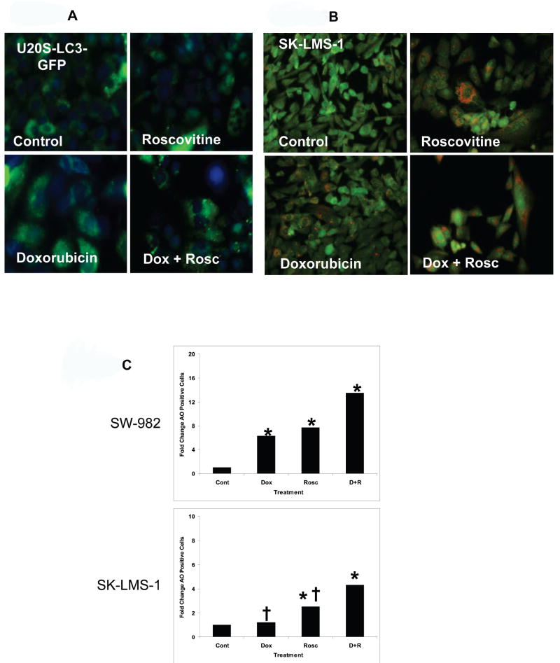
Doxorubicin is a genotoxic chemotherapy agent used in treatment of a wide variety of cancers. Significant clinical side effects, including cardiac toxicity and myelosuppression, severely limit the therapeutic index of this commonly used agent and methods which improve doxorubicin efficacy could benefit many patients. Because doxorubicin cytotoxicity is cell cycle specific, the cell cycle is a rational target to enhance its efficacy. We examined the direct, cyclin-dependent kinase inhibitor roscovitine as a means of enhancing doxorubicin cytotoxicity. This study showed synergistic cytotoxicity between doxorubicin and roscovitine in three sarcoma cell lines: SW-982 (synovial sarcoma), U2OS-LC3-GFP (osteosarcoma), and SK-LMS-1 (uterine leiomyosarcoma), but not the fibroblast cell line WI38. The combined treatment of doxorubicin and roscovitine was associated with a prolonged G(2)-M cell cycle arrest in the three sarcoma cell lines. Using three different methods for detecting apoptosis, our results revealed that apoptotic cell death did not account for the synergistic cytotoxicity between doxorubicin and roscovitine. However, morphologic changes observed by light microscopy and increased cytoplasmic LC3-GFP puncta in U20S-LC3-GFP cells after the combined treatment suggested the induction of autophagy. Induction of autophagy was also shown in SW-982 and SK-LMS-1 cells treated with both doxorubicin and roscovitine by acridine orange staining. These results suggest a novel role of autophagy in the enhanced cytotoxicity by cell cycle inhibition after genotoxic injury in tumor cells. Further investigation of this enhanced cytotoxicity as a treatment strategy for sarcomas is warranted.


Figures

Figure 1
Figure 1
Synergistic effect of doxorubicin and roscovitine in sarcoma cells. (A) HTCA was used to compare the cytotoxic effects of doxorubicin alone, roscovitine alone and doxorubicin followed by roscovitine in SW-982, U2OS-LC3-GFP, SK-LMS-1 and WI38 cell lines (X-axis: doxorubicin (Dox) μM; Y-axis: roscovitine (Rosc) μM; Z-axis: percent cell death). (B) Isobologram analysis demonstrated synergistic interactions in all three sarcoma cell lines. Isobologram analysis and graphs were obtained using CalcuSyn software which performs drug dose-effect calculation using the median effect method described by Chou and Talalay (20). (C and D) Clonogenic assays also demonstrated a synergistic effect between doxorubicin and roscovitine in the three cell lines (Cont = untreated, D = doxorubicin, R = roscovitine, D + R = doxorubicin plus roscovitine). Representative of 3 experiments.
Figure 2
Figure 2
Effect of doxorubicin and roscovitine on cell cycle. (A) SW-982 cells, U2OS-LC3-GFP cells and SK-LMS-1 cells were treated with either doxorubicin (Dox) 0.005μM (SW-982 and U20S-LC3-GFP) or 0.01 μM (SK-LMS-1) for 24 hours, roscovitine (Rosc) 10μM (SW-982 and U20S-LC3-GFP) or 20 μM (SK-LMS-1) for 48 hours, the combination of doxorubicin and roscovitine (D+R) or untreated (Cont). At the completion of drug treatment (T1) and 72-96 hours after the completion of drug treatment (T2), cells were harvested and the cell cycle distribution was determined using fluorescence-activated cell sorting (B). Cell cycle distribution histograms of SK-LMS-1. Representative of 3 experiments.
Figure 3
Figure 3
Morphological changes observed by light microscopy. (A) U2OS-LC3-GFP cells and (B) SK-LMS-1 cells were treated with either doxorubicin 0.005μM (U20S-LC3-GFP) or 0.01μM (SK-LMS-1) for 24 hours, roscovitine 10μM (U20S-LC3-GFP) or 20 μM (SK-LMS-1) for 48 hours, the combination of doxorubicin and roscovitine (Dox+Rosc) or left untreated (Control). At 6 days, the cells were observed by light microscopy. Arrows indicate enlarged cells with prominent micofibrils and multinucleated cells seen in combination treatment.
Figure 4
Figure 4
(A) Fluorescence-activated cell sorting-based apoptosis analyses. SW-982 (upper panel), U20S-LC3-GFP (middle panel) and SK-LMS-1 cells (lower two panels) were treated with either doxorubicin (Dox) 0.005μM (SW-982 and U20S-LC3-GFP) or 0.01 μM (SK-LMS-1) for 24 hours, roscovitine (Rosc) 10μM (SW-982 and U20S-LC3-GFP) or 20 μM (SK-LMS-1) for 48 hours, the combination of doxorubicin and roscovitine (D+R) or untreated (Cont). At 6 days, the cells were harvested and stained with either Annexin V-APC or TUNEL and subjected to fluorescence-activated cell sorting. (*) indicates significant difference as compared with untreated cells (p<0.05); (†) indicates significant difference compared with D+R (p<0.05). Representative of 3 individual experiments. (B) Effect of combined treatment with doxorubicin and roscovitine on apoptosis. SW-982, U20S-LC3-GFP and SK-LMS-1 cells were treated with either doxorubicin (Dox) 0.005 μM (SW-982 and U20S-LC3-GFP) or 0.01 μM (SK-LMS-1) for 24 hours, roscovitine (Rosc) 10 μM (SW-982 and U20S-LC3-GFP) or 20 μM (SK-LMS-1) for 48 hours, the combination of doxorubicin and roscovitine (D+R) or left untreated. Western blot analysis using antibody specific for Parp (full-length and cleaved) was performed at 0 and 72 hours after treatment. Pos Cont: SW-982 cells treated with 4 μM camptothecin for 12 hours. Representative of 3 individual experiments.
Figure 5
Figure 5
Induction of autophagy by combined treatment with doxorubicin and roscovitine. (A) U20S-LC3-GFP cells were treated with either doxorubicin 0.005 μM for 24 hours, roscovitine for 48 hours, the combination of doxorubicin and roscovitine (Dox+Rosc) or left untreated (Control). Cells were observed under fluorescence microscopy to detect the presence of LC3-GFP puncta, which are indicative of autophagy. Representative of 3 separate experiments. (B) SK-LMS-1 cells were treated with either doxorubicin 0.01 μM for 24 hours, roscovitine 20 μM for 48 hours, the combination of doxorubicin and roscovitine (Dox+Rosc) or left untreated (Control). 72 hours after treatment, cells were stained with acridine orange (AO) and observed with fluorescence microscopy to detect the presence of AO puncta. (C) SW-982 and SK-LMS-1 cells were treated with either doxorubicin (Dox) 0.005 μM (SW-982) or 0.01 μM (SK-LMS-1) for 24 hours, roscovitine (Rosc) 10μM (SW-982) or 20 μM (SK-LMS-1) for 48 hours, the combination of doxorubicin and roscovitine (D+R) or left untreated (Cont). 72 hours after treatment cells were stained with acridine orange (AO) and quantified by fluorescence-activated cell sorting. (*) indicates significant difference as compared with untreated cells (p<0.05); (†) indicates significant difference compared with D+R (p<0.05). Representative of 3 separate experiments.
Figure 6
Figure 6
Proposed mechanism of autophagy-mediated synergistic cell death by genotoxic chemotherapy and cyclin dependent kinase inhibition.

Similar articles

Cited by

References

    1. Zahm SH, Fraumeni JF., Jr The epidemiology of soft tissue sarcoma. Semin Oncol. 1997;24:504–14. - PubMed
    1. Gaynor JJ, Tan CC, Casper ES, et al. Refinement of clinicopathologic staging for localized soft tissue sarcoma of the extremity: a study of 423 adults. J Clin Oncol. 1992;10:1317–29. - PubMed
    1. Jaques DP, Coit DG, Hajdu SI, Brennan MF. Management of primary and recurrent soft-tissue sarcoma of the retroperitoneum. Ann Surg. 1990;212:51–9. - PMC - PubMed
    1. Singer S, Demetri GD, Baldini EH, Fletcher CD. Management of soft-tissue sarcomas: an overview and update. Lancet Oncol. 2000;1:75–85. - PubMed
    1. Verweij J, Lee SM, Ruka W, et al. Randomized phase II study of docetaxel versus doxorubicin in first- and second-line chemotherapy for locally advanced or metastatic soft tissue sarcomas in adults: a study of the european organization for research and treatment of cancer soft tissue and bone sarcoma group. J Clin Oncol. 2000;18:2081–6. - PubMed

Publication types

MeSH terms