. 2008 Dec;82(23):11526-35.
doi: 10.1128/JVI.01111-08. Epub 2008 Oct 1.

Excretion of human immunodeficiency virus type 1 through polarized epithelium by immunoglobulin A

Affiliations

Excretion of human immunodeficiency virus type 1 through polarized epithelium by immunoglobulin A

Alison Wright et al. J Virol. 2008 Dec.

Abstract

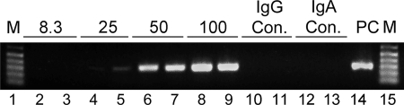
Human immunodeficiency virus (HIV) is transmitted primarily sexually across mucosal surfaces. After infection, HIV propagates initially in the lamina propria below the polarized epithelium and causes extensive destruction of mucosal T cells. Immunoglobulin A (IgA) antibodies, produced in the lamina propria and then transcytosed across the mucosal epithelium into the lumen, can be the first line of immune defense against HIV. Here, we used IgA monoclonal antibodies against HIV envelope proteins to investigate the abilities of polarized primate and human epithelial cells to excrete HIV virions from the basolateral to the apical surface via polymeric Ig receptor (pIgR)-mediated binding and the internalization of HIV-IgA immune complexes. African green monkey kidney cells expressing pIgR demonstrated HIV excretion that was dependent on the IgA concentration and the exposure time. Matched IgG antibodies with the same variable regions as the IgA antibodies and IgA antibodies to non-HIV antigens had no HIV excretory function. A mixture of two IgA anti-bodies against gp120 and gp41 showed a synergistic increase in the level of HIV excreted. The capacity for HIV excretion correlated with the ability of IgA antibodies to bind HIV and of the resulting immune complexes to bind pIgR. Consistent with the epithelial transcytosis of HIV-IgA immune complexes, the colocalization of HIV proteins and HIV-specific IgA was detected intracellularly by confocal microscopy. Our results suggest the potential of IgA antibodies to excrete HIV from mucosal lamina propria, thereby decreasing the viral burden, access to susceptible cells, and the chronic activation of the immune system.


Figures

FIG. 1.
FIG. 1.
Transepithelial excretion of HIV through polarized pIgR+ Vero cells mediated by different IgA antibodies. (A) Detection of HIV after fivefold dilutions (10−5 to 10−8) in lanes 2 to 8. Lanes 1 and 9 are DNA ladders (M, molecular size) identifying the 320-bp amplicon location. (B) HIV excretion ability of each IgA at 100 μg/ml. RT-PCR products from apical samples collected after 8 h are shown in lanes 2 to 13. Irrelevant IgA (IgA control [IgA con.]) is in lanes 12 and 13. A positive control (PC; 10−6 dilution of virus) is shown in lane 14 and in lane 8 in panel C. (C) HIV excretion by IgA MAbs at 300 μg/ml.
FIG. 2.
FIG. 2.
Relative abilities of IgA MAbs to bind to HIV and of HIV-IgA immune complexes to bind to pIgR. Results are expressed as means ± SD. (A) Binding of MAbs (100 μg/ml) to HIV (n = 3). (B) Binding of immune complexes (100 μg/ml MAb) to pIgR (n = 3).
FIG. 3.
FIG. 3.
Synergy of HIV excretion through pIgR+ Vero cells with a combination of D10A (anti-gp41) and D47A (anti-gp120). (A) Viral copy number in virions per milliliter (mean ± SD; n = 2) detected in the apical compartment after 8 h of excretion with D10A or D47A alone and in combination at a final MAb concentration of 100 μg/ml. (B) RT-PCR products comparing virus excretion levels with D10A or D47A at 50 and 100 μg/ml and the D10A/D47A mixture (50 and 50 μg/ml). Lanes 12 to 15 contain the IgG control (con.), a 100-μg/ml mixture of D10G and D47G, and the irrelevant IgA control. The positive control (PC) was as described in the legend to Fig. 1. M, molecular size markers.
FIG. 4.
FIG. 4.
HIV excretion through pIgR+ Vero cells at different IgA antibody concentrations. Apical supernatants were analyzed after 8 h of excretion of basolateral virus from medium containing equal amounts of D10A and D47A at total concentrations of 8.3 (lanes 2 and 3), 25 (lanes 4 and 5), 50 (lanes 6 and 7), and 100 (lanes 8 and 9) μg/ml. IgG and IgA controls (con.) were as described in the legend to Fig. 3. The positive control (PC) was as described in the legend to Fig. 1. M, molecular size markers.
FIG. 5.
FIG. 5.
HIV excretion at different times. Apical samples from pIgR+ Vero cells were analyzed after 2 (lanes 2 and 3), 4 (lanes 4 and 5), 8 (lanes 6 and 7), and 12 (lanes 8 and 9) h of basolateral exposure to HIV immune complexes with D10A/D47A at 100 μg/ml. IgG and IgA controls (con.) at 12 h (lanes 10 to 13) were as described in the legend to Fig. 3. The positive control (PC) was as described in the legend to Fig. 1. M, molecular size markers.
FIG. 6.
FIG. 6.
Infectivity of HIV excreted by nonneutralizing D10A through pIgR+ Vero cells. Shown are the levels (means ± SD; n = 2) of p24 produced by target T cells at different days postinfection during 8-h incubations.
FIG. 7.
FIG. 7.
HIV excretion through polarized pIgR+ cells of human epithelial cell lines. Apical HIV was detected 8 h after the exposure of the monolayers to basolateral HIV mixed with D10A/D47A (100 μg/ml total). Both cell lines, HEC-1A and HT-29, were capable of excreting HIV-IgA immune complexes. Specific IgG and irrelevant IgA controls (con.) were as described in the legend to Fig. 3. The positive control (PC) was as described in the legend to Fig. 1. M, molecular size markers.
FIG. 8.
FIG. 8.
Intracellular colocalization of IgA and HIV protein within polarized epithelial cells after the endocytosis of virus-IgA immune complexes observed by confocal immunofluorescence microscopy. Apical, middle, and basal horizontal sections through the cell monolayers after 8 h of excretion are shown for the red channel (HIV protein), the green channel (IgA), and merged red and green channels. A yellow to orange signal in the merged channel indicates colocalization. Vero C1008 pIgR+ cells and HT-29 pIgR+ cells are shown. Cells transcytosing irrelevant IgA showed no HIV, while cells transcytosing immune complexes of HIV and specific IgA antibody showed the colocalization of HIV protein and IgA at all levels. Bars, 10 μm.

Similar articles

Cited by

References

    1. Alfsen, A., P. Iniguez, E. Bouguyon, and M. Bomsel. 2001. Secretory IgA specific for a conserved epitope on gp41 envelope glycoprotein inhibits epithelial transcytosis of HIV-1. J. Immunol. 1666257-6265. - PubMed
    1. Alfsen, A., H. Yu, A. Magerus-Chatinet, A. Schmitt, and M. Bomsel. 2005. HIV-1-infected blood mononuclear cells form an integrin- and agrin-dependent viral synapse to induce efficient HIV-1 transcytosis across epithelial cell monolayer. Mol. Biol. Cell 164267-4279. - PMC - PubMed
    1. Allaway, G. P., A. M. Ryder, G. A. Beaudry, and P. J. Maddon. 1993. Synergistic inhibition of HIV-1 envelope-mediated cell fusion by CD4-based molecules in combination with antibodies to gp120 or gp41. AIDS Res. Hum. Retrovir. 9581-587. - PubMed
    1. Ambrose, Z., K. Larsen, J. Thompson, Y. Stevens, E. Finn, S. L. Hu, and M. L. Bosch. 2001. Evidence for early local viral replication and local production of antiviral immunity upon mucosal simian-human immunodeficiency virus SHIV89.6 infection in Macaca nemestrina. J. Virol. 758589-8596. - PMC - PubMed
    1. Amerongen, H. M., R. Weltzin, C. M. Farnet, P. Michetti, W. A. Haseltine, and M. R. Neutra. 1991. Transepithelial transport of HIV-1 by intestinal M cells: a mechanism for transmission of AIDS. J. Acquir. Immune Defic. Syndr. 4760-765. - PubMed

Publication types

Substances

LinkOut - more resources