. 2008 Dec;82(24):12335-45.
doi: 10.1128/JVI.01181-08. Epub 2008 Oct 1.

Characterization of simian immunodeficiency virus SIVSM/human immunodeficiency virus type 2 Vpx function in human myeloid cells

Affiliations

Characterization of simian immunodeficiency virus SIVSM/human immunodeficiency virus type 2 Vpx function in human myeloid cells

Caroline Goujon et al. J Virol. 2008 Dec.

Abstract

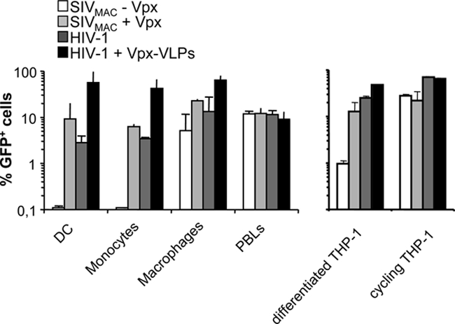
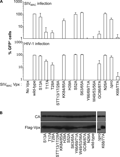
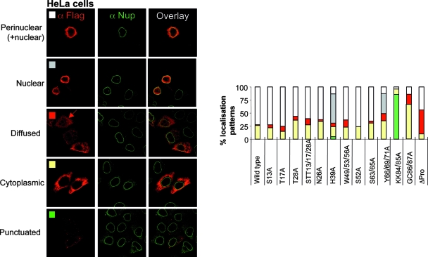
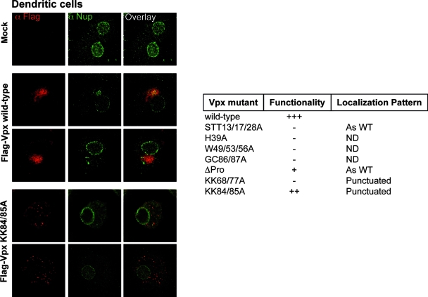
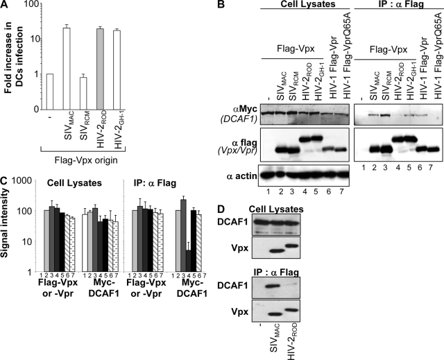
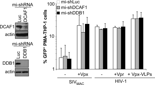
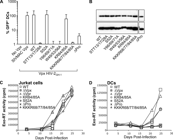
Human immunodeficiency virus type 2 (HIV-2)/simian immunodeficiency virus SIV(SM) Vpx is incorporated into virion particles and is thus present during the early steps of infection, when it has been reported to influence the nuclear import of viral DNA. We recently reported that Vpx promoted the accumulation of full-length viral DNA following the infection of human monocyte-derived dendritic cells (DCs). This positive effect was exerted following the infection of DCs with cognate viruses and with retroviruses as divergent as HIV-1, feline immunodeficiency virus, and even murine leukemia virus, leading us to suggest that Vpx counteracted an antiviral restriction present in DCs. Here, we show that Vpx is required, albeit to a different extent, for the infection of all myeloid but not of lymphoid cells, including monocytes, macrophages, and monocytoid THP-1 cells that had been induced to differentiate with phorbol esters. The intracellular localization of Vpx was highly heterogeneous and cell type dependent, since Vpx localized differently in HeLa cells and DCs. Despite these differences, no clear correlation between the functionality of Vpx and its intracellular localization could be drawn. As a first insight into its function, we determined that SIV(SM)/HIV-2 and SIV(RCM) Vpx proteins interact with the DCAF1 adaptor of the Cul4-based E3 ubiquitin ligase complex recently described to associate with HIV-1 Vpr and HIV-2 Vpx. However, the functionality of Vpx proteins in the infection of DCs did not strictly correlate with DCAF1 binding, and knockdown experiments failed to reveal a functional role for this association in differentiated THP-1 cells. Lastly, when transferred in the context of a replication-competent viral clone, Vpx was required for replication in DCs.


Figures

FIG. 1.
FIG. 1.
Schematic representation of the LVs used here. The LVs used in this study were pseudotyped with the VSVg envelope. HIV-1 and SIVMAC vectors were produced upon the transfection of packaging (8.91 and SIV15, respectively)-, transfer miniviral genome-, and VSVg-encoding DNAs into 293T cells. mi-shRNAs specific for DDB1, DCAF1, or luciferase (Luc) were expressed in the context of an HIV-1 vector from the pAPMmi-shRNA construct, which allows puromycin selection of transduced cells. The structure of the replication-competent viral clone HIV-2GL-AN is also shown. SD, splice donor; SA, splice acceptor; cPPT, central polypurine tract; RRE, Rev-responsive element; U3*, self-inactivating U3.
FIG. 2.
FIG. 2.
Vpx is required during the early steps of infection of myeloid blood cells and in differentiated THP-1 cells. Primary blood monocytes, macrophages, DCs, PHA/IL-2-stimulated PBLs, as well as cycling and differentiated THP-1 cells were compared for their susceptibilities to infection with SIVMAC vectors carrying or not carrying Vpx and for the positive effect of Vpx-VLP preincubation on infection with HIV-1. The different vectors were produced by the transfection of 293T cells and purified through a double-sucrose cushion. They were then normalized for their infectious titer on HeLa cells and used on the indicated cell type at an MOI of 1 for 2 h prior to extensive cell washing. Noninfectious Vpx-VLPs were produced in the absence of the viral genome and were quantified by exo-RT in comparison with standards of known infectivity. When indicated, Vpx-VLPs were added to DCs together with GFP-encoding HIV-1 vectors. The percentage of infected cells was determined 3 days later by flow cytometry or between days 5 and 7 in the case of monocytes. THP-1 cells were differentiated for 24 h with PMA, which induces their macrophage-like differentiation prior to infection. The graph presents data obtained from three independent experiments with cells obtained from different blood donors.
FIG. 3.
FIG. 3.
Identification of point mutations that modulate the functionality of Vpx in DCs. (A) Infectious GFP-encoding SIVMAC vectors incorporating WT or mutant FLAG-Vpx proteins were produced upon the cotransfection of 293T cells and purified through a double-sucrose cushion. The different viral preparations were normalized for their protein contents by exo-RT activity and then used to infect DCs in a single-round infection assay (SIVMAC infection) (top). Similarly, the ability of Vpx-VLPs to increase the infectivity of GFP-encoding HIV-1 vectors (HIV-1 infection) (bottom) was tested in a preincubation assay, as previously described (16). Briefly, Vpx-VLPs incorporating the indicated Vpx mutants were normalized by exo-RT and provided to DCs together with HIV-1. The percentage of infected GFP-positive cells was determined 3 days later by flow cytometry. Each graph presents data obtained from three independent experiments after normalization with WT SIVMAC Vpx. (B) Normalized amounts of SIVMAC virion particles were analyzed by WB using anti-FLAG and anti-capsid antibodies.
FIG. 4.
FIG. 4.
Vpx displays a heterogeneous subcellular localization pattern in HeLa cells. HeLa cells were transfected with WT and mutant FLAG-Vpx expression constructs and analyzed by confocal microscopy 24 h later with anti-FLAG and anti-Nup153 (a nucleoporin localized at the nuclear membrane) antibodies. Pictures representative of the different localization patterns observed here are shown together with a graph presenting their relative distributions. At least 100 cells were scored for each mutant in at least two independent experiments.
FIG. 5.
FIG. 5.
The intracellular localization of Vpx is cell type dependent. DCs were infected with HIV-1-derived LVs encoding WT or mutant FLAG-Vpx proteins. Four to seven days later, DCs were centrifuged on slides, fixed, and stained with anti-FLAG and anti-Nup153 antibodies. The pictures depict typical localization patterns observed here (n = 100 scored cells in at least two independent experiments). The table summarizes the localization pattern observed upon the expression of a larger panel of SIVMAC Vpx mutants. ND, not detected.
FIG. 6.
FIG. 6.
DCAF1 associates with Vpx proteins derived from the SIVSM/HIV-2 and SIVRCM lineages. (A) Normalized amounts of noninfectious VLPs incorporating Vpx proteins derived from SIVMAC, SIVRCM, or two viral clones of HIV-2, namely, HIV-2ROD and HIV-2GH-1, were used in a preincubation assay of DCs prior to infection with a constant amount of HIV-1 vectors encoding GFP. The graph presents the ratio of the relative increase in HIV-1 infection observed with or without the preincubation of DCs with Vpx-VLPs (17). All FLAG-Vpx proteins were efficiently incorporated into VLPs (not shown) (17). (B) 293T cells were transfected with the indicated expression constructs, lysed 36 h posttransfection, and immunoprecipitated with an anti-FLAG antibody. The immunoprecipitates were analyzed by WB using antibodies specific for the Myc and FLAG epitopes. Actin was used to normalize the different cellular lysates. (C) Quantification of the amount of viral proteins was obtained using the Odyssey infrared quantification system. The graph corresponds to data from an average of four independent experiments. (D) THP-1 cells were transduced with FLAG-vpx-carrying HIV-1 vectors and differentiated with PMA prior to immunoprecipitation with an anti-FLAG antibody and analysis by WB.
FIG. 7.
FIG. 7.
DCAF1 and DDB1 are dispensable for the effect of Vpx in differentiated THP-1 cells. DCAF1 and DDB1 (or luciferase [Luc] as a control) were silenced upon the transduction of cycling THP-1 cells with HIV-1 vectors carrying three different microRNA (mi)-based shRNAs per target gene followed by 1 week of selection of transduced cells with puromycin. THP-1 cells were then differentiated with PMA, analyzed by WB, and infected with the indicated viruses. The percentage of GFP-positive cells was measured 3 days later by flow cytometry. The graph presents data obtained with three independent experiments.
FIG. 8.
FIG. 8.
Mutagenesis of the HIV-2GH-1 Vpx protein and cell-type-specific phenotype of Vpx mutants during viral replication. (A) Point mutations previously examined in the context of SIVMAC Vpx were introduced into HIV-2GH-1 FLAG-Vpx and tested in a preincubation experiment with DCs. DCs were infected with Vpx-VLPs incorporating the different HIV-2 Vpx mutants together with gfp-carrying HIV-1 vectors. The percentage of GFP-positive cells was determined 3 days later by flow cytometry. The graph presents data obtained from two independent experiments. (B) The amount of Vpx protein incorporated into virion particles was assessed by WB. A few of these mutations were then transferred in the context of a replication-competent viral clone. (C and D) Jurkat cells (C) and DCs (D) were infected with normalized amounts of WT and Vpx mutant HIV-2GL-AN proviral clones together with a proviral clone bearing deletions in either vpr or vpx (43). Viral replication was measured by exo-RT in the supernatant of infected cultures at different time points after infection.

Similar articles

Cited by

References

    1. Accola, M. A., A. A. Bukovsky, M. S. Jones, and H. G. Göttlinger. 1999. A conserved dileucine-containing motif in p6gag governs the particle association of Vpx and Vpr of simian immunodeficiency viruses SIVmac and SIVagm. J. Virol. 739992-9999. - PMC - PubMed
    1. Akari, H., J. Sakuragi, Y. Takebe, K. Tomonaga, M. Kawamura, M. Fukasawa, T. Miura, T. Shinjo, and M. Hayami. 1992. Biological characterization of human immunodeficiency virus type 1 and type 2 mutants in human peripheral blood mononuclear cells. Arch. Virol. 123157-167. - PubMed
    1. Arfi, V., L. Rivière, L. Jarrosson-Wuillème, C. Goujon, D. Rigal, J.-L. Darlix, and A. Cimarelli. 2008. Characterization of the early steps of infection of primary blood monocytes by human immunodeficiency virus type 1. J. Virol. 826557-6565. - PMC - PubMed
    1. Belshan, M., L. A. Mahnke, and L. Ratner. 2006. Conserved amino acids of the human immunodeficiency virus type 2 Vpx nuclear localization signal are critical for nuclear targeting of the viral preintegration complex in non-dividing cells. Virology 346118-126. - PubMed
    1. Belshan, M., and L. Ratner. 2003. Identification of the nuclear localization signal of human immunodeficiency virus type 2 Vpx. Virology 3117-15. - PubMed

Publication types

MeSH terms

Substances