. 2008 Dec;19(12):5181-92.
doi: 10.1091/mbc.e08-02-0231. Epub 2008 Oct 1.

The scaffold protein POSH regulates axon outgrowth

Affiliations

The scaffold protein POSH regulates axon outgrowth

Jennifer Taylor et al. Mol Biol Cell. 2008 Dec.

Abstract

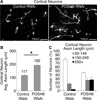
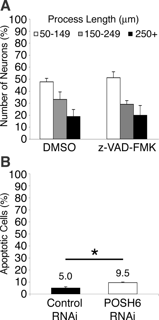
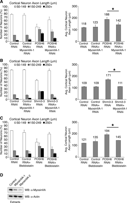
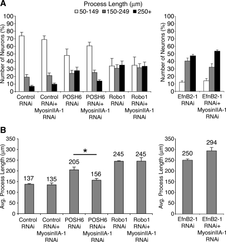
How scaffold proteins integrate signaling pathways with cytoskeletal components to drive axon outgrowth is not well understood. We report here that the multidomain scaffold protein Plenty of SH3s (POSH) regulates axon outgrowth. Reduction of POSH function by RNA interference (RNAi) enhances axon outgrowth in differentiating mouse primary cortical neurons and in neurons derived from mouse P19 cells, suggesting POSH negatively regulates axon outgrowth. Complementation analysis reveals a requirement for the third Src homology (SH) 3 domain of POSH, and we find that the actomyosin regulatory protein Shroom3 interacts with this domain of POSH. Inhibition of Shroom3 expression by RNAi leads to increased process lengths, as observed for POSH RNAi, suggesting that POSH and Shroom function together to inhibit process outgrowth. Complementation analysis and interference of protein function by dominant-negative approaches suggest that Shroom3 recruits Rho kinase to inhibit process outgrowth. Furthermore, inhibition of myosin II function reverses the POSH or Shroom3 RNAi phenotype, indicating a role for myosin II regulation as a target of the POSH-Shroom complex. Collectively, these results suggest that the molecular scaffold protein POSH assembles an inhibitory complex that links to the actin-myosin network to regulate neuronal process outgrowth.


Figures

Figure 1.
Figure 1.
RNAi-mediated reduction of POSH expression leads to enhanced process outgrowth. (A) Inhibition of endogenous POSH by POSH siRNAs. Extracts were prepared from puromycin-selected P19 cells transiently transfected with POSH or control SIBR siRNA expression vectors and a GFP/puromycin expression vector. POSH1, POSH2, and POSH6 RNAi vectors express a single siRNA directed against a unique sequence in POSH. POSH1+2 expresses both POSH1 and POSH2 siRNAs from a single transcript. The RNAi control vector expresses an siRNA that targets luciferase; targeting luciferase has no functional consequences in P19 cells and this serves as a control for off-target effects. Endogenous POSH was detected in extracts by western blotting with an anti-POSH antibody (top). Western blotting for actin, loading control (bottom). (B–F) POSH RNAi enhances process outgrowth in P19 neurons. P19 cells were transiently transfected with expression vectors for the neural bHLH proteins to drive the differentiation program, GFP to mark the transfected cells, and RNAi vectors. Transfected cells were plated to laminin-coated dishes; neurons were fixed 72 h after transfection and stained for neural markers of differentiation. Photographs of neurons were captured with a digital camera and the length of the longest process per cell measured. One hundred cells were analyzed for each RNAi construct per condition per experiment in three independent experiments. (B) Representative images depicting the change in process length between control and POSH RNAi neurons. P19 neurons were transfected with Ngn2, GFP, and control or POSH6 RNAi vectors, fixed, and then stained for neuronal β-tubulin. (C) Average process length (±SD) of POSH RNAi neurons is enhanced relative to control (*p < 0.001, Student's t test). (D) Neurons were also classified on the basis of axon length: 50–149, 150–249, and 250 μm and greater (+), ±SD. The majority of control neurons have short processes (50–149 μm), whereas the population of neurons with long axons (250+ μm) was increased with POSH RNAi (p < 0.0002, Wilcoxon's rank sum test). (E) Representative images of P19 neurons transfected with Ngn1, GFP, and control or POSH1+2 RNAi vectors, fixed, and stained for MAP2. Thus, two different RNAi vectors expressing siRNAs targeting different POSH sequences (POSH6 or POSH1+2) give similar phenotypes, and similar phenotypes are observed with different bHLH proteins (Ngn2 or Ngn1) driving the differentiation program in P19 cells. (F) Representative images of P19 cells transfected with Ngn2, GFP, and control synthetic siRNA duplexes or POSH6 synthetic siRNA duplexes. Similar POSH RNAi phenotypes are induced by synthetic siRNAs (F) or siRNAs expressed in vivo (vector expressed) (B–E).
Figure 2.
Figure 2.
POSH regulates process outgrowth in primary cortical neurons. UI4-control or UI4-POSH RNAi expression vectors were introduced into cortical primary neurons prepared from E14.5 mouse embryos by nucleofection. In the UI4 vectors, a single vector-derived transcript mediates both RNAi and GFP marker expression. Nucleofected cells were plated to laminin–poly-l-lysine–coated dishes, and neurons were fixed and stained for GFP to enhance signal strength 72 h after nucleofection. Photographs of neurons were captured with a digital camera and the length of the longest process per cell measured. (A) Representative images depicting the change in axon length between control and POSH RNAi neurons 72 h after nucleofection. (B) Average process length of POSH RNAi primary cortical neurons is enhanced relative to control (*p < 0.043, Student's t test). (C) Neurons were also classified on the basis of axon length: the majority of control neurons have short processes (50–149 μm), whereas the population of neurons with long axons (250+ μm) was increased with POSH RNAi (p < 0.0002, Wilcoxon's rank sum test).
Figure 3.
Figure 3.
Apoptotic mechanisms do not negatively regulate process length in P19 neurons. (A) Inhibition of cell death with z-VAD-FMK has little effect on process length of P19 control neurons. P19 cells were transfected with Ngn2 and control GFP expression vectors. Transfected cells were plated to laminin-coated dishes. The caspase inhibitor z-VAD-FMK (20 μM) or DMSO (vehicle control) was added 24 h after transfection. Neurons were fixed and stained for GFP 72 h after transfection and process length determined. (B) The number of apoptotic cells is increased in POSH RNAi neurons. A decrease in apoptotic cell number would be expected if POSH RNAi enhanced process length indirectly by promoting survival. P19 cells transfected with Ngn2 and control or POSH RNAi expression vectors were plated to laminin coated dishes, and 72 h after transfection, they were fixed and stained for active caspase-3 and GFP. The percentage of apoptotic cells is the number of transfected active caspase-3–positive cells divided by the number of transfected cells × 100. (*p < 0.015, Student's t test).
Figure 4.
Figure 4.
POSH RING and SH3-3 domains are required for complementation of the POSH RNAi process outgrowth phenotype. (A) Domain structure of POSH and POSH mutants. RING: Really interesting new gene, a conserved feature of a subfamily of E3 ubiquitin ligases. SH3, Src homology 3 protein–protein interaction domain. RBD, Rac binding domain. (B and C) Complementation analysis in P19 neurons indicates a requirement for the POSH RING domain (B) and the POSH SH3-3 domain (C). P19 cells were transfected with Ngn2 and an RNAi complementation vector, which simultaneously allows for RNAi, complementation analysis, and marker expression from a single transcript. POSH6 RNAi targets the 3′-UTR of endogenous POSH, allowing for complementation by expression of the POSH coding sequence, which lacks 3′-UTR sequences. POSH6 RNAi+POSH denotes a vector that expresses POSH6 siRNAs, full-length POSH, and GFP. POSH6 RNAi+POSHΔRING denotes a vector that expresses POSH6 siRNAs, a POSH mutant deleted for the RING domain, and GFP. Vectors expressing full-length or various deletion mutants of POSH (shown in A) were transfected into P19 cells along with Ngn2. Neurons were fixed and stained for GFP, which marks the transfected neurons 70–75 h after transfection. Process length was measured on 100 cells per condition per experiment for two to three independent experiments. The distribution of process lengths (50–149, 150–249, and 250+ μm; left) as well as average process lengths (right) is shown. Pairwise comparisons that are statistically significant (Wilcoxon's rank sum test, left; Student's t test, right): control RNAi, POSH6 RNAi; control RNAi, POSH6RNAi+POSHΔRING; POSH6 RNAi, POSH6RNAi+POSH; POSH6 RNAi+POSH, POSH6RNAi+POSHΔ3/4; POSH6 RNAi, POSH6 RNAi+POSHΔ4.
Figure 5.
Figure 5.
POSH and Shroom3 coassociate. (A) A yeast two-hybrid screen identified Shroom3 as an interacting partner for POSH. Alignment showing the domain structure of Shroom3L (aa 1-1986) and Shroom3S (aa 177-1986), the location of the Shroom3 two-hybrid isolates, and the number of times each class of isolate was recovered from the screen. PDZ, PSD-95/Dgl/ZO-1 (aa 1-106). ASD1/2, Apx/Shroom domain 1/2 (aa 927-1028 and aa 1659–1983, respectively). ABD, F-actin binding domain (aa 754-952). EVH1 BS, Enabled/vasodilator-stimulated phosphoprotein homology 1 domain binding site (aa 1533-1539). PDZ BS, PDZ domain binding site (aa 1983-1986). (B) Direct interaction between POSH and Shroom3. GST-POSH SH3-3 or GST and His epitope-tagged Shroom3 POSH binding domain (PBD) fusion proteins were purified from bacteria, allowed to bind in vitro, then His-Shroom3 was immunoprecipitated (IP). His-Shroom3 and coassociated GST proteins were detected by Western blot analysis (WB) with antibodies directed against the epitope tags after SDS-PAGE. (C) Endogenous POSH and Shroom3 coassociate. Shroom3 was immunoprecipitated from E16.5 mouse embryonic brain extracts (left) or IMR32 human neuroblastoma cells (right). An irrelevant goat polyclonal antibody was used as a specificity control (Ctrl). After SDS-PAGE, Western blot analysis was used to detect Shroom3 and coassociated POSH.
Figure 6.
Figure 6.
Shroom3 regulates axon outgrowth in cortical primary neurons. (A) RNAi-mediated reduction of Shroom3. Extracts were prepared from puromycin selected P19 cells transiently transfected with Shroom3 or control RNAi expression vectors and a GFP/puromycin expression vector. Shrm3-3 targets a sequence in the 3′-UTR region of Shroom3, and is effective in reducing endogenous Shroom3 expression. Endogenous Shroom was detected in extracts by Western blotting with an anti-Shroom3 antibody (top). Western blotting for actin, loading control (bottom). (B and C) RNAi-mediated reduction of Shroom3 enhances axon outgrowth of primary cortical neurons. Control, POSH1+2, or Shrm3-3 RNAi expression vectors were introduced into cortical primary neurons prepared from E14.5 mouse embryos by nucleofection; a GFP expression vector was included to identify the transfected cells. Process length was measured on fixed, stained GFP-labeled neurons 3 and 4 d after nucleofection. (B) Shroom3 RNAi increases the percentage of neurons with long processes relative to control (p < 0.0002, two-tailed Wilcoxon's rank sum test). (C) Shroom3 RNAi also increases the average process length relative to control neurons (*p < 0.0001, Student's t test).
Figure 7.
Figure 7.
Functional domains of Shroom3: requirement for ASD1/actin binding and ASD2/myosin regulatory domains. (A) Expression of dominant-negative Shroom3 ASD1/actin binding domain enhances axon outgrowth in primary cortical neurons. Primary cortical neurons were nucleofected with expression vectors for the ASD1/actin binding domain of Shroom3 or a vector control. The ASD1/ABD domain of Shroom3 functions as a dominant negative to block binding of endogenous Shroom3 to actin. GFP-positive neurons were fixed and stained 72 h after nucleofection, and process length was measured. The population of neurons with long axons (250 μm and greater) (p < 0.0004, Wilcoxon's rank sum test) as well as average process length (*p < 0.0001, Student's t test) was increased relative to control neurons upon expression of the ASD1/ABD domain, suggesting that the interaction of Shroom3 with actin is required for Shroom3 to function in process outgrowth inhibition. (B) Shroom3 RNAi complementation analysis: requirement for ASD2 domain. Primary cortical cells were nucleofected with expression vectors for wild-type or mutant Shroom3, GFP, and a Shroom3 RNAi vector. The Shroom3 RNAi vector expresses Shrm3-3, an siRNA that targets the 3′-UTR of endogenous Shroom3. The expression vectors for wild-type or mutant Shroom3 include only coding regions and, therefore, are not targeted by the RNAi construct. GFP-labeled neurons were fixed and stained 3 d after nucleofection, and process length was measured. Expression of wild-type Shroom3S (Shrm), but not Shroom3S deleted for the ASD2 domain (ShrmΔASD2), complements the Shroom3 RNAi-enhanced process outgrowth phenotype in primary cortical neurons, indicating a functional requirement for the Shroom3 ASD2 domain during process outgrowth inhibition. Left, p < 0.003, Wilcoxon's rank sum test, Shrm3 RNAi versus Shrm3 RNAi+Shrm. Right, *p < 0.001, Student's t test.
Figure 8.
Figure 8.
Interference of ROCK interaction with Shroom3 leads to enhanced process outgrowth. P19 cells were transfected with a control vector, a POSH6 RNAi vector, or a vector expressing the Shroom3 binding region of hROCK1, denoted R1-C1. 54 h after transfection, differentiated P19 cells were treated with and without 10 μM of the ROCK inhibitor Y-27632 for 19 h. Cells were fixed, stained, and process length was measured. Ectopic expression of R1-C1 leads to enhanced process outgrowth compared with control neurons, similar to enhanced process outgrowth induced by RNAi-mediated reduction of POSH function. Ectopic expression of R1-C1 increases the population of neurons with long axons (250 μm) and decreases the population of neurons with short axons (50–149 μm), and average process length is increased as well (B). Blocking ROCK function in control neurons with Y-27632 also enhances process outgrowth, increasing the population of neurons with long axons and average process length. R1-C1–expressing or POSH RNAi neurons exhibit enhanced process outgrowth upon addition of Y-27632, but the response to the ROCK inhibitor is attenuated. This suggests that ROCK regulates process length in both a POSH–Shroom-dependent and -independent manner. (A) Distribution of process length. Wilcoxon's rank sum test: control ± Y-27632, control versus R1-C1, control versus POSH RNAi, p < 0.0002; R1-C1 ± Y-27632, POSH RNAi ± Y-27632, p < 0.003. (B) Average process length. * and control versus R1-C1, p < 0.0001, Student's t test.
Figure 9.
Figure 9.
Inhibition of myosin II decreases process length in POSH and Shroom3 RNAi neurons. (A and B) RNAi-mediated reduction in myosin IIA function reverses the POSH (A) or Shroom3 (B) RNAi-mediated enhanced process outgrowth phenotype in primary cortical neurons. Primary cortical progenitors were nucleofected with control, POSH, or Shroom3 RNAi vectors, with and without an RNAi vector that reduces myosin IIA function. Cells were cultured for 72 h on poly-l-lysine-laminin–coated coverslips, fixed, stained, imaged, and process length of GFP-labeled neurons was determined. Reduction of myosin IIA function shifts the distribution of axon lengths to control levels and decreases average process length to the levels of the control neurons for both POSH RNAi (A) and Shroom3 RNAi neurons (B). (C) Treatment of primary cortical neurons with the myosin II inhibitor blebbistatin reverses the POSH RNAi phenotype. Embryonic primary cortical progenitors were nucleofected with control or POSH RNAi vectors and cultured for 72 h on poly-l-lysine-laminin. At 72 h, the cultures were treated with DMSO (vehicle control) or blebbistatin, a pharmacological inhibitor of myosin II function (50 μM) for 2 h. Process length was determined on GFP-labeled fixed, stained neurons. Blebbistatin treatment alters the process length distribution of POSH RNAi neurons, shifting process distribution to levels of control neurons (C, left). Blebbistatin treatment also decreases the average process length of POSH RNAi neurons, to control levels (C, right). (A–C) Pairwise comparisons [POSH6 RNAi, POSH6 RNAi+myosin IIA RNAi; Shrm3-3 RNAi, Shrm3-3 RNAi+myosin IIA RNAi, POSH6 RNAi, POSH6 RNAi+blebbistatin] are statistically significant: Wilcoxon's rank sum test (left, A–C; p < 0.0001) and Student's t test (right, A–C; *p < 0.0001). (D) RNAi-mediated reduction of myosin IIA. Extracts were prepared from puromycin selected P19 cells transiently transfected with myosin IIA or control RNAi expression vectors and a GFP/puromycin expression vector. Myosin IIA-1/2 are two independent RNAi constructs targeting different sequences. Endogenous myosin IIA was detected in extracts by Western blotting with an anti-myosin IIA–specific antibody (top). Western blotting for actin, loading control (bottom).
Figure 10.
Figure 10.
RNAi-mediated reduction of myosin IIA selectively attenuates POSH RNAi long process outgrowth phenotype and does not attenuate the long process outgrowth phenotype of RNAi-mediated decrease in the axon guidance cues Robo1 and EphrinB2 (EfnB2). P19 cells were transfected with expression vectors for Ngn2 and RNAi vectors targeting POSH, Robo, EphrinB2, with and without a second RNAi vector targeting myosin IIA. Cells were cultured on laminin for 72 h, fixed, stained, and process length of GFP-labeled transfected neurons was determined. Robo and Ephrin B2 RNAi neurons exhibit enhanced process length, consistent with their roles as regulators of process outgrowth. No significant changes in distribution of process lengths (A) or average process lengths (B) are evident when myosin IIA function is reduced in the Robo or EphrinB2 RNAi neurons, in contrast to POSH RNAi neurons (*p < 0.0001, Student's t test).

References

    1. Alroy I., et al. The trans-Golgi network-associated human ubiquitin-protein ligase POSH is essential for HIV type 1 production. Proc. Natl. Acad. Sci. USA. 2005;102:1478–1483. - PMC - PubMed
    1. Brown J., Bridgman P. C. Role of myosin II in axon outgrowth. J. Histochem. Cytochem. 2003;51:421–428. - PubMed
    1. Chantler P. D., Wylie S. R. Elucidation of the separate roles of myosins IIA and IIB during neurite outgrowth, adhesion and retraction. IEE Proc. Nanobiotechnol. 2003;150:111–125. - PubMed
    1. Chung K. H., Hart C. C., Al-Bassam S., Avery A., Taylor J., Patel P. D., Vojtek A. B., Turner D. L. Polycistronic RNA polymerase II expression vectors for RNA interference based on BIC/miR-155. Nucleic Acids Res. 2006;34:e53. - PMC - PubMed
    1. Dietz M. L., Bernaciak T. M., Vendetti F., Kielec J. M., Hildebrand J. D. Differential actin-dependent localization modulates the evolutionarily conserved activity of Shroom family proteins. J. Biol. Chem. 2006;281:20542–20554. - PubMed

Publication types

MeSH terms

LinkOut - more resources