Comparative Study
. 2008 Oct 1;28(40):9953-68.
doi: 10.1523/JNEUROSCI.0880-08.2008.

Role of GluR1 in activity-dependent motor system development

Affiliations
Comparative Study

Role of GluR1 in activity-dependent motor system development

Lei Zhang et al. J Neurosci. .

Abstract

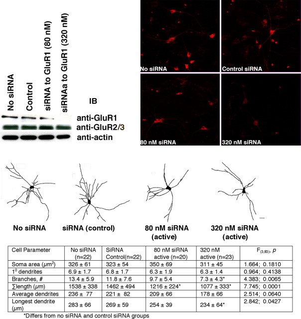
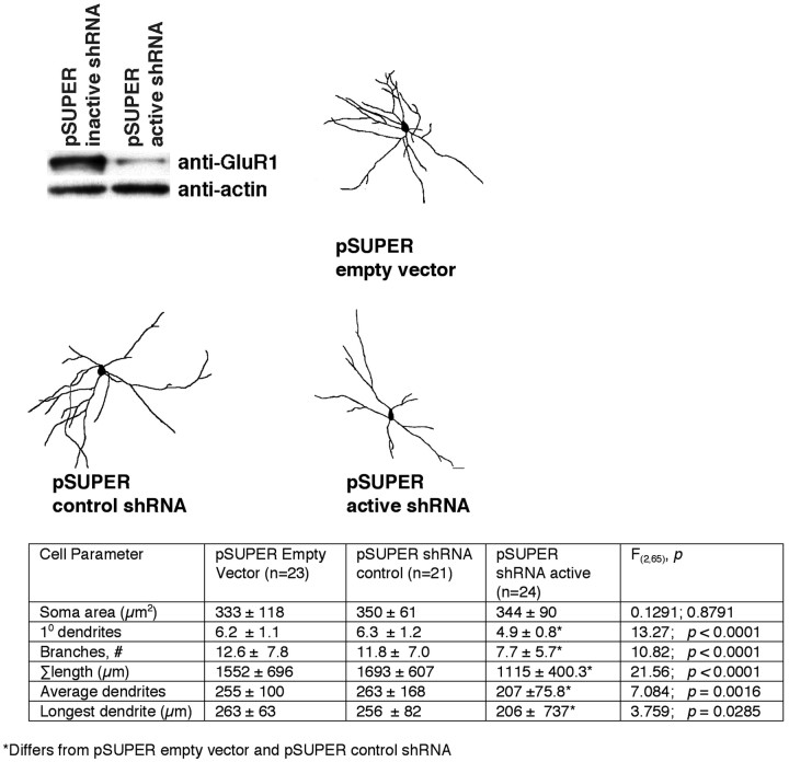
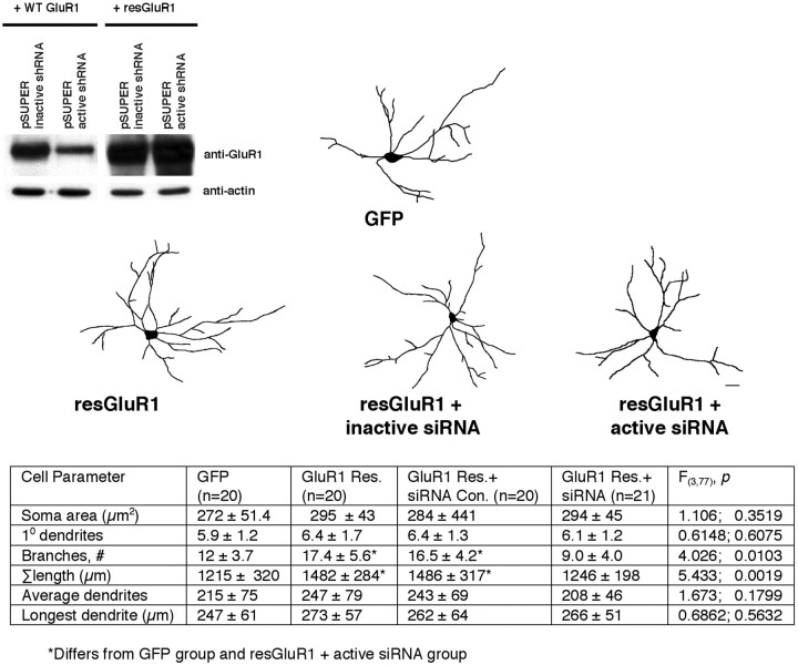
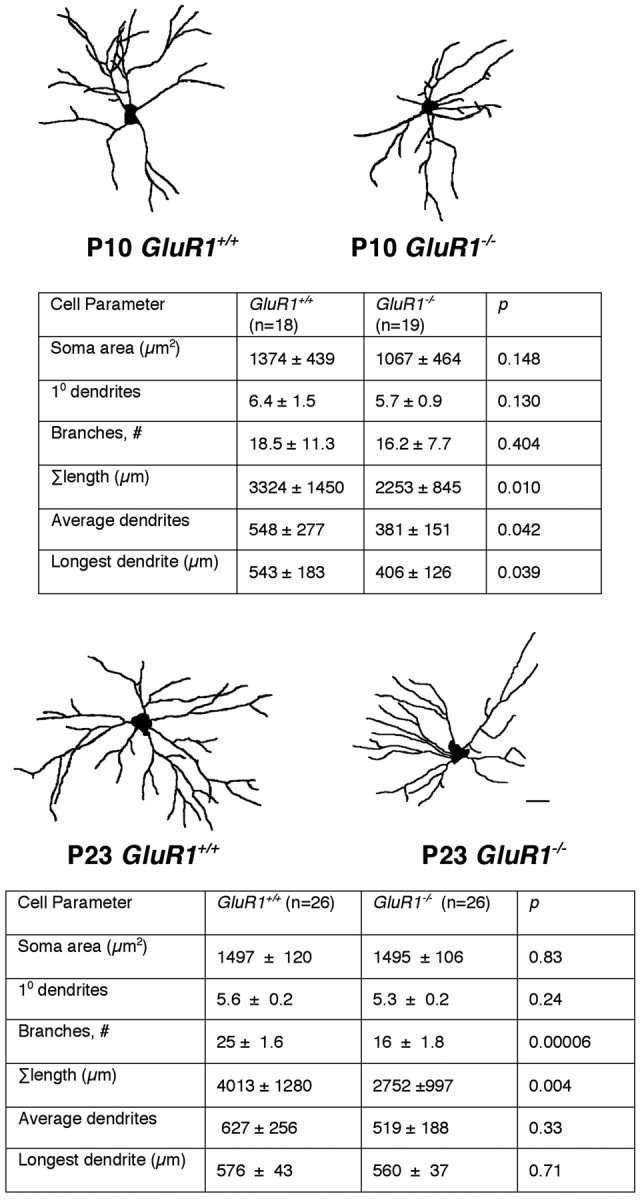
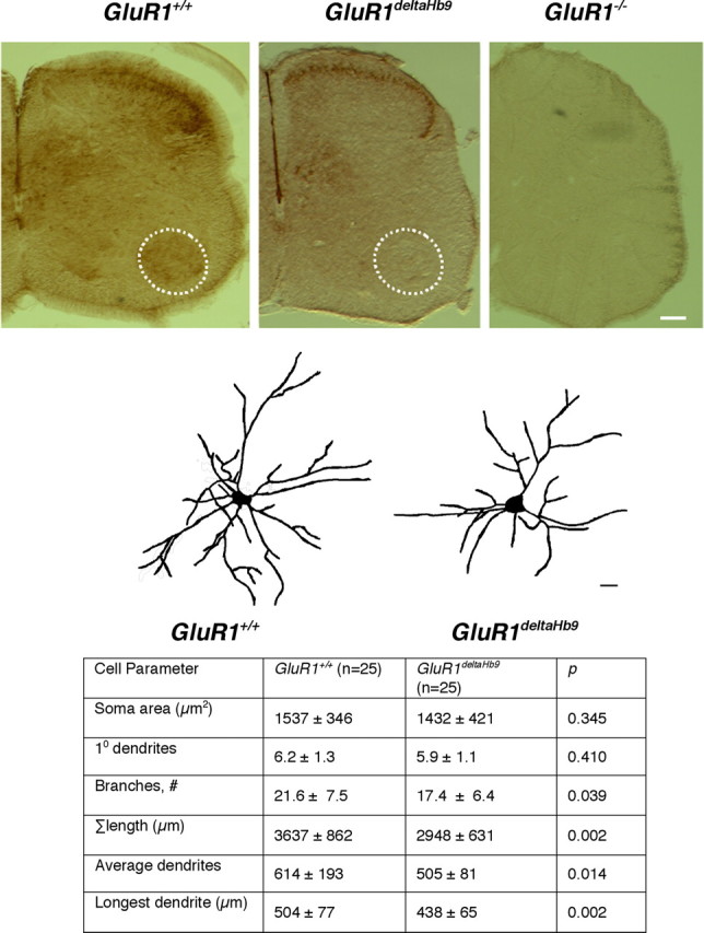
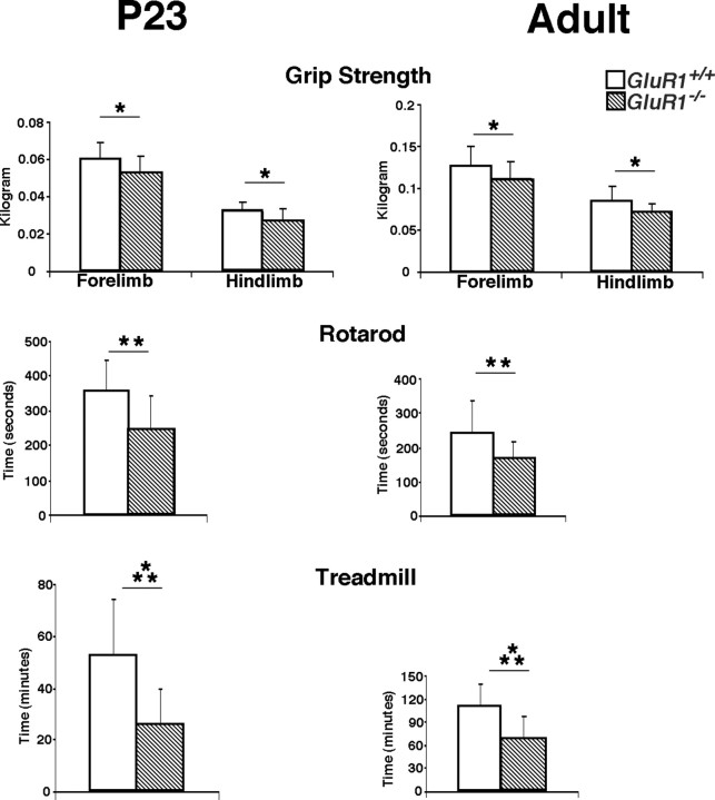
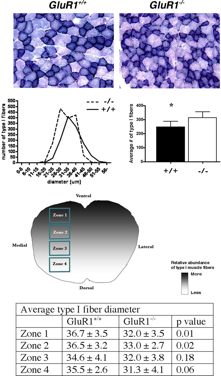
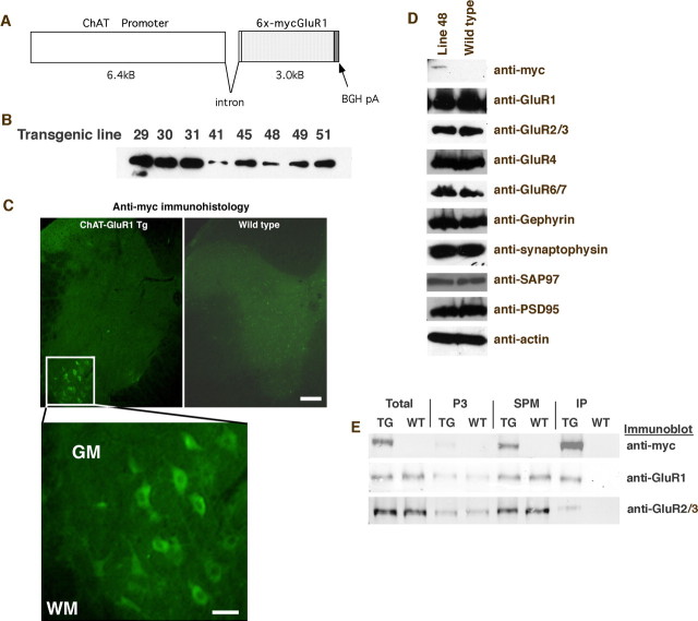
Activity-dependent specification of neuronal architecture during early postnatal life is essential for refining the precision of communication between neurons. In the spinal cord under normal circumstances, the AMPA receptor subunit GluR1 is expressed at high levels by motor neurons and surrounding interneurons during this critical developmental period, although the role it plays in circuit formation and locomotor behavior is unknown. Here, we show that GluR1 promotes dendrite growth in a non-cell-autonomous manner in vitro and in vivo. The mal-development of motor neuron dendrites is associated with changes in the pattern of interneuronal connectivity within the segmental spinal cord and defects in strength and endurance. Transgenic expression of GluR1 in adult motor neurons leads to dendrite remodeling and supernormal locomotor function. GluR1 expression by neurons within the segmental spinal cord plays an essential role in formation of the neural network that underlies normal motor behavior.


Figures

Figure 1.
Figure 1.
Reduction in GluR1 expression in neurons with siRNA-penetratin inhibits dendrite growth. Spinal cord cultures were treated with vehicle (no siRNA), a scrambled-sequence siRNA (control), or active siRNA at 80 or 320 nm (linked to penetratin) for 5 d. The top left shows Western blots of lysates from treated cultures. The active siRNA at 320 nm (and to a lesser degree at 80 nm) leads to a specific reduction in the abundance of GluR1, but not GluR2/3 or actin. To the right are immunocytological images of GluR1-stained neurons in cultures treated with the labeled reagents. Below are representative camera lucida drawings of neurons from cultures treated with active siRNA (2 different concentrations), the control siRNA, or vehicle. Scale bar, 20 μm. Quantitative morphometry is displayed in the table below the camera lucida images. In the cell parameter row, n in each column refers to the number of cells drawn. By ANOVA, there are statistically significant reductions of total dendrite length in the 80 and 320 nm active siRNA treatment groups compared with the no siRNA and control siRNA groups. There is a statistically significant reduction in branches and length of longest dendrite in the 320 nm active siRNA group compared with the other groups. There are no statistically significant differences between the no siRNA versus control siRNA groups.
Figure 2.
Figure 2.
Reduction in GluR1 expression in neurons with pSUPER shRNA inhibits dendrite growth. HEK293 cells were transfected with pSUPER-scrambled shRNA (control) or pSUPER-active shRNA plus the cDNA for GluR1 in a mammalian expression vector (pGW). Western blots of lysates 2 d after transfection (top left) demonstrate the specific reduction in GluR1 abundance cells expressing the active shRNA. To the right and below are representative camera lucida drawings of neurons from spinal cord cultures treated with pSUPER-empty vector, pSUPER-scrambled shRNA (control), or pSUPER-active shRNA for 5 d. Scale bar, 20 μm. Quantitative morphometry is displayed in the table below the camera lucida images. In the cell parameter row, n in each column refers to the number of cells drawn. By ANOVA, there are statistically significant reductions in the number of primary dendrites, branches, overall tree size, average dendrites, and longest dendrites in the pSUPER active shRNA group compared with other two groups.
Figure 3.
Figure 3.
Expression of GluR1 in neurons enhances dendrite growth, but only if neighboring neurons also express GluR1. HEK293 cells were transfected with an expression vector containing wild-type (WT) GluR1 or resGluR1 in which the coding sequence has been altered to prevent perfect binding of the active siRNA to the mRNA. Cotransfected with these plasmids was either pSUPER with the inactive shRNA or pSUPER with the active shRNA. In the upper left, Western blots of lysates from transfected cells are shown. The active shRNA knocks down the expression of WT but not resGluR1. There is no effect on the expression of actin. To the right and below are representative camera lucida images of neurons expressing GFP alone or in combination with resGluR1, resGluR1 plus inactive siRNA, or resGluR1 plus active siRNA. Quantitative morphometry is displayed in the table below the camera lucida images. In the cell parameter row, the n in each column refers to the number of cells drawn. By ANOVA, there is a statistically significant increase in branches and overall tree size in the resGluR1 and resGluR1 plus inactive siRNA groups compared with GFP or resGluR1 plus active siRNA groups. There are no statistical differences between the GFP versus the resGluR1 plus active siRNA group.
Figure 4.
Figure 4.
Comparison of motor neuron dendrites from GluR1 +/+ versus GluR1 −/− animals at P10 and P23. Representative camera lucida drawings from mice of specified age and genotype are displayed above the tables. Scale bar, 30 μm. At P10, there is a statistically significant (by Student's t test) reduction in overall tree size and longest dendrite and average dendrite length in the GluR1 −/− animals compared with GluR1 +/+ animals. At P23, there is a statistically significant (by Student's t test) reduction in branches and overall tree size in the GluR1 −/− animals compared with GluR1 +/+ animals.
Figure 5.
Figure 5.
A comparison of the number and distribution of premotor interneurons in GluR1 −/− versus GluR1 +/+ mice. A diagram of the Rexed's lamina in the lumbar spinal cord and a low-power image of PRV-GFP-positive cells at P10. Three-dimensional bar graphs summarize the quantitative results (axes: number of cells × Rexed's lamina × lumbar segment). Red columns indicate no differences between the GluR1 −/− and GluR1 +/+ animals. The light blue column indicates a statistically significant increase in the number of neurons in the GluR1 +/+, and the dark blue column indicates a statistically significant decrease in the number of neurons in the GluR1 −/− tissues. The greatest differences between genotypes were found in the number of premotor interneurons contralateral to the injection site, especially in Rexed's lamina VIII. Bottom, The primary data are presented in table format. Ipsi, Ipsilateral; contra, contralateral.
Figure 6.
Figure 6.
Comparison of motor neuron dendrites from GluR1 +/+ versus GluR1deltaHb9 animals at P23. The top three panels are representative photomicrographs of P10 spinal cords immunostained with anti-GluR1 from GluR1 +/+, GluR1deltaHb9 and GluR1 −/− mice. Motor neurons residing in Rexed's layer IX (dashed circle in the ventral horn) are stained in the GluR1 +/+ animals but not in the GluR1deltaHb9 and GluR1 −/− mice. There are no other clear differences between the GluR1 +/+ and the GluR1deltaHb9 mice. As expected, there is no immunoreactivity in the GluR1 −/− mice. Scale bar, 125 μm. Middle panels, Representative camera lucida drawings from P23 mice of a specified genotype are displayed above the table. Scale bar, 25 μm. Bottom, There is a statistically significant (by Student's t test) reduction in branches, overall tree size, longest dendrite, and average dendrite length in the GluR1deltaHb9 animals in comparison with GluR1 +/+ animals.
Figure 7.
Figure 7.
Comparison of motor behavior in GluR1 +/+ versus GluR1 −/− animals. The two genotypes were studied at P23 and adulthood on three motor tasks: grip strength (forelimb and hindlimb), rotarod, and treadmill. Note the change in y-axis scale at different ages. *p < 0.05; **p < 0.01; ***p < 0.001. The GluR1 +/+ animals (open bars) perform better than the GluR1 −/− animals (striped bars) in a statistically significant manner in all tests.
Figure 8.
Figure 8.
Comparison of gastrocnemius muscle from GluR1 +/+ and GluR1 −/− mice. Top pictures show stained muscle from animals of each genotype and qualitatively demonstrate the reduction in size and increase in number of darkly stained (type I) fibers in the GluR1 −/− versus GluR1 +/+ animals. Below, a frequency-distribution histogram reveals a leftward shift in the GluR1 −/− versus GluR1 +/+ animals, indicating more smaller type I fibers in the GluR1 −/− animals. The bar graph to the right demonstrates a statistically significant increase in type I fibers in the GluR1 −/− versus the GluR1 +/+ animals. *p < 0.05. Below is a diagram of a cross section of the gastrocnemius muscle and the relative distribution of type I fibers displayed. There are more type I fibers in the ventral (top) versus the dorsal (bottom) portion of muscle, and the location of the four zones we analyzed are shown in boxes. The table below shows the comparison of fiber I fiber diameter of GluR1 +/+ versus GluR1 −/− mice in zones 1–4. Type I fibers were statistically significantly small in the GluR1 −/− animals in the zones enriched with type I fibers (zones 1 and 2).
Figure 9.
Figure 9.
A transgenic mouse that expresses GluR1 in adult motor neurons. A, The construct used to make the transgenic (TG) mouse uses 6.4 kB of the ChAT promoter, followed by an intron, the 6-myc-tagged GluR1(Q)flip cDNA followed by the BGH polyadenylation sequence. B, Eight mouse lines incorporated the transgene into the germ line, and Western blots of spinal cord tissue for the myc tag showed varying levels of transgene expression. Because of the relatively low level of expression in line 48, these mice were chosen for further study. C, Immunohistology of 3-month-old line 48 mice with anti-myc antibody showed that the transgene is expressed in motor neurons. Scale bars: top, 150 μm; bottom, 50 μm. D, Western blot analysis of spinal cord lysates from wild-type (WT) and line 48 mice showed no large scale changes in the expression of numerous proteins. E, Subcellular fractionation of spinal cord lysate from line 48 and wild-type mice. Myc immunoreactivity is found only in line 48 mice in the total lysate, the P3, and the SPM fraction. Myc immunoreactivity is enriched in synaptic membranes. Both GluR1 and GluR2/3 immunoreactivity are similarly seen in total lysate, the P3, and the SPM fraction with SPM enrichment. Immunoprecipitation of the SPM fraction using anti-myc brought down the myc-tagged protein from line 48 but not wild-type homogenates. The immunoprecipitated material is enriched for GluR1 (consistent with IP of the myc-GluR1 construct) and, to a lesser extent, GluR2/3. WM, White matter; GM, gray matter.

Similar articles

Cited by

References

    1. Altman J, Sudarshan K. Postnatal development of locomotion in the laboratory rat. Animal Behav. 1975;23:896–920. - PubMed
    1. Ausoni S, Gorza L, Schiaffino S, Gundersen K, Lømo T. Expression of myosin heavy chain isoforms in stimulated fast and slow rat muscles. J Neurosci. 1990;10:153–160. - PMC - PubMed
    1. Bannerman DM, Deacon RM, Brady S, Bruce A, Sprengel R, Seeburg PH, Rawlins JN. A comparison of GluR-A-deficient and wild-type mice on a test battery assessing sensorimotor, affective, and cognitive behaviors. Behav Neurosci. 2004;118:643–647. - PubMed
    1. Bar-Peled O, O'Brien RJ, Morrison JH, Rothstein JD. Cultured motor neurons possess calcium-permeable AMPA/kainate receptors. Neuroreport. 1999;10:855–859. - PubMed
    1. Blackstone CD, Levey AI, Martin LJ, Price DL, Huganir RL. Immunological detection of glutamate receptor subtypes in human central nervous system. Ann Neurol. 1992;31:680–683. - PubMed

Publication types

Substances

LinkOut - more resources