Review
. 2008 Nov;23(6):599-606.
doi: 10.1097/HCO.0b013e328312c2f5.

Optimal imaging strategies to assess coronary blood flow and risk for patients with coronary artery disease

Affiliations
Review

Optimal imaging strategies to assess coronary blood flow and risk for patients with coronary artery disease

Michael Jerosch-Herold et al. Curr Opin Cardiol. 2008 Nov.

Abstract

Purpose of review: This review is meant as a balanced summary of the current state of cardiac magnetic resonance (CMR) perfusion imaging in assessing alterations in myocardial blood flow due to coronary artery disease (CAD). We aim to provide first an accessible technical overview of first-pass CMR perfusion imaging and contrast it with other conventional perfusion imaging modalities, and then address the potential advantages of CMR for a qualitative assessment of perfusion defects, as well as quantitative blood flow measurements. Most recent results from clinical trials on the utility of CMR perfusion and novel directions will be explored.

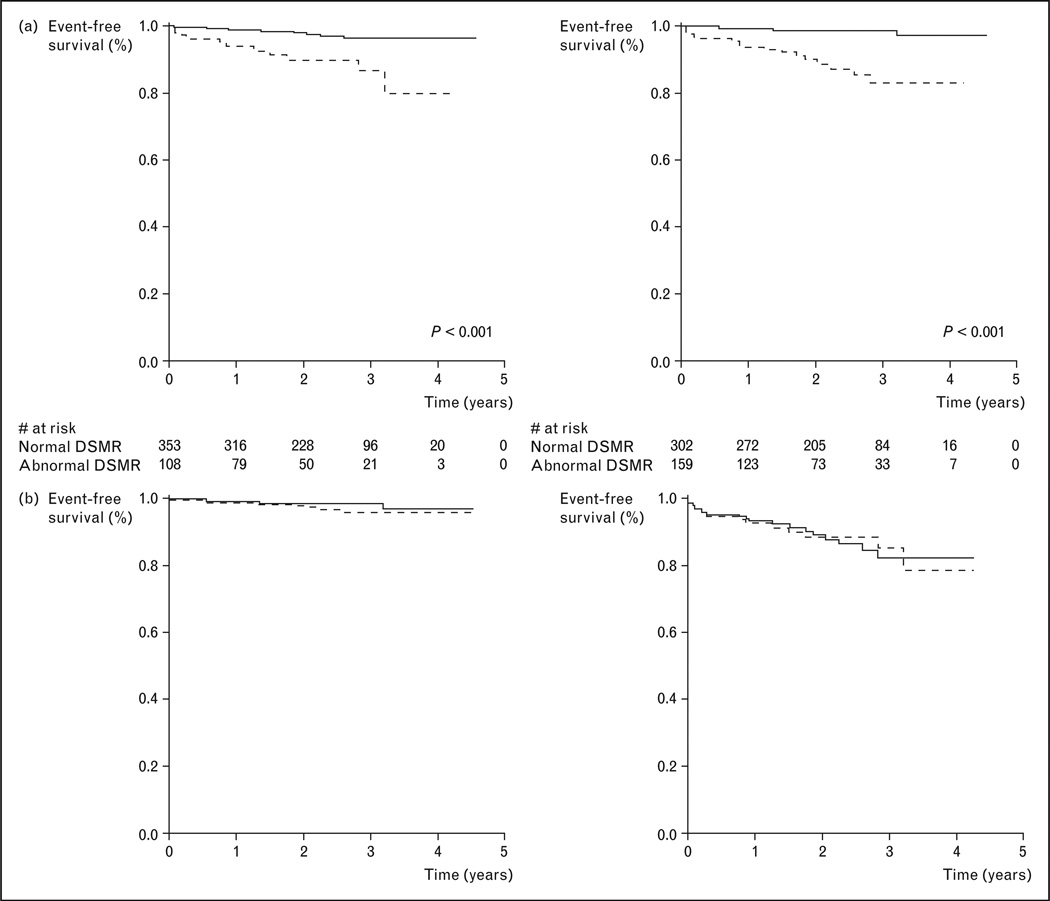
Recent findings: Recent results of the first multicenter multivendor CMR perfusion study demonstrated superior diagnostic utility in detecting CAD by CMR compared with conventional nuclear single-photon emission computed tomography. Several large clinical trials provide additional evidence indicating the strong prognostic implications when CMR perfusion was performed in a clinical setting in patients with an intermediate clinical likelihood of CAD. A negative adenosine stress CMR perfusion study conferred a favorable 3-year prognosis towards nonfatal myocardial infarction or cardiac death.

Summary: CMR perfusion imaging during the first pass of gadolinium-based contrast agents has undergone many technical improvements and levels of clinical validation. Rapidly increasing clinical use worldwide over the last years in diagnosing chest pain syndromes supports the role of CMR in a comprehensive and efficient noninvasive assessment of altered myocardial physiology in CAD.


Figures

Figure 1
Figure 1. Schematic illustration of cardiac magnetic resonance data acquisition in myocardial perfusion imaging during the first-pass transit of a tight intravenous bolus injection of gadolinium-based contrast agent
Images are acquired with an electrocardiogram-triggered, multislice imaging protocol, for example, in the short-axis orientation as in this example, and at a rate equal to the heart rate, to achieve adequate temporal resolution. A visual or quantitative analysis of the signal intensity changes in the myocardium allows the identification of hypoperfused areas. The signal intensity changes shown in the graph of this figure were determined for a myocardial region and a region in the center of the left ventricle (LV), with the latter serving as arterial reference or input function. For a quantitative analysis, this arterial input is used as ‘reference’ for analysis of the myocardial enhancement.
Figure 2
Figure 2. Depiction of a clinical case of a 55-year-old man who experienced recurrent chest pain several years after suffering a ST-elevation myocardial infarction
Only matching short-axis location of adenosine stress CMR perfusion (left), diastolic frame of cine function (middle), and late enhancement for infarction (right) were shown. The patient had evidence of a full-thickness anterior myocardial infarction as demonstrated by late enhancement (arrows in the right image) with thinned anterior wall (arrows in middle image), during first-pass perfusion, there was an extensive subendocardial perfusion defect matching the location of the infarction but also extended into surrounding segments with preserved myocardial thickness and noninfarcted segments (arrows in the left image). This patient had returned as a result of the attempt to stent the proximal LAD had failed and this was confirmed on subsequent X-ray angiography. Matching locations of the different components of CMR and the high spatial resolution of CMR perfusion contributed to this study. CMR, cardiac magnetic resonance; LAD, left anterior descending artery.
Figure 3
Figure 3. Case example from the MR-IMPACT study
A 47-year-old patient is shown 2 months after successful stenting of the LAD and experienced mild angina. The perfusion CMR study during hyperemia (at 0.1 mmol/kg gadolinium – DTPA) demonstrates a perfusion deficit in the subendocardium of the lateral wall (b and c; arrow head) appreciated by all three readers. SPECT in this patient was positive for the presence of CAD for one reader only. Coronary X-ray angiography demonstrated a significant stenosis in the circumflex coronary artery. Perfusion in the anterior wall was assessed correctly by both techniques (normal perfusion) despite a stent in the LAD. CAD, coronary artery disease; DTPA, diethylenetriaminepentaacetic acid; LDA, left anterior descending artery; MR-IMPACT, Magnetic Resonance Imaging for Myocardial Perfusion Assessment in Coronary Artery Disease Trial; SPECT, single-photon emission computed tomography. Reproduced with permission from [13].
Figure 4
Figure 4. Kaplan–Meier survival curves illustrating the strong prognostic value of CMR perfusion imaging in a study of 513 patients who were followed for a median of 2.3 years
Dobutamine stress CMR function and adenosine stress perfusion achieved similar prognostic values. (a) ——, Normal DSMR; -----, abnormal DSMR; ——, normal magnetic resonance perfusion; -----, abnormal magnetic resonance perfusion. (b) -----, Normal DSMR; ——, normal magnetic resonance perfusion; -----, abnormal DSMR; ——, abnormal magnetic resonance perfusion. CMR, cardiac magnetic resonance; DSMR, dobutamine stress magnetic resonance imaging. Reproduced with permission from [23].

Similar articles

Cited by

References

    1. Wischgoll T, Meyer J, Kaimovitz B, et al. A novel method for visualization of entire coronary arterial tree. Ann Biomed Eng. 2007;35:694–710. - PubMed
    1. Fayad ZA, Fuster V, Fallon JT, et al. Noninvasive in vivo human coronary artery lumen and wall imaging using black-blood magnetic resonance imaging. Circulation. 2000;102:506–510. - PubMed
    1. Worthley SG, Helft G, Fayad ZA, et al. Cardiac gated breath-hold black blood MRI of the coronary artery wall: an in vivo and ex vivo comparison. Int J Cardiovasc Imaging. 2001;17:195–201. - PubMed
    1. Maintz D, Ozgun M, Hoffmeier A, et al. Selective coronary artery plaque visualization and differentiation by contrast-enhanced inversion prepared MRI. Eur Heart J. 2006;27:1732–1736. - PubMed
    1. Bassingthwaigthe JB, Raymond GR, Chan JIS. Principles of tracer kinetics. In: Zaret BL, Beller GA, editors. Nuclear cardiology: state of the art and future directions. St. Louis: Mosby-Year Book; 1993. pp. 3–23.